
Describe SMARCB1 deficient tumors
SMARCB1 deficient tumors INI1 loss
SMARCB1 INI1 rhabdoid tumor pathology
SMARCB1 INI1 loss rhabdoid tumor histology immunohistochemistry
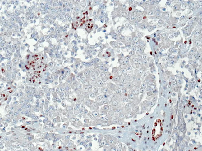
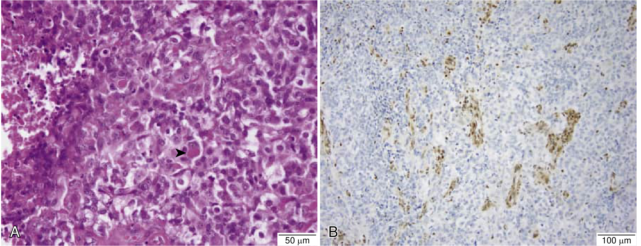
Immunohistochemistry for INI1 (SMARCB1) was performed on a renal tumor tissue section. The scan shows complete loss of nuclear INI1 expression in the malignant rhabdoid cells, with intact INI1 staining in admixed stromal cells and in benign renal tubular epithelium serving as internal positive controls. The tumor cells display classic rhabdoid morphology, including large polygonal cells with abundant eosinophilic or amphophilic cytoplasm, vesicular nuclei, prominent nucleoli, and high mitotic activity embedded in a fibrous to myxoid stroma. Immunostaining pattern helps distinguish SMARCB1-deficient rhabdoid tumors from other renal neoplasms. This INI1 loss is characteristic of rhabdoid tumors and forms part of a broader SMARCB1-deficient tumor spectrum, which includes atypical teratoid/rhabdoid tumors (ATRT) and extrarenal rhabdoid/teratoid tumors; however, roughly twenty percent of INI1-deficient rhabdoid tumors lack detectable SMARCB1 gene alterations, implying alternative inactivation mechanisms. Clinically, INI1 deficiency portends aggressive behavior and necessitates integrated diagnostic workup, including molecular studies. The image demonstrates the diagnostic significance of INI1 immunostaining for accurate classification, prognostication, and management decisions in pediatric and adult renal tumors. This biomarker supports differential diagnosis when histology is ambiguous and guides genetic counseling and potential targeted therapeutic considerations in SMARCB1-related neoplasms. Correlation with patient age, tumor stage, and ancillary tests enhances utility.
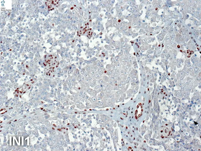
Immunohistochemistry image of a renal rhabdoid tumor (RTK) demonstrating loss of nuclear INI1 (SMARCB1) staining in tumor cells with preserved INI1 in endothelial and inflammatory cells, providing an internal positive control. Morphologically, the lesion shows sheets of rhabdoid cells with large polygonal shapes, eccentrically placed nuclei, prominent nucleoli, and abundant cytoplasm; paranuclear inclusions may be present. The tumor exhibits an overlapping immunophenotype: vimentin positivity with pan-c cytokeratin and EMA expression, compatible with the dual mesenchymal-epithelial nature of RTKs. In contrast, the tumor cells are negative for smooth muscle actin (SMA), desmin, and S‑100 protein. Myogenin and neuroendocrine markers can be variably expressed in some cases, underscoring potential immunophenotypic overlap with other renal neoplasms. INI1 loss is a defining feature of SMARCB1-deficient tumors, though rare RTKs retain INI1 and SMARCA4 (BRG-1) inactivation can occur with BRG-1 loss. Clinically, INI1 deficiency strongly supports a diagnosis of RTK and carries prognostic implications due to aggressive behavior. The image also notes that renal medullary carcinoma can display INI1 loss, highlighting overlapping but distinct SWI/SNF-deficient entities. This IHC pattern aids in differential diagnosis from Wilms tumor and other renal neoplasms, informs molecular testing strategy, and guides research into targeted therapies targeting the SWI/SNF complex.
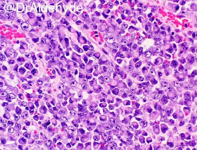
Histopathology slide of a rhabdoid tumor showing the classic INI1/SMARCB1-deficient rhabdoid phenotype on hematoxylin-eosin stained tissue. Bright-field microscopy reveals loosely cohesive sheets of monomorphic tumor cells with eccentrically placed vesicular nuclei, prominent nucleoli, and intracytoplasmic eosinophilic inclusions. Cells may adopt a plasmacytoid or rhabdomyosarcomatous appearance. The pattern is highly characteristic but not entirely specific, and SMARCB1/INI1 loss by immunohistochemistry supports the diagnosis. Rhabdoid tumors are aggressive neoplasms most common in infancy and childhood, typically arising in the kidney or central nervous system (atypical teratoid/rhabdoid tumor), but extrarenal and extra-CNS sites have been described. Clinically relevant associations include rapid progression and poor prognosis, requiring prompt multimodal therapy. In differential diagnosis, focal rhabdoid features can complicate other renal neoplasms such as Wilms tumor, mesoblastic nephroma, and renal cell carcinoma, making accurate IHC and molecular testing essential. Diagnostic significance centers on recognizing INI1-deficient rhabdoid morphology, which guides prognosis and informs genetic counseling. This image serves educational purposes for pathologists, clinicians, and trainees, enabling histologic-pathologic correlation, differential diagnosis, and study of SMARCB1-related tumorigenesis across pediatric and, occasionally, adult populations. Correlate with radiology and clinical data for comprehensive diagnosis and treatment planning; consider SMARCB1 sequencing in ambiguous cases. This enhances diagnostic accuracy and patient management.
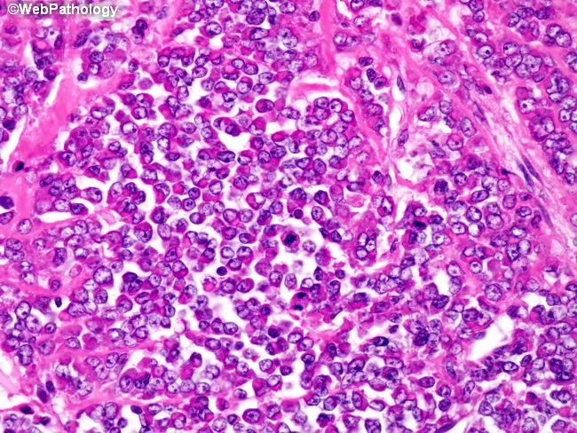
Histopathology image of a pediatric renal rhabdoid tumor captured with light microscopy on an H&E-stained renal tumor biopsy. The section shows high cellularity with sheets and clusters of tumor cells embedded in scant, fibrous stroma. Tumor cells are oval to polygonal with markedly eosinophilic to amphophilic cytoplasm, and nuclei are large, round to elongated with prominent nucleoli. Occasional rhabdoid features include eccentric, vesicular nuclei and prominent, glassy cytoplasm; mitotic activity is brisk with frequent atypical mitoses. There may be necrotic foci. The overall pattern is highly infiltrative, sometimes with perivascular pseudo-rosettes described in some renal rhabdoid tumors. The image may reflect loss of INI1 (SMARCB1) expression on immunohistochemistry, a hallmark of rhabdoid tumor, though not visible on H&E alone. Clinically, renal rhabdoid tumors are aggressive and often present with advanced disease including lymph node, liver, and lung metastases; prognosis is poor with 5-year survival around 15-25%, and less than 20% presenting at stage I. Treatment typically involves multimodal therapy: surgery, chemotherapy, and radiotherapy; investigational SMARCB1-targeted epigenetic therapies are under clinical trial. This histology is essential for differential diagnosis against Wilms tumor with rhabdoid features, renal medullary carcinoma, and other renal sarcomas, guiding patient management and prognosis. Important educational tool for pathology.
SMARCB1-Deficient Tumors
The Gene and Protein
The Spectrum of Tumors
1. Malignant Rhabdoid Tumor of the Kidney (RTK)
- Epidemiology: The most aggressive and lethal childhood renal tumor; accounts for ~2% of pediatric renal neoplasms. Median age at diagnosis is under 16 months.
- Molecular basis: Characterized by loss-of-function of SMARCB1/INI1/SNF5/BAF47 at 22q11.2; both alleles are inactivated. Germline alterations found in ~1/3 of patients, indicating hereditary predisposition.
- Histology: Solid, monotonous sheets of large cells with abundant cytoplasm, eccentric oval/kidney-shaped nuclei, vesicular chromatin, prominent nucleoli, and characteristic eosinophilic paranuclear cytoplasmic inclusions. Intense vimentin immunoreactivity; may also express cytokeratin and desmin.
- IHC hallmark: Complete loss of nuclear INI1 staining in tumor cells, with retained staining in background stromal/vascular cells (internal positive control).
- Clinical features: Palpable abdominal mass, hematuria, heterogeneous solid renal mass with necrosis/hemorrhage on CT; hilum typically involved.
- Spread: Lung metastasis in 56% at diagnosis; up to 15% develop CNS lesions.
- Prognosis: Overall poor; younger age (<1 year) is a major adverse factor. No standard chemotherapy has proven highly effective.
2. Atypical Teratoid/Rhabdoid Tumor (AT/RT)
- Definition: An embryonal CNS neoplasm, WHO Grade IV.
- Age: Mostly infants and young children; one of the most aggressive human tumors. Average survival 6-8 months.
- Common misdiagnosis: Frequently misidentified as medulloblastoma due to prominent small round blue cell component - but the distinction is biologically critical because AT/RT does not respond to standard medulloblastoma therapy.
- Defining histological cell: The rhabdoid cell - enlarged, eccentric oval to kidney-shaped nucleus, vesicular chromatin, eosinophilic paranuclear inclusion (highlighted by vimentin IHC).
- Polyphenotypic immunoprofile: Coexpresses EMA, smooth muscle actin (SMA), vimentin, and various neuroepithelial markers - a diagnostic red flag pointing away from medulloblastoma.
- Genetics: Monosomy 22 or 22q deletions with SMARCB1 mutation in the majority. A minority harbor SMARCA4 (BRG1) mutations with retained SMARCB1 expression.
- Diagnosis confirmed by: Loss of nuclear SMARCB1 on IHC (monoclonal antibodies now in widespread clinical use).
- Location: Posterior fossa most common in infants; supratentorial in older children.
3. Extrarenal Malignant Rhabdoid Tumor (EMRT)
- Rhabdoid tumors arising outside the kidney (soft tissue, liver, mediastinum, and other sites) with identical morphology and SMARCB1 deletion.
- The chromosomal alteration (22q11 loss, SMARCB1) is considered a characteristic genetic defect for rhabdoid tumors of all sites.
- May present as synchronous renal + CNS tumors (rhabdoid tumor predisposition syndrome in germline carriers).
4. Epithelioid Sarcoma
- Types: Classic (distal) and proximal (large-cell) variants.
- Distal type: Nodular proliferation of spindle-to-epithelioid cells with eosinophilic cytoplasm and small uniform nuclei; central necrosis within nodules creates a pseudogranulomatous pattern.
- Proximal type: Large epithelioid cells with vesicular nuclei, prominent nucleoli; rhabdoid morphology more common.
- IHC: Positive for keratins, EMA, CD34 (unusual combination); negative for S100, HMB45, CD31.
- Molecular: Distinct deletions/mutations of SMARCB1/INI1 at 22q11-12 → loss of INI1 IHC expression is characteristic of both variants.
- Behavior: Highly infiltrative, along tendons/nerves/fascia; skip lesions common. Local recurrence rate is high. Metastases in 40-50% (lung, lymph nodes). 5-year OS: 60-80%; 10-year OS: 40-60%.
- Treatment advance: Tazemetostat (EZH2 inhibitor) - an oral drug approved for INI1-loss epithelioid sarcoma based on an international Phase 2 basket trial, showing improved outcomes in metastatic/locally advanced disease.
5. SMARCB1-Deficient Renal Medullary Carcinoma (RMC)
- An aggressive renal cell carcinoma uniquely associated with sickle cell hemoglobinopathies (especially sickle cell trait, HbSC) - particularly in teenagers and young adults.
- Previously called "renal medullary carcinoma"; now classified as SMARCB1-deficient RCC in the Harrison's framework.
- Growth patterns include cribriform, reticular, sarcomatoid, adenoid, and microcystic.
- Molecular: focal SMARCB1 deletions, +8q, 22q abnormalities.
6. Other SMARCB1-Deficient Entities
| Tumor | Key Features |
|---|---|
| Schwannomatosis | Germline SMARCB1 mutations; multiple schwannomas without vestibular component; no NF2 germline mutation |
| Cribriform neuroepithelial tumor (CRINET) | Rare CNS tumor; SMARCB1-deficient |
| Sinonasal undifferentiated carcinoma (some) | Small subset SMARCB1-deficient |
| SMARCB1-deficient undifferentiated thoracic tumors | Emerging category in thorax/mediastinum |
| SWI/SNF-deficient undifferentiated pancreatic carcinoma | Clinicopathologically distinct; very aggressive |
Unifying Molecular Mechanism
- Removes a brake on EZH2 (the catalytic subunit of PRC2 polycomb complex), allowing it to trimethylate H3K27 and silence many tumor-suppressor loci.
- Dysregulates cyclin D1 and CDK4/6 pathways (cell cycle entry).
- Impairs p16/CDKN2A and RB pathway regulation.
- Alters MYC target gene expression (especially in AT/RT).
Diagnostic IHC Summary
| Marker | Finding |
|---|---|
| INI1/SMARCB1 IHC | Complete nuclear loss in tumor cells; internal positive control in vessels/stroma |
| Vimentin | Strongly positive |
| EMA, keratins | Variably positive (especially AT/RT, epithelioid sarcoma) |
| CD34 | Positive in epithelioid sarcoma |
| SMA | Positive in AT/RT |
Treatment Principles
- Surgery: Radical resection is cornerstone for all solid tumors in this group.
- Chemotherapy: Intensive multi-agent regimens (e.g., vincristine, actinomycin D, doxorubicin for RTK); responses are often poor.
- Radiation: Used adjuvantly in some protocols.
- EZH2 inhibition (Tazemetostat): FDA-approved for locally advanced or metastatic epithelioid sarcoma with INI1 loss. Under investigation for other SMARCB1-deficient tumors including AT/RT. The Lancet Oncology 2020 Phase 2 trial established efficacy.
- Germline testing: Recommended for patients (especially children) with any SMARCB1-deficient tumor given the ~30% germline rate in RTK; germline carriers have rhabdoid tumor predisposition syndrome.
Key Recent Literature
- SWI/SNF-deficient tumors of the CNS: An update - Hasselblatt M et al., Clin Neuropathol 2024 (PMID 37969088) - reviews AT/RT and related CNS entities with updated WHO 2021 classification.
- Cytopathologic and histopathologic characteristics of SMARCB1-deficient neoplasm - Sanchez SI et al., Hum Pathol 2025 (PMID 41224025) - correlates morphology with IHC and molecular findings across the spectrum.
- SWI/SNF complex-deficient undifferentiated carcinoma of the pancreas - Yavas A et al., Mod Pathol 2024 (PMID 39094734) - characterizes an emerging entity.