
Microbiology of dental plaque
Reading File
Dental plaque is a structured biofilm containing hundreds of bacterial species, organized in a specific succession pattern. Let me search the medical library for comprehensive coverage.
Searching sources...
Finding Sources
Finding Sources
Reading File
Reading File
Reading File
Reading File
Loading Image
I now have comprehensive content from two authoritative microbiology textbooks. Let me compile the full answer.
Microbiology of Dental Plaque
What Is Dental Plaque?
Dental plaque is an adherent bacterial biofilm that forms on the tooth surface, composed almost entirely of bacteria derived from the resident oral microbiota. It is the most prevalent and densest human biofilm. Plaque is the primary cause of dental caries, gingivitis, and periodontitis - which together make it the central pathologic agent of oral disease.
- Sherris & Ryan's Medical Microbiology, 8th Ed.
- Jawetz, Melnick & Adelberg's Medical Microbiology, 28th Ed.
The Dental Pellicle: Foundation of Plaque
Before bacteria colonize, a dental pellicle forms on the tooth surface. This is a thin, acellular organic film composed of proteins and glycoproteins derived from saliva and oral secretions (including statherin, sialylated mucins, alpha-amylase, salivary agglutinin, and proline-rich proteins). Plaque biofilm evolves in relation to the pellicle, not the mineralized tooth itself.
Stages of Plaque Formation
Plaque forms in a sequential, layered process at two levels:
- Anatomic location - starts supragingival, then extends subgingival
- Bacterial succession - early colonizers followed by late colonizers
Stage 1: Early Colonizers (Gram-positive bacteria)
These organisms use ionic, hydrophobic, and lectin-like (carbohydrate-binding) surface interactions to adhere to pellicle components.
| Organism | Role |
|---|---|
| Streptococcus sanguis | Prototype early colonizer |
| S. mutans, S. mitis, S. salivarius, S. oralis, S. gordonii | Additional early streptococci |
| Haemophilus parainfluenzae | Early colonizer |
| Actinomyces naeslundii | Early colonizer, bridges to late colonizers |
| Lactobacilli | Present in early plaque |
Stage 2: Late Colonizers (Gram-negative anaerobes)
If early colonizers are undisturbed, late colonizers appear within 2-4 days. These are primarily Gram-negative anaerobes and spirochetes.
| Organism | Notes |
|---|---|
| Fusobacterium nucleatum | Central "bridging" organism in the biofilm |
| Porphyromonas gingivalis | Key periodontal pathogen |
| Prevotella species | Subgingival plaque |
| Veillonella atypica | Gram-negative anaerobic coccus |
| Treponema denticola | Anaerobic spirochete |
| Actinobacillus actinomycetemcomitans | Late colonizer, aggressive periodontitis |
| Propionibacterium acnes | Bridges early and late zones |
| Campylobacter, Capnocytophaga, Eikenella | Predominantly subgingival |
In total, mature dental plaque is thought to harbor 300-400 bacterial species.
Dental Plaque Biofilm - Colonization Diagram
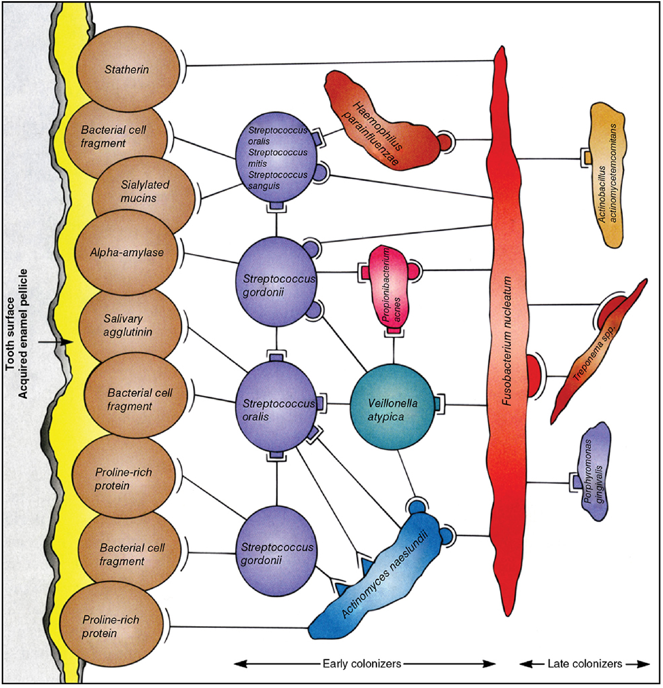
Dental plaque biofilm: Early colonizers (streptococci, Actinomyces) bind to enamel pellicle proteins; late colonizers (Fusobacterium, Porphyromonas, Treponema) attach to the early colonizer layer. Fusobacterium nucleatum acts as the central bridging organism.
Adhesion Mechanisms: How the Biofilm Holds Together
- Ionic and hydrophobic interactions between bacteria and pellicle
- Lectin-like surface structures (carbohydrate-binding proteins) on bacterial surfaces
- High-molecular-weight extracellular glucan polymers (produced mainly by S. mutans and Actinomyces) act as a cement binding the biofilm together
- Sucrose is used by S. mutans to synthesize dextrans and levans (polyglycans) via transferase enzymes - these contribute to aggregation and accumulation on tooth surface and serve as nutrient reserves for other plaque bacteria
Supragingival vs. Subgingival Plaque
| Feature | Supragingival | Subgingival |
|---|---|---|
| Location | Above gum line | Below gum line, in sulcus/periodontal pockets |
| Predominant organisms | Gram-positive aerobes/facultatives | Gram-negative anaerobes + spirochetes |
| Structure | No distinct nonadherent zone | Thin adherent layer (tooth-attached) + nonadherent zone (facing epithelium) |
| Disease association | Dental caries, gingivitis | Periodontitis, necrotizing periodontal disease |
Subgingival plaque additionally includes Campylobacter, Capnocytophaga, and Eikenella species in addition to the late colonizers described above.
Preferred Locations for Plaque Accumulation
Plaque accumulates in non-self-cleansing areas not swept clean by chewing:
- Pits and fissures of teeth
- Along gingival margins
- Interproximal (between teeth)
These are exactly the locations where caries, gingivitis, and periodontitis are most common and severe.
Plaque and Dental Caries
Streptococcus mutans is the primary cariogenic organism:
- Plaque bacteria metabolize dietary sugars (glucose, fructose, sucrose, lactose, maltose) through glycolysis, generating organic acids
- Acids demineralize tooth enamel - this is the first step of caries
- Foods with high sucrose content that adhere to teeth (long oral clearance) are most cariogenic
- S. mutans has a metabolic advantage - it metabolizes sucrose more efficiently than other oral bacteria
- Dentin decomposition follows enamel breakdown
Plaque and Periodontal Disease
Porphyromonas gingivalis plays a key role in periodontitis by impairing innate immunity, altering the growth and development of the entire biofilm and triggering a breakdown in the homeostatic host-microbiota relationship in the periodontium. It is primarily the host inflammatory response (not the bacteria directly) that causes destruction of periodontium and eventual tooth loss.
Archaeal DNA (genus Methanobrevibacter) has also been correlated with severity of periodontal disease, though a causative association has not been established.
Systemic Implications
Oral biofilm organisms can cause systemic disease when displaced from the mouth:
- Infective endocarditis - Streptococcus species from plaque
- Bacteremia in granulocytopenic patients - Capnocytophaga species (fusiform, Gram-negative, gliding anaerobes) and Rothia dentocariosa (pleomorphic, aerobic, Gram-positive rods)
Control of Dental Plaque
| Method | Mechanism |
|---|---|
| Mechanical removal (brushing, flossing) | Physical disruption of biofilm - primary method |
| Chlorhexidine (0.12% rinse) | Bis-biguanide antiseptic; binds tooth surfaces, inhibits plaque |
| Fluoride | Enhances acid resistance of enamel |
| Triclosan, phenolic compounds, essential oils | Adjunct antimicrobial additives in toothpaste/rinse |
| Limiting sucrose intake | Reduces substrate for acid production and glucan synthesis |
| Scaling/curettage | Removes calculus (calcified plaque) |
Dental plaque cannot be effectively removed by chemical/enzymatic means alone, and prophylactic antibiotics for plaque prevention are not clinically justified (though patients on long-term antibiotics for other reasons show lower caries rates).
Sources:
- Sherris & Ryan's Medical Microbiology, 8th Ed., Chapter 41
- Jawetz, Melnick & Adelberg's Medical Microbiology, 28th Ed., Chapter 10
Microbiology of dental plaque in bds 2nd year university exam
Finding Sources
Searching the Web
BDS 2nd year microbiology dental plaque exam questions university syllabus
Reading File
Finding Sources
Reading File
Reading File
Loading Image
Reading File
Reading File
Loading Image
I now have everything needed to write a complete, exam-focused BDS 2nd year answer. Here it is:
Microbiology of Dental Plaque
BDS 2nd Year Exam Answer (Long Essay / 8-10 marks format)
Definition
Dental plaque is an adherent bacterial biofilm that forms on the tooth surface, composed almost entirely of bacteria derived from the normal resident microbiota of the oral cavity. It is the most prevalent and densest human biofilm, and the primary etiological agent of dental caries and periodontal disease.
Dental Pellicle - The Foundation
Before bacteria colonize, an acquired enamel pellicle forms on the tooth surface. It is:
- A thin, acellular, organic film
- Composed of proteins and glycoproteins from saliva and oral secretions
- Contains: statherin, sialylated mucins, alpha-amylase, salivary agglutinin, proline-rich proteins
Key point: Plaque forms in relation to the pellicle, NOT on the mineralized tooth surface directly.
Stages of Plaque Formation
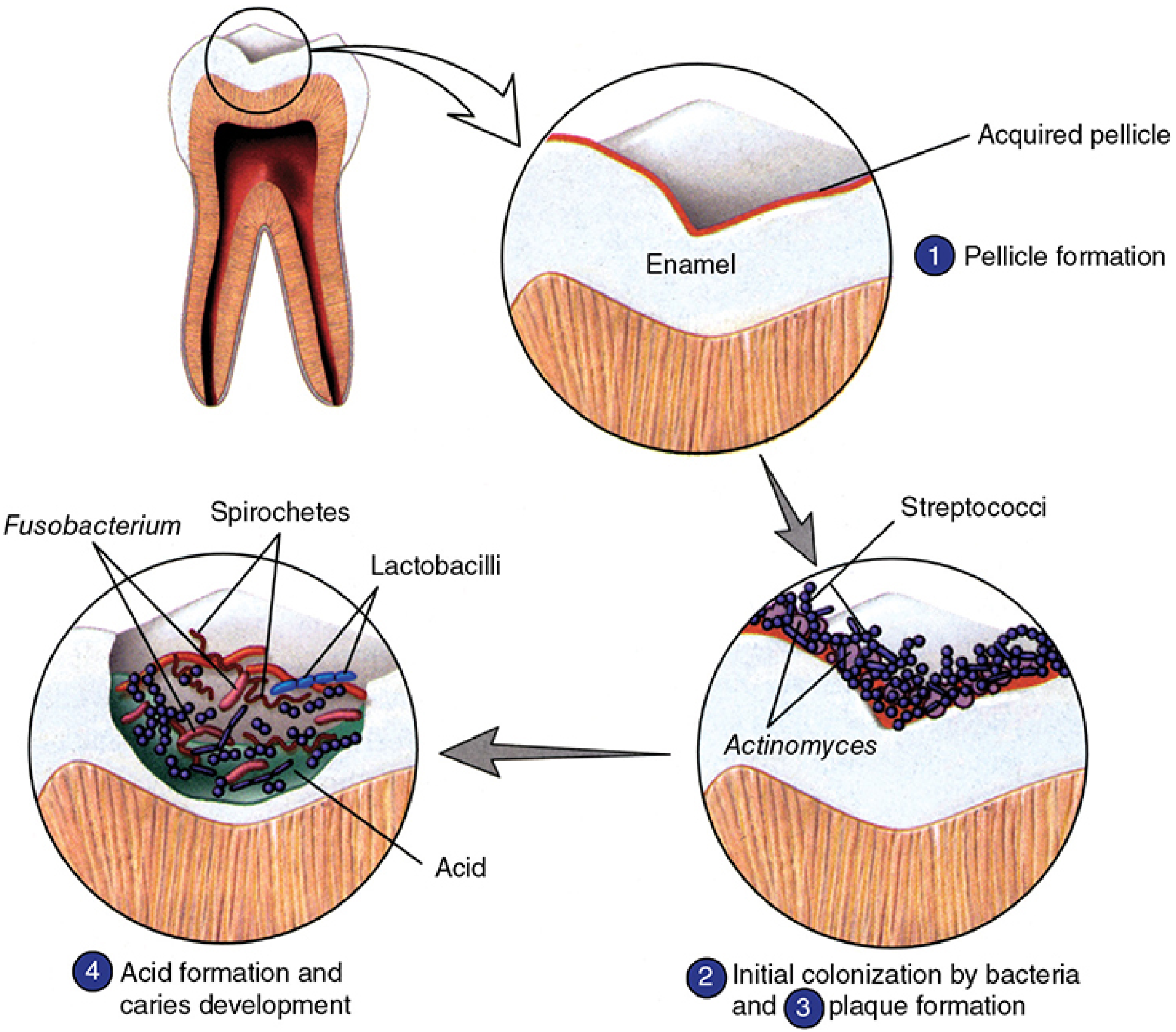
Plaque formation occurs at two levels:
- Anatomic level - supragingival first, then extends subgingivally
- Bacterial succession level - early colonizers followed by late colonizers
Microbial Succession in Dental Plaque
Stage 1 - Early Colonizers (Gram-positive, days 1-4)
These organisms bind to pellicle using ionic, hydrophobic, and lectin-like (carbohydrate-binding) interactions.
| Organism | Notes |
|---|---|
| Streptococcus sanguis | Prototype early colonizer |
| S. mutans | Most cariogenic; produces dextrans |
| S. mitis, S. salivarius, S. oralis, S. gordonii | Other early streptococci |
| Haemophilus parainfluenzae | Gram-negative early colonizer |
| Actinomyces naeslundii | Acts as bridge to late colonizers |
| Lactobacilli | Contribute to acidogenesis |
Stage 2 - Late Colonizers (Gram-negative anaerobes, after day 4)
If early colonizers are undisturbed for 2-4 days, late colonizers appear. These are primarily Gram-negative anaerobes and spirochetes that bind to the early colonizer layer.
| Organism | Significance |
|---|---|
| Fusobacterium nucleatum | Central "bridge" organism - connects early and late colonizers |
| Porphyromonas gingivalis | Key pathogen in chronic periodontitis |
| Prevotella spp. | Subgingival plaque pathogen |
| Veillonella atypica | Anaerobic Gram-negative coccus |
| Treponema denticola | Anaerobic spirochete |
| Aggregatibacter actinomycetemcomitans | Aggressive periodontitis |
| Campylobacter, Capnocytophaga, Eikenella | Subgingival flora |
Mature dental plaque contains 300-400 bacterial species in total.
Biofilm Diagram
Adhesion Mechanisms
| Mechanism | Details |
|---|---|
| Ionic & hydrophobic interactions | Early colonizers bind to pellicle proteins |
| Lectin-like surface structures | Carbohydrate-binding proteins on bacterial surfaces |
| Glucan polymers (dextrans, levans) | Produced by S. mutans via glucosyltransferase; act as cement holding biofilm together |
Sucrose is the key substrate - S. mutans uses it to synthesize extracellular dextrans and levans, which increase adhesion and serve as stored carbohydrate reserves for other plaque bacteria.
Supragingival vs. Subgingival Plaque
| Feature | Supragingival | Subgingival |
|---|---|---|
| Location | Above gum line | Below gum line in sulcus/periodontal pockets |
| Predominant flora | Gram-positive facultatives | Gram-negative strict anaerobes + spirochetes |
| Structure | No distinct nonadherent zone | Adherent layer (tooth side) + nonadherent zone (epithelial side) |
| Disease caused | Dental caries, gingivitis | Periodontitis, necrotizing periodontal disease |
Role of Dental Plaque in Dental Caries
Pathogenesis of caries:
- S. mutans and other plaque bacteria metabolize dietary sugars (sucrose > glucose > fructose > lactose) via glycolysis
- Produce organic acids (mainly lactic acid)
- At pH below 5.5 - enamel hydroxyapatite is demineralized
- Lesion progresses inward: Enamel → Dentin → Pulp
Why S. mutans is most cariogenic:
- Metabolizes sucrose more efficiently than other oral bacteria
- Produces dextransucrase (glucosyltransferase) - synthesizes insoluble dextran for biofilm adherence
- Synthesizes intracellular glycogen - prolongs acidogenesis even after dietary carbohydrate is exhausted
- Other contributors: S. sobrinus, S. salivarius, Lactobacillus acidophilus, L. casei, Actinomyces viscosus, A. naeslundii
Three factors needed for caries (Keyes triad concept):
- Susceptible host/tooth
- Appropriate cariogenic microflora
- Fermentable carbohydrate substrate
Role of Dental Plaque in Periodontal Disease
Gingivitis (reversible):
- Develops within 2 weeks of failure to clean teeth
- Inflammatory infiltrate (PMNs, lymphocytes, plasma cells) in gingival connective tissue
- Collagen loss from inflamed connective tissue
- No direct bacterial invasion of tissue in early stages
Chronic Periodontitis (irreversible):
- Caused by subgingival plaque in the gingival crevice/sulcus
- Key organisms: Porphyromonas gingivalis, Treponema denticola, Aggregatibacter actinomycetemcomitans (the "red complex")
- P. gingivalis impairs innate immunity, altering the entire biofilm and triggering dysregulated host inflammation
- It is primarily the host inflammatory response (not bacteria directly) that destroys periodontium and causes tooth loss
- Virulence factors of periodontal pathogens include: collagenase, proteases, leukotoxin, endotoxin (LPS), fimbriae
Control of Dental Plaque
| Method | Mechanism |
|---|---|
| Mechanical removal (brushing, flossing) | Primary method - physically disrupts biofilm |
| Chlorhexidine (0.12% rinse) | Bis-biguanide; binds tooth surface, inhibits plaque growth |
| Fluoride (water, toothpaste, topical) | Replaces OH⁻ with F⁻ in hydroxyapatite → fluorapatite → acid-resistant; also inhibits demineralization and promotes remineralization |
| Scaling and root planing | Removes calculus (calcified plaque) from subgingival surfaces |
| Dietary modification | Reduce sucrose intake; limit sticky retentive foods |
| Triclosan, phenolic compounds, essential oils | Adjuncts in toothpastes and rinses |
Dental plaque cannot be effectively removed by chemical/enzymatic means alone. Prophylactic antibiotics for plaque prevention are not clinically justified.
Important Exam Points (Quick Recall)
- Most prevalent human biofilm = dental plaque
- Prototype early colonizer = Streptococcus sanguis
- Most cariogenic organism = Streptococcus mutans
- Bridge/central organism in biofilm = Fusobacterium nucleatum
- Key periodontal pathogen = Porphyromonas gingivalis
- Late colonizers appear in = 2-4 days
- Total species in mature plaque = 300-400
- Extracellular polymers produced by = S. mutans (dextrans via glucosyltransferase)
- Critical pH for enamel demineralization = below 5.5
- Most effective anti-caries measure = fluoridation
Sources: Sherris & Ryan's Medical Microbiology, 8th Ed., Ch. 41; Jawetz, Melnick & Adelberg's Medical Microbiology, 28th Ed., Ch. 10
This is a shared conversation. Sign in to Orris to start your own chat.