
13 yrs old boy with dengue igM positive Explain about dengue symptoms mechanism monitoring in hospital
dengue fever pathophysiology rash clinical features
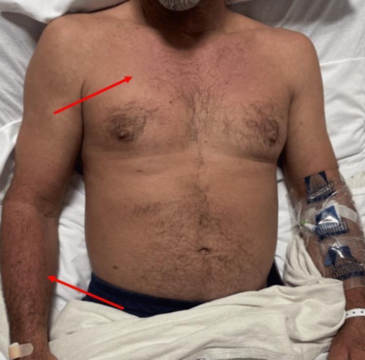
Clinical photograph of a male patient's torso and upper extremities in a hospital setting, illustrating a diffuse, patchy erythematous rash. The skin lesions are most prominent across the upper chest, mid-abdomen, and bilateral arms, with red arrows highlighting the erythematous distribution on the right pectoral region and left forearm. The morphology is consistent with a dissipating maculopapular or morbilliform eruption often seen in systemic viral infections such as Dengue fever. Notable clinical features include the absence of visible scaling and a generalized, non-confluent pattern. On the patient's left forearm, an indwelling intravenous (IV) catheter is secured with a transparent adhesive dressing and tape, and a white hospital identification band is visible on the wrist. This visual evidence supports the clinical assessment of infectious exanthems in the context of systemic illness and thrombocytopenia.
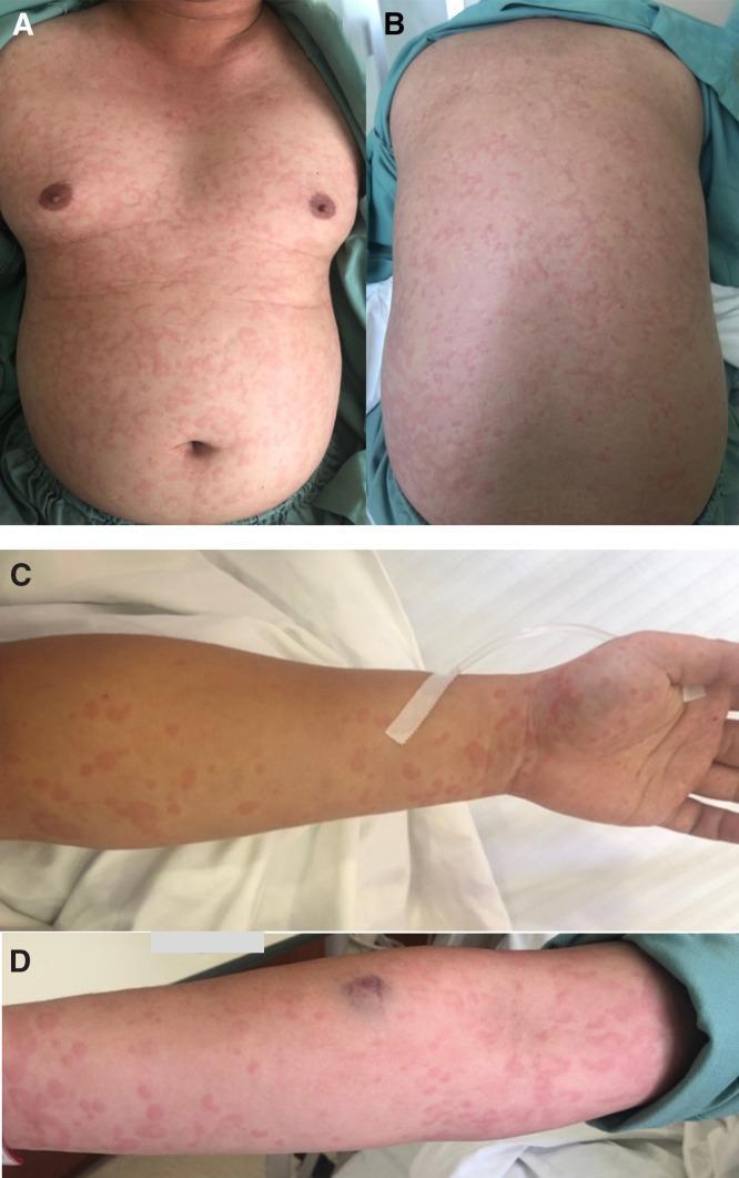
Clinical photograph series showing skin manifestations of dengue fever in a 25-year-old male. Panels A and B display the anterior (chest/abdomen) and posterior (back) trunk, revealing a generalized, pruritic eruption. The rash is characterized by erythematous wheals that are partially confluent, creating a map-like or mottled appearance with areas of normal skin interspersed. Panels C and D show similar lesions on the upper extremities, demonstrating raised, irregularly shaped, pink-to-red wheals across the forearms and wrists. A singular ecchymosis (bruise) is visible on the distal forearm in panel D. The morphology is consistent with urticarial-like lesions frequently observed in the early stages of viral exanthems. The images serve as an educational reference for recognizing atypical dermatological presentations of dengue virus infection, distinguishing them from classic petechial rashes.
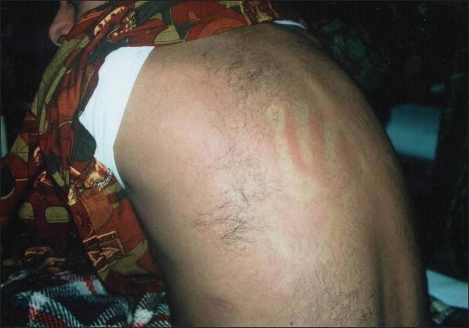
This clinical photograph displays a diffuse exanthematous rash on a patient's torso, characteristic of the later stages of Dengue Fever. The image reveals generalized, confluent erythema that covers a broad anatomical area. A hallmark diagnostic feature is visible: the presence of multiple, small, rounded 'islands of sparing'—pale, unaffected areas of skin that stand out against the erythematous background, often referred to as 'white islands in a sea of red.' Additionally, there is evidence of dermatographism or linear erythematous streaks, likely resulting from pressure or scratching. This cutaneous manifestation typically appears 3–6 days after the onset of fever and represents a key clinical sign for differentiating Dengue Fever from other tropical febrile illnesses. The rash serves as an important educational example of viral exanthems and their unique morphological patterns in infectious disease and dermatology.
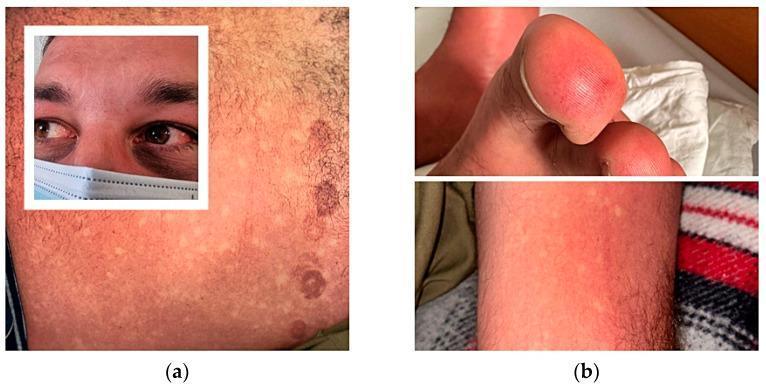
This composite of clinical photographs illustrates the classic dermatological and ocular manifestations of Dengue fever. Panel (a) displays a characteristic confluent erythematous, blanching maculopapular rash on the chest, often described as 'islands of white in a sea of red' due to distinct patches of skin sparing. Additionally, circular ecchymotic lesions are visible on the lateral chest wall, corresponding to pressure sites from ECG electrodes. An inset focuses on the patient's face, showing bilateral subconjunctival hemorrhages and periorbital ecchymoses. Panel (b) illustrates peripheral vascular involvement, showing cutaneous vasculitis of the fingers and toes, characterized by intense erythema and swelling. The lower panel in (b) further demonstrates the diffuse erythematous rash on the lower limb with smaller focal spots of skin sparing. These findings are hallmarks of Dengue Hemorrhagic Fever (DHF) and emphasize the systemic vascular permeability and coagulopathy associated with the virus.
dengue warning signs hemorrhagic fever shock syndrome platelet
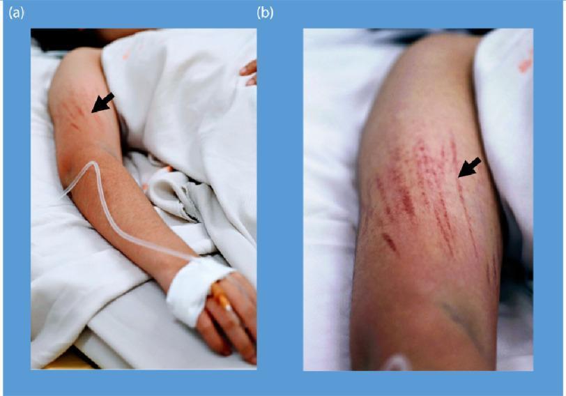
Clinical photograph of a patient's arm demonstrating bleeding manifestations characteristic of Dengue Shock Syndrome (DSS). The image is divided into two panels: (a) shows the full arm in a clinical setting with an intravenous (IV) line secured to the hand, and (b) provides a close-up view of the upper arm. Both panels highlight linear petechiae (indicated by black arrows), which are small, red to purplish hemorrhagic spots arranged in distinct linear patterns. These lesions represent microvascular fragility and are shown following the inflation of a blood pressure cuff (Hess test or capillary fragility test), a common diagnostic maneuver for assessing vascular permeability in dengue fever. The presence of an IV line and hospital bedding contextualizes the severity of the illness, illustrating the systemic plasma leakage and hemostatic dysfunction associated with severe viral hemorrhagic fevers. Key educational concepts include endothelial dysfunction, capillary fragility, and clinical signs of thrombocytopenia or vasculopathy.
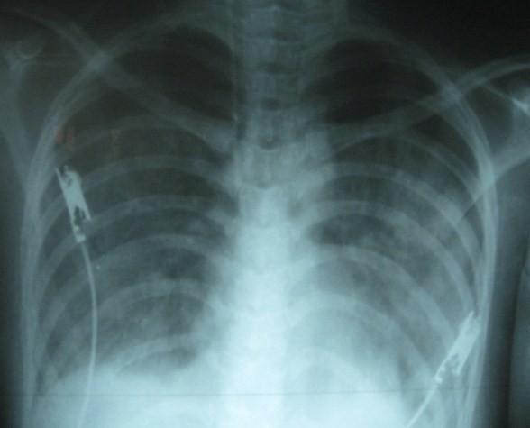
This diagnostic image is an anteroposterior (AP) chest X-ray demonstrating severe pulmonary edema and pleural effusion in a 19-year-old female patient with Dengue Shock Syndrome. The lung fields show bilateral, diffuse, fluffy opacities and increased interstitial markings, characteristic of pulmonary edema. There is significant blunting of the right costophrenic angle, indicating a right-sided pleural effusion. The cardiac silhouette appears moderately enlarged, and the normal clear visualization of pulmonary vasculature is obscured by interstitial and alveolar infiltrates. ECG electrodes and leads are visible on the chest wall, reflecting an acute clinical monitoring setting. This imaging serves as a critical educational example of 'third space' fluid accumulation and capillary leak syndrome secondary to viral hemorrhagic fever, illustrating the rapid progression of respiratory distress during fluid resuscitation in Dengue Shock Syndrome.
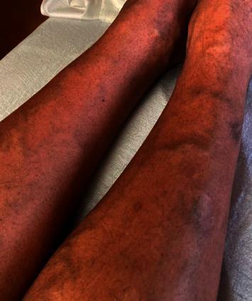
This clinical photograph shows the bilateral lower extremities of a 47-year-old male presenting with a diffuse dermatologic manifestation of thrombocytopenia. The skin demonstrates widespread, non-blanching petechiae, appearing as numerous pinpoint-sized (1-2 mm), reddish-purple macules. These lesions are densely scattered across the anterior and lateral aspects of the shins and calves. In addition to the petechial rash, several larger, coalescent purpuric lesions and subtle ecchymoses are visible, indicating small-vessel bleeding into the dermis. The background skin exhibits generalized erythema. This visual finding is characteristic of severe thrombocytopenia, in this clinical context associated with a viral hemorrhagic fever such as dengue. The image serves as an educational example of the cutaneous signs of low platelet counts and the systemic progression of infectious diseases like dengue fever in a hospital setting.
Dengue in a 13-Year-Old Boy (IgM Positive)
1. The Virus - Background
- Red Book 2021, p. 520
- Sherris & Ryan's Medical Microbiology, p. 646
2. Mechanism / Pathophysiology
Viral Entry and Replication
Antibody-Dependent Enhancement (ADE) - Key Mechanism for Severe Disease
- First infection with one serotype produces lifelong immunity against that serotype only
- Cross-reactive antibodies from the first infection are non-neutralizing against other serotypes
- On second infection with a different serotype, these cross-reactive antibodies actually enhance viral entry into macrophages via Fc receptors - this is ADE
- ADE dramatically increases viral load and triggers a cytokine storm, leading to severe dengue (DHF/DSS)
- This is why severe dengue is more common in children in endemic areas (who may have had prior infection) and in infants born to dengue-immune mothers
Plasma Leakage - The Critical Pathology
- Rising hematocrit (hemoconcentration)
- Hypovolemic shock
- Pleural effusion, ascites
Thrombocytopenia Mechanism
-
Virus directly infects megakaryocytes (platelet precursors), reducing platelet production
-
Cross-reactive antibodies against platelets cause immune-mediated platelet destruction
-
Endothelial activation consumes platelets
-
Result: platelet count can drop below 20,000/µL
-
Robbins Pathologic Basis of Disease, p. 336-337
3. Clinical Symptoms - Three Phases
Phase 1: Febrile Phase (Days 1-3)
| Symptom | Notes |
|---|---|
| Sudden high fever (39-40°C) | Abrupt onset, lasts 2-7 days |
| Severe headache | Frontal/retro-orbital (behind eyes) - classic |
| Myalgia / arthralgia | "Breakbone fever" - intense bone and joint pain |
| Facial erythema, flushing | |
| Nausea / vomiting | |
| Maculopapular rash | Appears days 2-5 |
| Mild bleeding | Petechiae, nosebleed, gum bleed |
| Leukopenia | WBC typically falls |
Phase 2: Critical Phase (Days 3-7, around defervescence)
- Abdominal pain or tenderness
- Persistent vomiting (3+ episodes in 1 hour)
- Clinical fluid accumulation (pleural effusion, ascites)
- Mucosal bleeding
- Lethargy, restlessness, behavioral change
- Liver enlargement >2 cm
- Rapid decline in platelet count with rising hematocrit
Phase 3: Convalescence (Days 7-10)
-
Gradual improvement, fluid reabsorption
-
Secondary rash may appear - "white islands in a sea of red" (areas of sparing amid erythema)
-
Fatigue and bradycardia common
-
Risk of fluid overload as third-space fluid is reabsorbed
-
Red Book 2021, p. 520-521
4. Dengue Severity Classification (WHO 2009)
| Category | Criteria |
|---|---|
| Dengue without warning signs | Fever + 2 of: nausea/vomiting, rash, aches, leukopenia, positive tourniquet test |
| Dengue with warning signs | Above + any warning sign (see Phase 2 above) |
| Severe dengue | Severe plasma leakage (shock/respiratory distress), severe bleeding, or severe organ involvement (AST/ALT ≥1000 IU/L, impaired consciousness, organ failure) |
5. Clinical Images
6. Hospital Monitoring Protocol
Daily Monitoring (Minimum)
| Parameter | Frequency | Why |
|---|---|---|
| Temperature | Every 4-6 hours | Track febrile and defervescence phase |
| Pulse rate & BP | Every 4-6 hours (hourly if warning signs) | Detect early shock |
| Respiratory rate | Every 4-6 hours | Detect pleural effusion |
| Urine output | Every 6-8 hours | Target >0.5 mL/kg/hr; drop indicates shock |
| Capillary refill time | Every 4-6 hours | Peripheral perfusion |
Laboratory Monitoring
| Test | Frequency | Target / Action |
|---|---|---|
| CBC with platelets | Every 12-24 hours (daily); every 6-8 hrs if <50,000 | Platelets <20,000 or rapid fall = danger; Hematocrit rise >20% above baseline = plasma leakage |
| Hematocrit (PCV) | Every 6-12 hours in critical phase | Rising Hct = plasma leak; falling Hct = dilution from reabsorption or occult bleeding |
| Liver function (AST/ALT) | Every 24-48 hours | AST/ALT ≥1000 = severe dengue criterion |
| Serum electrolytes | Daily | Hyponatremia common |
| Blood urea / creatinine | Every 24-48 hours | Renal impairment in shock |
| Coagulation (PT/aPTT) | If bleeding occurs | DIC screen |
| Blood glucose | As needed | Hypoglycemia in hepatitis |
Clinical Monitoring at Bedside
- Tourniquet test (positive = ≥20 petechiae in 1-inch square): assess capillary fragility on admission
- Check for abdominal tenderness and measure liver span daily
- Auscultate chest for pleural effusion (reduced air entry at bases)
- Monitor for abdominal distension (ascites)
- Watch for mucosal bleeding (gums, nose), melena, hematemesis
Fluid Management Monitoring
- Avoid aspirin and NSAIDs (increase bleeding risk) - use paracetamol only for fever
- During febrile phase: encourage oral hydration; IV fluids if unable to tolerate orally
- During critical phase: IV crystalloids (Ringer's lactate / normal saline), titrate to clinical response
- Monitor for fluid overload during convalescence (reabsorption phase) - watch for new respiratory distress, rising pulse, falling Hct
- Refractory shock: may need colloids or blood products
Indications for ICU / Escalation
- Systolic BP <90 mmHg or pulse pressure ≤20 mmHg
- Platelet <20,000/µL with active bleeding
- Hematocrit rise >20% from baseline
- Impaired consciousness
- Respiratory distress / SpO2 <95%
- Persistent vomiting preventing oral hydration
7. Investigations Summary for This 13-Year-Old
| Investigation | Status/Purpose |
|---|---|
| Dengue IgM | Already positive - confirms infection from day 3-5 |
| Dengue NS1 antigen | Useful in first 7 days (may be negative now if late) |
| Dengue IgG | If positive alongside IgM = secondary infection (higher risk of severe dengue) |
| RT-PCR | Most specific; detectable days 1-7 only |
| CBC daily | Essential monitoring |
| LFT | Baseline and daily if unwell |
| Chest X-ray / USG abdomen | If respiratory symptoms or abdominal pain (detect effusion, ascites) |
8. Treatment Summary
- Paracetamol for fever (avoid aspirin, ibuprofen, diclofenac)
- Oral hydration aggressively during febrile phase
- IV fluids (crystalloids) during critical phase if clinically deteriorating
- Blood transfusion / platelets only for significant bleeding or very low platelets with clinical instability (not prophylactically)
- Close monitoring as described above - early recognition of shock saves lives
Early recognition of shock and intensive supportive therapy reduces mortality from severe dengue from ~5-10% down to less than 1%. - Red Book 2021, p. 522
Key Points for This Child
- 13 years old - at age where secondary infection is plausible if in endemic area; higher risk of severe dengue
- IgM positive confirms active infection; check if IgG is also positive (secondary infection = higher risk)
- The critical phase (days 3-7) is when the child needs the closest monitoring even if fever seems to be resolving
- Discharge only after: fever-free for 24-48 hours, platelet count rising (>50,000), no warning signs, adequate oral intake, stable hematocrit