
Braddyarrhythmias
bradyarrhythmia management guidelines
bradyarrhythmia ECG AV block sinus node dysfunction
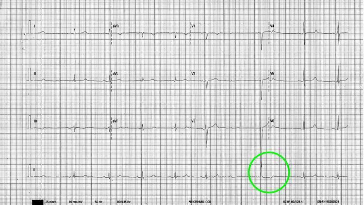
A 12-lead electrocardiogram (ECG) tracing on standard grid paper, demonstrating sinus node dysfunction and atrioventricular (AV) conduction abnormalities. The initial portion of the tracing shows a regular sinus rhythm at approximately 60 beats per minute, characterized by visible P waves preceding each narrow QRS complex. A green circle highlights a specific clinical finding in lead II and V6: a sudden pause caused by a blocked P wave (sinus arrest or high-grade exit block) followed by a narrow-complex junctional escape beat. This visual demonstrates common electrophysiological manifestations of sinus node dysfunction and high-grade AV block. The ECG includes limb leads (I, II, III), augmented limb leads (aVR, aVL, aVF), precordial leads (V1-V6), and a continuous Lead II rhythm strip at the bottom. The content is suitable for medical education regarding cardiac arrhythmias, specifically sinus arrest and escape rhythms.
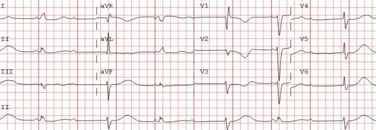
This diagnostic image is a standard 12-lead electrocardiogram (ECG) displayed on a red grid background, illustrating a complex cardiac arrhythmia. The tracing demonstrates sinus bradycardia with a high-grade atrioventricular (AV) block. Key diagnostic features include the presence of clear P waves that occur at a regular atrial rate; however, not every P wave is followed by a QRS complex, indicating failure of conduction through the AV node. Specifically, the tracing shows patterns of 2:1 Mobitz type II second-degree AV block. There is an observed irregular ventricular rhythm with occasional ventricular escape beats characterized by wider QRS morphologies compared to conducted beats. Notable features include a consistent PR interval in the conducted beats and a prolonged QTc interval. All 12 leads (I, II, III, aVR, aVL, aVF, V1-V6) are visible, providing a comprehensive view of the cardiac conduction disturbance. This ECG is representative of a clinical scenario involving syncope and high-grade heart block in an elderly patient, emphasizing the educational concepts of AV nodal disease and bradyarrhythmia management.
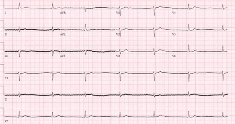
This diagnostic image is a 12-lead electrocardiogram (ECG) demonstrating junctional bradycardia. The tracing shows a slow, regular ventricular rate with narrow QRS complexes (duration within normal limits, <120 ms). A key diagnostic feature is the absence of clear, consistent sinus P waves preceding each QRS complex, suggesting an escape rhythm originating from the atrioventricular junction. In some leads, such as II and aVF, P waves are either absent or dissociated, consistent with the clinical context of a progressive bradyarrhythmia. The precordial leads (V1-V6) show preserved R-wave progression. There is no evidence of acute ST-segment elevation or significant T-wave inversions. This ECG illustrates a junctional escape rhythm, which may occur in the setting of sinus node dysfunction, high-grade AV block, or metabolic disturbances. The educational focus is on identifying non-sinus bradycardias and distinguishing junctional rhythms by their narrow QRS morphology and lack of preceding P waves.
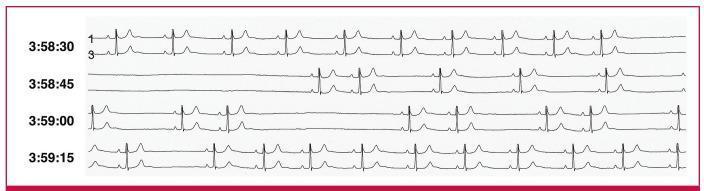
This diagnostic image shows a continuous 24-hour Holter electrocardiogram (ECG) rhythm strip illustrating severe sinus node dysfunction. The tracing is organized into four sequential time segments labeled from 3:58:30 to 3:59:15. The first segment shows a stable sinus rhythm with identifiable P waves, narrow QRS complexes, and prominent T waves. The second segment (3:58:45) captures a prolonged asystolic pause lasting approximately seven seconds, characterized by a complete absence of P-QRS-T complexes without any ventricular escape beats, indicative of sinus arrest. Subsequent segments (3:59:00 and 3:59:15) demonstrate a return to sinus rhythm interrupted by shorter, intermittent pauses. These shorter pauses are consistent with sino-atrial (SA) exit block, where P waves are absent at expected intervals. The image serves as an educational tool for distinguishing between types of sinus node pauses and illustrates the clinical presentation of symptomatic bradyarrhythmia requiring pacemaker intervention.
Bradyarrhythmias
Classification
1. Sinus Node Dysfunction (SND)
- Sinus bradycardia (<60 bpm; clinically significant when <45 bpm; <40 bpm is more specific for true SND)
- Sinus pause / sinus arrest
- Sinoatrial (SA) exit block (1st, 2nd, 3rd degree)
- Tachy-brady syndrome (bradycardia-tachycardia syndrome)
- Chronotropic incompetence (failure to increase heart rate appropriately with exercise)
- Atrial standstill
2. Atrioventricular (AV) Block
- First-degree AV block
- Second-degree: Mobitz Type I (Wenckebach), Mobitz Type II
- High-grade AV block (2:1, 3:1)
- Third-degree (complete) AV block
- His-Purkinje block / bifascicular / trifascicular block
- Paroxysmal AV block
Epidemiology
- Bradycardia (HR <60 bpm) prevalence: ~15% in adult males, ~7% in adult females
- Prevalence rises to 50-80% in endurance athletes
- During sleep: sinus bradycardia <40 bpm in 20-25% of healthy adults; Wenckebach block in 5-10%
- Incident SND: ~0.8 per 1000 person-years, highly age-related
- SND accounts for >50% of pacemakers implanted in the United States
- Only ~0.26% of U.S. adults ultimately receive a pacemaker for bradycardia
- Goldman-Cecil Medicine, p. 557; Fuster and Hurst's The Heart, 15th Ed.
Pathobiology / Physiology
Sinus Node
- Resting membrane potential: -50 to -60 mV (less negative than atrial cells)
- Phase 0 is mediated by slow inward calcium current (I-CaL), not fast sodium current
- Absence of inward rectifier potassium current (I-Kr)
- Phase 4 automaticity driven by the interaction of two "clocks":
- Membrane clock: decay of outward K+ repolarizing currents + activation of the "funny current" (I-f, activated by hyperpolarization, carried by Na+)
- Calcium clock: spontaneous rhythmic Ca2+ release from the sarcoplasmic reticulum
AV Conduction
Causes of SND
Intrinsic (structural sinus node disease)
| Cause | Notes |
|---|---|
| Idiopathic degenerative disorder | Most common - progressive fibrosis + loss of functional myocytes; also reduced connexin-45 and ion channel remodeling |
| Atrial arrhythmias / AF | Chronic overdrive suppression; downregulation of I-f and calcium clock; "tachy-brady syndrome" |
| Ischemic heart disease | Inferior/posterior MI (affects SN artery); ~15-20% of acute MI |
| Heart failure | Neurohormonal activation + negative chronotropic drugs |
| Surgical trauma | Mustard, Senning, Glenn, Fontan procedures; also heart transplantation |
| Genetic abnormalities | HCN4 mutations, SCN5A, etc. |
Extrinsic (no structural SN disease)
- Enhanced vagal tone (athletes, vasovagal states)
- Medications: beta-blockers, calcium channel blockers, cardiac glycosides, antiarrhythmics (most class I and III), clonidine, lithium, donepezil
- Hypothyroidism, electrolyte abnormalities
- Sleep apnea
Causes of AV Block
| Category | Examples |
|---|---|
| Inherited | Progressive cardiac conduction disease (SCN5A, TRPM4, KCNJ2, PRKAG2), laminopathies, congenital complete AV block (autoimmune - anti-Ro/anti-La), long QT with functional block |
| Degenerative | Idiopathic age-related fibrosis (Lev's / Lenegre's disease); aortic valve calcification |
| Ischemic | Inferior MI (usually supra-Hisian, often transient); anterior MI (infra-Hisian, often permanent) |
| Infiltrative | Sarcoidosis, amyloid, hemochromatosis |
| Infectious | Lyme carditis, endocarditis, Chagas disease, myocarditis |
| Surgical | Cardiac valve surgery, TAVR (risk up to 34% with some valves), catheter ablation |
| Drugs | Beta-blockers, CCBs, digitalis, Class I/III antiarrhythmics, clonidine, donepezil |
| Collagen vascular disease | |
| Metabolic | Hyperkalemia, hypermagnesemia, hyperthyroidism |
ECG Manifestations
Sinus Node Dysfunction
| Finding | Description |
|---|---|
| Sinus bradycardia | HR <60 bpm with normal P-wave morphology and PR interval |
| Sinus pause/arrest | Absent P waves; pause is not a multiple of prior P-P interval |
| SA exit block | Absent P waves, but pause = multiple of prior P-P cycle (2nd degree: type I has progressive shortening of P-P before dropped beat; type II has fixed P-P before dropped) |
| Tachy-brady syndrome | Alternating bradycardia and atrial tachyarrhythmia (often AF) |
| Chronotropic incompetence | Failure to achieve 85% of MPHR during exercise |
| Atrial standstill | Complete absence of atrial activity |
AV Block
| Type | ECG Feature |
|---|---|
| 1st degree | PR interval >200 ms; all P waves conduct |
| 2nd degree - Mobitz I (Wenckebach) | Progressive PR prolongation until a P wave is blocked; QRS width usually normal; infranodal location uncommon |
| 2nd degree - Mobitz II | Fixed PR interval with sudden non-conduction of P wave; QRS usually wide; infra-Hisian location; higher risk of progression to complete block |
| 2:1 block | Every other P wave blocked; cannot distinguish Mobitz I vs II without 3:2 or other periods |
| High-grade AV block | 2 or more consecutive blocked P waves |
| 3rd degree (complete) | P waves and QRS completely dissociated; escape rhythm from junction (narrow, 40-60 bpm) or ventricle (wide, 20-40 bpm) |
| AV dissociation | AV rate ≥ ventricular rate (not necessarily blocked - junctional tachycardia with sinus bradycardia can cause this) |
Clinical Presentation
- Fatigue, exercise intolerance (most common, especially with chronotropic incompetence)
- Presyncope / syncope (Stokes-Adams attacks in complete heart block)
- Palpitations (especially tachy-brady syndrome)
- Dyspnea, heart failure exacerbation
- Cognitive slowing in the elderly
Diagnostic Evaluation
| Test | Role |
|---|---|
| 12-lead ECG | First-line; identifies type of bradyarrhythmia |
| Ambulatory monitoring (Holter, event recorder, implantable loop recorder) | Correlation of symptoms with rhythm; 24-48h Holter for frequent symptoms, implantable loop recorder (ILR) for infrequent syncope |
| Exercise testing | Assesses chronotropic competence; rate increase with exercise suggests vagal/extrinsic cause |
| Electrophysiology study (EPS) | Measures HV interval (>70 ms suggests infra-Hisian disease); sinus node recovery time; assesses risk of complete block in bundle branch block |
| Tilt-table testing | Evaluates vasovagal syncope |
| Blood tests | TSH, electrolytes, Lyme serology, ANA/anti-Ro (if congenital block suspected) |
| Echocardiography | Structural heart disease evaluation |
Management
Acute / Emergency Management
- Identify and treat reversible causes (withhold offending drugs, correct electrolytes, treat infection/ischemia)
- Atropine 0.5-1 mg IV (up to 3 mg total) - for AV nodal block and sinus node dysfunction; less effective for infra-Hisian block
- Temporary transvenous pacing - for hemodynamically significant bradycardia not responding to atropine
- Transcutaneous pacing as a bridge
- Dopamine or epinephrine infusion if pacing unavailable
- Glucagon for beta-blocker toxicity; calcium chloride for CCB toxicity
Long-Term / Permanent Pacing (2018 ACC/AHA/HRS Guidelines)
For SND (Pacing Indications):
| Class | Recommendation |
|---|---|
| I (Indicated) | Symptomatic SND directly attributable to SND |
| I | Symptomatic sinus bradycardia due to required drug therapy with no alternative |
| IIa (Reasonable) | Tachy-brady syndrome with symptomatic bradycardia |
| IIa | Symptomatic chronotropic incompetence - rate-responsive pacing |
| IIb (May consider) | Trial of oral theophylline for possible SND to assess symptom correlation |
| III (Harm) | Asymptomatic sinus bradycardia or sleep-related pauses from elevated vagal tone |
Choice of Pacing Mode for SND:
- Persistent AF: rate-responsive ventricular pacemaker (VVIR)
- No AV disease: rate-responsive single-chamber atrial pacemaker (AAIR)
- With AV disease or unknown: rate-responsive dual-chamber pacemaker (DDDR - preferred in most)
- Atrial-based pacing (vs. VVI) reduces risk of AF by ~20% and may reduce stroke by ~19%
- Long-term RV apical pacing causes LV dysfunction via dyssynchrony - minimize unnecessary RV pacing (use AV hysteresis, His-bundle pacing, or CRT if RV pacing >40% expected)
For AV Block (Pacing Indications):
| Degree | Indication |
|---|---|
| 1st degree | Pacing not indicated (unless markedly prolonged causing symptoms) |
| Mobitz I (Wenckebach) | Pacing if symptomatic; generally benign if nodal |
| Mobitz II | Pacing indicated even if asymptomatic (high risk of progression) |
| 2:1 block | Pacing based on level (His-bundle EPS may help) |
| Complete (3rd degree) block | Permanent pacing always indicated |
ECG Examples
Key Clinical Pearls
- Inferior MI causes AV block that is usually supra-Hisian (AV nodal), tends to be transient, and responds to atropine.
- Anterior MI with AV block implies His-Purkinje involvement, is often permanent, and has higher mortality.
- Lyme carditis is a reversible infectious cause of complete AV block - always test in endemic areas.
- TAVR carries a significant risk of new heart block (up to 34% with some valve types), especially in patients with pre-existing RBBB or 1st degree AV block.
- Tachy-brady syndrome: AF termination produces post-conversion pauses due to sinus node suppression - anticoagulation and pacemaker implantation are both often required.
- In complete heart block, the width and rate of the escape rhythm indicates the level: narrow QRS + 40-60 bpm = junctional (safer), wide QRS + 20-40 bpm = ventricular (unstable).
- The "funny current" (I-f) is the target of ivabradine (rate-lowering agent) - its name comes from the unusual property of being activated by hyperpolarization rather than depolarization.