
Ear pathway physology short answer easy words
Reading File
Finding Sources
Finding Sources
Reading File
Reading File
Reading File
Reading File
Now I have all the information needed. Let me also fetch the auditory pathway diagram for the visual aid.
Loading Image
Here is a clear, easy-to-understand explanation of the ear pathway physiology - from sound entering the ear all the way to the brain.
Ear (Auditory) Pathway - Physiology
Step 1: Sound Enters the Outer Ear
- Sound waves travel through the ear canal (external auditory meatus)
- They hit the eardrum (tympanic membrane), which vibrates back and forth
Step 2: Middle Ear - The Amplifier
The eardrum is connected to 3 tiny bones (ossicles):
| Bone | Nickname | What it does |
|---|---|---|
| Malleus | Hammer | Attached to eardrum, picks up vibration |
| Incus | Anvil | Passes vibration along |
| Stapes | Stirrup | Pushes against the oval window |
These bones amplify the sound - they act like a mechanical lever to make the signal stronger before it enters the fluid-filled inner ear.
The eustachian tube connects the middle ear to the throat and keeps air pressure equal on both sides of the eardrum. It opens when you swallow or yawn.
Step 3: Inner Ear (Cochlea) - Sound Becomes Electricity
- The stapes pushes on the oval window, sending waves through the fluid inside the cochlea (snail-shaped organ)
- Inside the cochlea is the organ of Corti, which sits on the basilar membrane
- The fluid waves move the basilar membrane, which bends tiny hair cells
- When hair cells bend, they open ion channels → potassium (K+) ions rush in → the cell depolarizes → an electrical nerve signal is created
Key idea: Different parts of the basilar membrane respond to different sound frequencies. High-pitched sounds vibrate near the base; low-pitched sounds vibrate near the apex (tip). This is called tonotopic organization.
Step 4: Auditory Nerve (CN VIII)
- The electrical signal from hair cells travels to the spiral ganglion (the first relay station)
- From there, nerve fibers form the cochlear branch of the 8th cranial nerve (vestibulocochlear nerve)
- This nerve carries the signal to the brainstem
Step 5: Brainstem Relay Stations
The signal passes through several relay stations in the brainstem:
- Cochlear nuclei (dorsal + ventral) - in the medulla - first stop
- Superior olivary complex - here, signals from both ears combine (this helps locate where sound is coming from)
- Inferior colliculus (midbrain) - center for auditory reflexes (e.g., the startle response)
At these brainstem levels, most fibers cross to the opposite side of the brain, so each ear is mostly processed by the opposite (contralateral) hemisphere.
Step 6: Thalamus
- Signals travel up to the medial geniculate body (MGB) in the thalamus
- This is the "relay switch" that passes the signal onward to the cortex
Step 7: Auditory Cortex (Final Destination)
- The MGB sends signals to the primary auditory cortex (A1), located in the superior temporal gyrus (temporal lobe)
- This is where you consciously hear and interpret the sound
- Low tones are processed anterolaterally; high tones are processed posteromedially
Further processing (speech, music, meaning) happens in surrounding association areas, including:
- Wernicke's area (left side) - understanding language/speech
- Right auditory cortex - pitch, melody, sound intensity
The Full Pathway at a Glance
Sound waves
   ↓
Outer ear (ear canal)
   ↓
Eardrum vibrates
   ↓
3 Bones: Malleus → Incus → Stapes
   ↓
Oval window → Cochlea fluid waves
   ↓
Basilar membrane → Hair cells bend → Electrical signal
   ↓
Spiral ganglion → Cochlear nerve (CN VIII)
   ↓
Cochlear nuclei (medulla)
   ↓
Superior olive (both ears merge here)
   ↓
Inferior colliculus (midbrain)
   ↓
Medial geniculate body (thalamus)
   ↓
Primary auditory cortex (temporal lobe)
   ↓
YOU HEAR THE SOUND!
Pathway Diagram
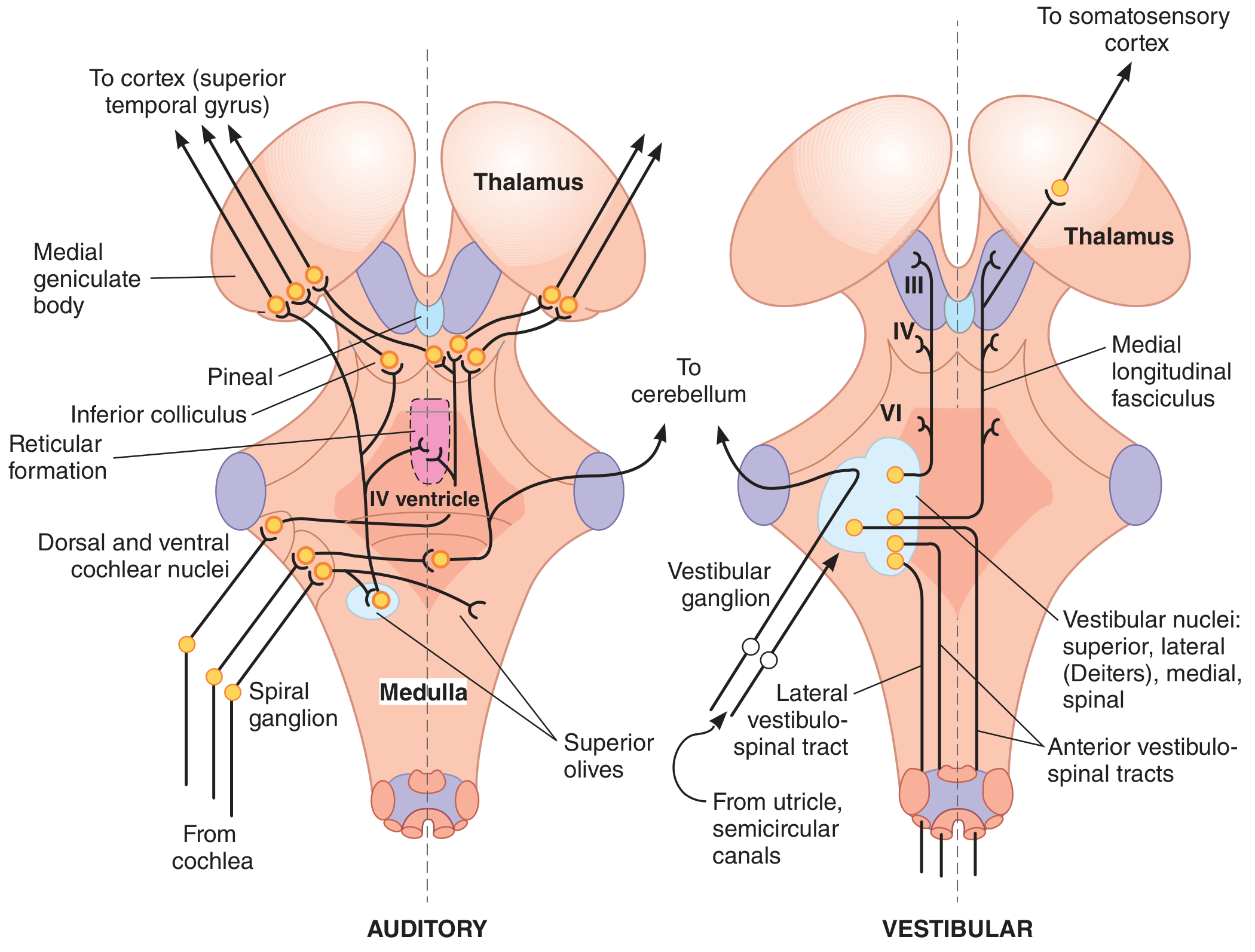
Auditory pathway (left side): from cochlea → spiral ganglion → cochlear nuclei → superior olives → inferior colliculus → medial geniculate body → auditory cortex. - Ganong's Review of Medical Physiology, 26th Ed.
Quick Summary Table
| Location | Structure | Function |
|---|---|---|
| Outer ear | Ear canal + Eardrum | Captures and vibrates with sound |
| Middle ear | Malleus, Incus, Stapes | Amplifies and transmits vibrations |
| Inner ear | Cochlea + Hair cells | Converts sound to electrical signals |
| Nerve | CN VIII (cochlear) | Carries signal to brain |
| Medulla | Cochlear nuclei | First brain relay |
| Pons | Superior olive | Sound localization, both ears |
| Midbrain | Inferior colliculus | Auditory reflexes |
| Thalamus | Medial geniculate body | Relay to cortex |
| Cortex | Temporal lobe (A1) | Conscious hearing + speech |
This is a shared conversation. Sign in to Orris to start your own chat.