
Haemophilic joint
haemophilic arthropathy joint X-ray radiograph
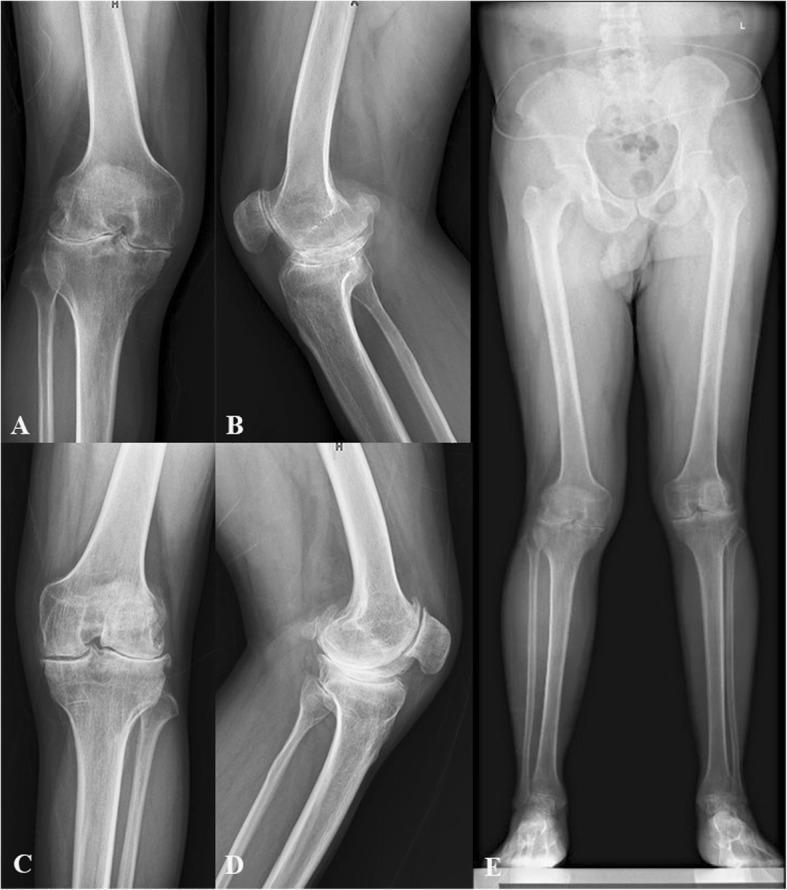
This composite of five diagnostic X-ray radiographs demonstrates severe, bilateral haemophilic arthropathy in a patient scheduled for total knee arthroplasty (TKA). Panels A and C show anteroposterior (AP) views of the right and left knees, respectively, revealing advanced tricompartmental degeneration characterized by severe, asymmetric joint space narrowing, subchondral sclerosis, and significant marginal osteophyte formation. Irregularity of the articular surfaces and squaring of the femoral condyles, typical of chronic hemarthrosis-related damage, are present. Panels B and D provide lateral views, highlighting anterior osteophytes, patellofemoral joint space loss, and posterior subluxation tendencies. Panel E is a full-length, weight-bearing standing AP radiograph of the lower limbs and pelvis, which visualizes the global alignment and mechanical axes. It confirms the symmetric severity of the knee pathology and allows for the assessment of lower limb deformities, such as valgus or varus malalignment, occurring secondary to the extensive articular cartilage and bone destruction. This imaging series illustrates the end-stage clinical manifestations of haemophilic joint disease.
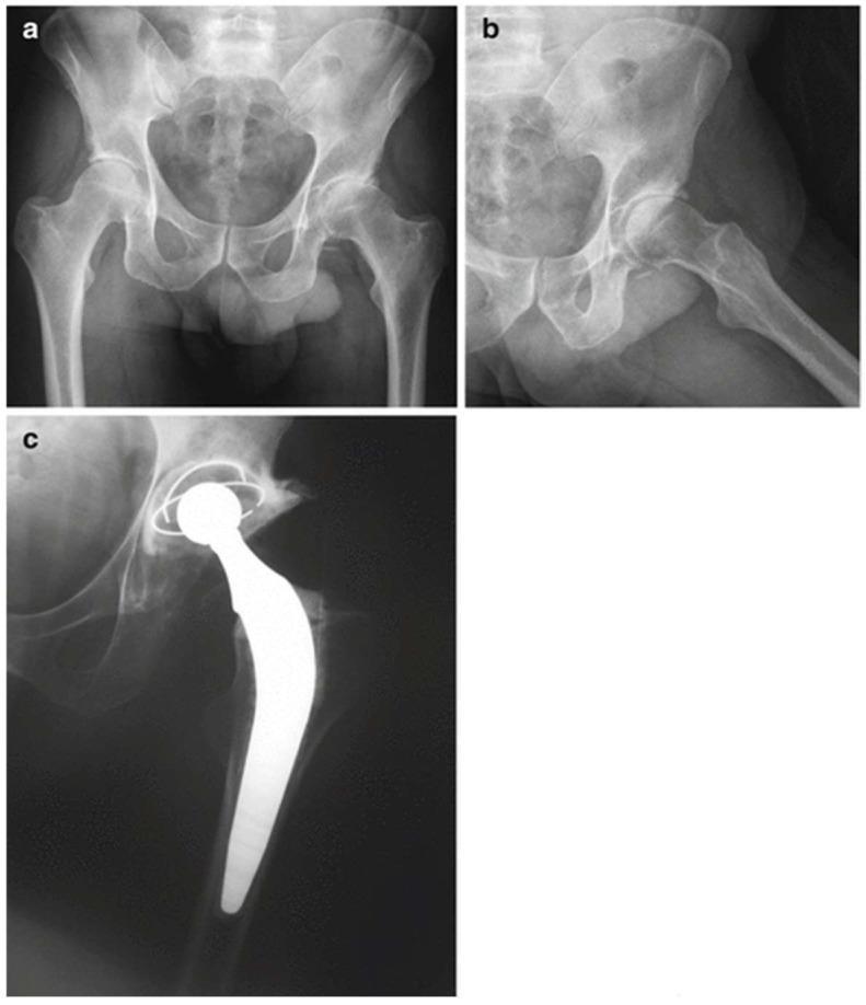
This composite of diagnostic X-ray radiographs documents a total hip replacement (THR) procedure in a patient with haemophilia. Panel (a) shows a preoperative anteroposterior view of the pelvis, revealing significant narrowing of the left hip joint space and altered morphology of the femoral head consistent with advanced haemophilic arthropathy or secondary osteoarthritis. Panel (b) provides a lateral preoperative view focusing on the left hip, highlighting the degenerative changes and bony remodeling. Panel (c) is a postoperative radiograph demonstrating the successful implantation of a total hip prosthesis. The image displays a highly radio-opaque metallic femoral stem anchored within the femoral canal, a spherical femoral head component, and an acetabular cup with visible marker wires. The alignment of the prosthetic components restores the joint space and structural integrity of the hip. This educational series illustrates the transition from severe joint pathology to surgical reconstruction in the context of specialized orthopedic management for bleeding disorders.
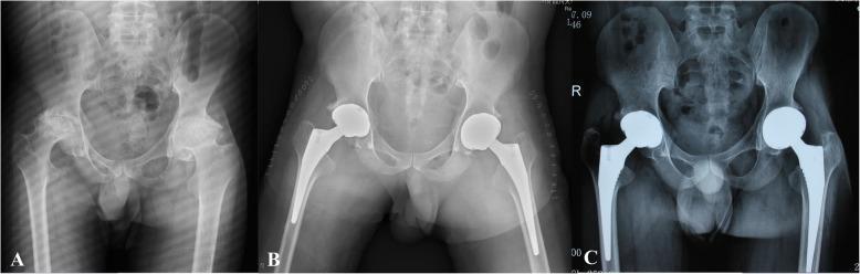
This composite diagnostic image features three anteroposterior (AP) pelvic X-rays (labeled A, B, and C) illustrating a case of haemophilic arthropathy and its surgical management with bilateral total hip arthroplasty (THA). Panel A shows the preoperative state, characterized by severe bilateral degenerative changes in the hip joints. Visible features include significant joint space narrowing, subchondral sclerosis, and irregular femoral head morphology, typical of advanced haemophilic arthropathy. Panel B displays the immediate postoperative appearance after bilateral THA. Radiopaque prosthetic components are visible on both sides, consisting of acetabular cups, femoral heads, and femoral stems securely positioned within the medullary canals. Panel C is a one-year follow-up radiograph with a digitally enhanced overlay of the prosthetic hardware. The image demonstrates stable implant positioning without evidence of progressive radiolucent lines (> 2 mm), indicating successful osseointegration and maintenance of alignment. The series serves as an educational comparison between end-stage joint disease and successful surgical intervention in a patient with Haemophilia A.
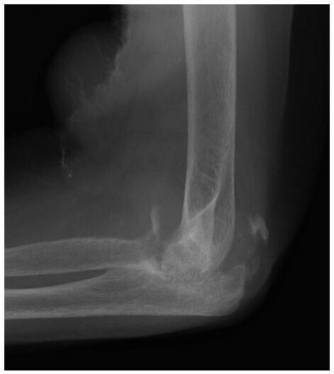
This lateral x-ray radiograph of the left elbow demonstrates advanced features of neuropathic arthropathy (Charcot joint). The primary findings include significant destruction and resorption of the proximal radius and ulna, leading to marked joint disorganization and architectural deformity. There is prominent soft tissue swelling suggestive of extensive joint effusion or distension. Within this distended joint capsule, multiple radiopaque loose bodies or osseous debris are visible, scattered away from the primary joint surfaces. Subchondral sclerosis and increased bone density are present in the periarticular regions. These findings represent the mixed hypertrophic and atrophic patterns typical of neuropathic joints, frequently secondary to underlying conditions like diabetic neuropathy or syringomyelia. The clinical relevance lies in distinguishing these catastrophic degenerative changes from osteomyelitis or acute trauma in patients with diminished protective sensation.
haemophilic arthropathy joint pathology management
hemophilic arthropathy
Haemophilic Joint (Haemophilic Arthropathy)
Background and Pathophysiology
- Tissue factor expression is relatively low in synovium, making thrombin generation dependent on the intrinsic tenase complex (factors VIII and IX)
- Tissue factor pathway inhibitor is relatively high in the joint
- Inadequate thrombin-activatable fibrinolysis inhibitor (TAFI) activation means local fibrinolysis is increased
- Neovascularization triggered after a first bleed creates fragile new vessels prone to repeated injury
Joints Affected
- Knees, ankles, elbows (most common - large synovial joints)
- Hips, shoulders (less common)
- Small joints of hands and feet (rare)
Clinical Stages
1. Acute Hemarthrosis
- The first joint bleed typically occurs at ~1.8 years of age (when the child starts walking)
- Prodrome: tingling sensation and tightness within the joint
- Followed by: rapid swelling, loss of range of motion (ROM), pain, warmth
- Flexion is the most comfortable position; disuse causes secondary muscle spasm
- Blood stays liquid in the joint due to absence of intrinsic clotting factors and tissue thromboplastin in synovium
- Blood is resorbed over ≥1 week; full recovery within ~2 weeks with factor replacement
- Fever >38.3°C warrants concern for septic arthritis (rare but serious)
2. Synovitis (Chronic)
- Synovium cannot fully remove blood remnants
- Iron accumulates as hemosiderin deposits in synovium and cartilage
- Iron triggers inflammatory response, stimulating synoviocyte proliferation
- Pro-inflammatory cytokines (including IL-1, TNF, VEGF) released
- New fragile blood vessels form (neovascularization) - predisposing to further bleeds
3. Established Haemophilic Arthropathy
| Process | Features |
|---|---|
| Synovial inflammation | Pannus formation, hemosiderin deposits, villous hypertrophy |
| Cartilage degeneration | Direct iron toxicity, lysosomal enzyme release, protease activity |
| Bone remodeling | Subchondral cysts, erosions, overgrowth, osteoporosis |
Progressive synovial fibrosis paradoxically reduces hemarthrosis frequency in end-stage disease.
Associated Musculoskeletal Complications
- Common sites: iliopsoas and flexor compartment of forearm
- Iliopsoas bleed: pain in lower abdomen/groin/lower back, pain on hip extension but not rotation (distinguishes from hemarthrosis)
- Risk of compartment syndrome - a life-threatening emergency
- Hip flexion contracture from iliopsoas involvement; femoral neuropathy from nerve compression
- Form when periosteum or bone is involved
- In children: distal to elbows/knees, from direct trauma, often responsive to conservative treatment
- In adults: femur and pelvis, usually refractory to treatment
- Appear as expanding, painful bony swellings
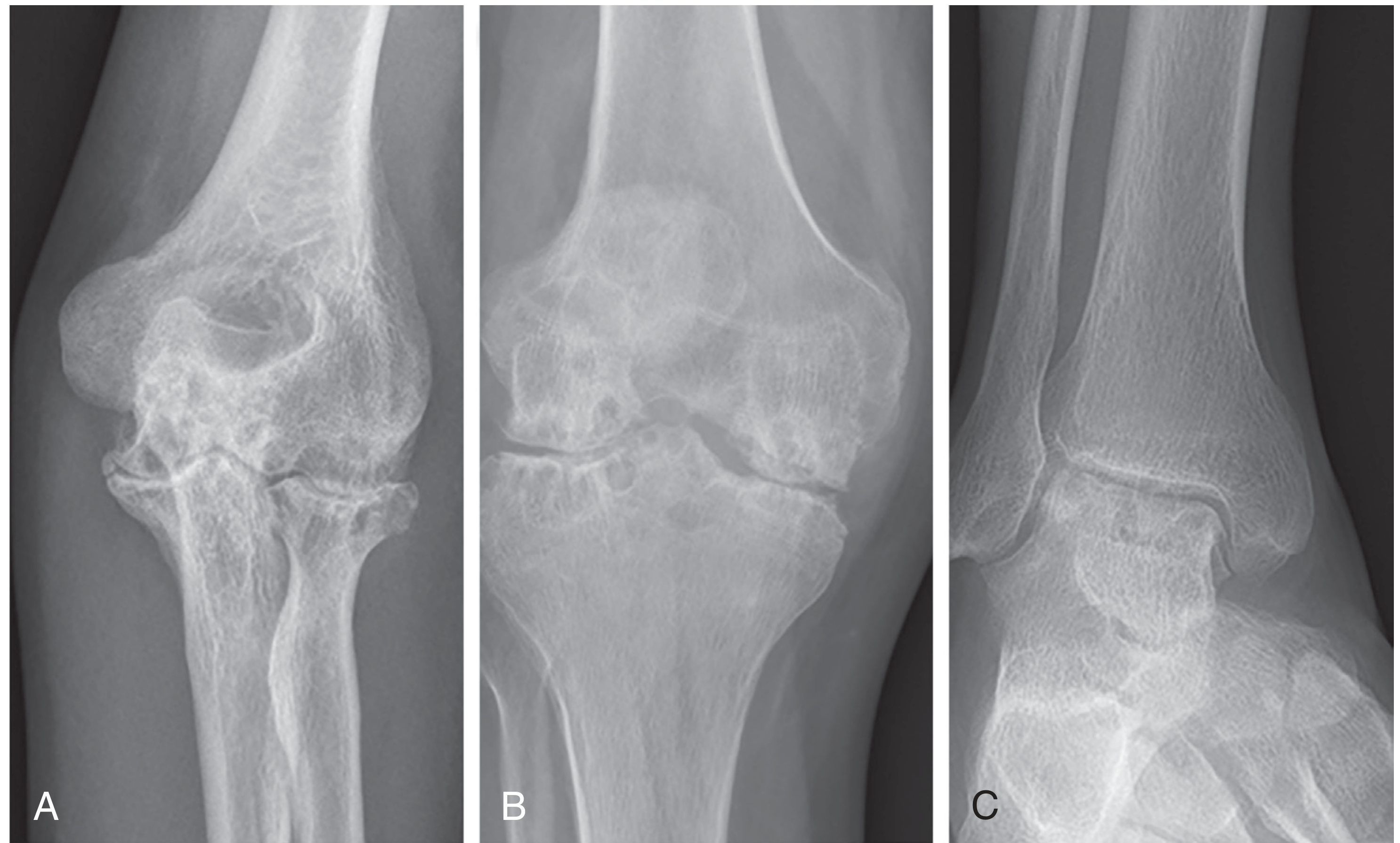
Radiographic Findings (Progression)
| Stage | Findings |
|---|---|
| Early | Joint capsule distension only |
| Intermediate | Juxtaarticular osteopenia, marginal erosions, subchondral cysts |
| Late | Joint space narrowing, bony overgrowth (similar to OA), calcified blood collections |
Diagnosis
Treatment
Acute Hemarthrosis
- Immediate factor VIII or IX infusion at first sign of bleeding
- Splint in gentle forced extension to prevent contracture
- Analgesia: avoid non-selective NSAIDs (inhibit platelets); COX-2 inhibitors are safer
- In patients with inhibitors (anti-factor antibodies): recombinant activated factor VIIa or activated prothrombin complex concentrate
Chronic Synovitis
- Clotting factor replacement (long-term) to break the hemarthrosis-synovitis cycle
- Radiosynovectomy with yttrium-90 silicate or phosphorus-32 colloid: effective, preferred when surgery is not advised
- Synovectomy (arthroscopic preferred over open): limits future bleeding and fibrosis; open procedure risks decreased ROM
Established Arthropathy - Conservative
- Analgesics, physiotherapy (muscle strengthening, stretching, joint stability)
- Manual traction, TENS
- Hydrotherapy (functional training with minimal weight-bearing)
- Orthotics/shoe adaptations, casting
Established Arthropathy - Surgical
| Procedure | Notes |
|---|---|
| Total joint replacement (knee, hip) | Effective for pain and function; restricted ROM may limit knee outcomes |
| Ankle arthrodesis | Very effective for pain and stopping bleeding; loss of mobility |
| Total ankle replacement | Preserves motion; concerns about aseptic loosening and infection |
| Joint distraction | Novel joint-preserving technique; 5mm distraction for 6-8 weeks; promising results for ankle |
Prevention
- Firestein & Kelley's Textbook of Rheumatology, Chapter 124 (Haemophilic Arthropathy)
- Harrison's Principles of Internal Medicine 22E (2025), Chapter 386 (Haemophilic Arthropathy)
Post op pain management in orthopaedics
postoperative pain management orthopedic surgery multimodal analgesia
nerve block regional analgesia total knee arthroplasty hip
Postoperative Pain Management in Orthopaedics
Why Pain Management Matters in Orthopaedics
- Sympathetic activation - hypertension, tachycardia, dysrhythmias
- Impaired early mobilisation and rehabilitation
- Risk of chronic pain, central sensitization, and complex regional pain syndrome (CRPS)
- Poor patient satisfaction (patient perception of pain control affects satisfaction more than the quantity of opioid prescribed)
Assessment of Pain
- Characteristics: onset, duration, location, quality, intensity
- Past and current management strategies
- Impact on daily activities and patient goals
- Numeric Rating Scale (NRS): 0-10 (most widely used)
- Visual Analog Scale (VAS): 10-cm horizontal scale
- Verbal/visual descriptors for non-communicating patients (grimacing, restlessness, vocalisation, vital signs)
- Within 30 minutes after parenteral drug administration
- Within 1 hour after oral drug administration
- With any new or changed pain
| Score | State | Action |
|---|---|---|
| S | Sleep, easy to arouse | Acceptable; may increase dose |
| 1 | Awake and alert | Acceptable |
| 2 | Slightly drowsy, easily aroused | Acceptable |
| 3 | Frequently drowsy, drifts off | Unacceptable - decrease dose 25-50%, monitor closely |
| 4 | Somnolent, minimal response | Stop opioid, consider naloxone, notify anaesthesiologist |
Respiratory depression in opioid users can be as high as 48%; sedation precedes respiratory depression and is the more reliable early warning sign.
Preoperative Considerations
- Screen all patients for opioid risk (abuse, misuse, diversion potential) before prescribing
- Patients on chronic NSAIDs (RA/OA): note risks of GI bleeding, renal toxicity, platelet dysfunction
- Preoperative regional anaesthesia ("block room") can be performed in a preoperative holding area to avoid OR delays
- Patients with prior opioid use or chronic pain have altered central pain processing and require tailored regimens
- The psychological state of the patient (catastrophizing) affects pain perception and outcomes; identify and address preoperatively
The Multimodal Analgesia Principle
- Reduces total opioid consumption (opioid-sparing)
- Minimises opioid side effects (nausea, sedation, respiratory depression, addiction risk)
- Improves functional recovery and facilitates physiotherapy
- Reduces length of hospital stay
1. Pharmacological Agents
A. Paracetamol (Acetaminophen)
- First-line non-opioid analgesic; also antipyretic
- IV form allows administration in perioperative NPO period
- Safe in most adults and children; use with caution in hepatic disease
- Core component of all multimodal regimens
B. NSAIDs
- Act by inhibiting cyclooxygenase (COX) → reduced prostaglandin synthesis → analgesia + anti-inflammatory
- Examples: ibuprofen, naproxen, meloxicam, ketorolac
- Ketorolac (IV/IM): highly effective for musculoskeletal and post-fracture pain; recent evidence supports use even after fracture surgery without significantly increased nonunion risk when used short-term
- COX-2 inhibitors (celecoxib): equivalent analgesia, do not impair platelet function (safer perioperatively), but carry cardiovascular, renal, and GI concerns
- Historic concern about NSAIDs impairing bone healing is now considered of less significance for short-term use
- Non-selective NSAIDs should be avoided if possible in patients with bleeding risk (platelet dysfunction)
C. Opioids
- Act on mu, delta, kappa receptors in brain, spinal cord, and peripheral nociceptors
- Also stimulate mesolimbic dopamine system (addiction potential)
- Remain essential for moderate-to-severe acute pain
- Risks: respiratory depression, sedation, constipation, nausea, addiction, possible delayed bone healing (animal data, not robustly shown in humans)
- Tramadol: weak opioid agonist + serotonin/noradrenaline reuptake inhibitor; useful for moderate pain (step-down from stronger opioids)
- Key principle: use at the lowest effective dose for the shortest duration, with a clear prescription limit (e.g. no refills, maximum 2 prescriptions)
D. Gabapentinoids (Gabapentin / Pregabalin)
- Block voltage-gated calcium channels → reduce neurotransmitter release
- Useful as adjuncts for neuropathic pain components
- Opioid-sparing effect: a 2025 systematic review (PMID 41239206) confirms effectiveness in orthopedic surgeries for postoperative pain and opioid-sparing
- Short-term use post-discharge (e.g. gabapentin 100 mg TID × 5 days) is favoured
- Side effects: sedation, dizziness - exercise caution especially in elderly
E. Muscle Relaxants
- Cyclobenzaprine, metaxalone, methocarbamol
- Central action to reduce muscle spasm
- Useful where muscle spasm contributes significantly to pain (e.g. spinal surgery, pelvic injuries)
F. Corticosteroids (Dexamethasone)
- Perioperative single dose reduces postoperative pain and nausea
- Used in selected cases
2. Regional / Neuraxial Techniques
Neuraxial (Spinal / Epidural) Anaesthesia
- Spinal (intrathecal) opioids (morphine, hydromorphone): provide extended postoperative analgesia; widely used with enhanced recovery protocols
- Epidural analgesia: useful after bilateral TKA; provides excellent pain control but more side effects (pruritus, nausea/vomiting, urinary retention, orthostatic hypotension) than peripheral nerve blocks
- Neuraxial techniques are associated with lower rates of all-cause infections, AKI, pulmonary/thromboembolic complications, and falls compared with general anaesthesia alone in hip/knee arthroplasty
Peripheral Nerve Blocks (PNBs)
| Procedure | Preferred Block |
|---|---|
| Shoulder surgery | Interscalene brachial plexus block |
| Upper limb / elbow | Supraclavicular / infraclavicular block |
| Distal radius fracture | Brachial plexus block |
| Total hip arthroplasty (THA) | Lumbar plexus / fascia iliaca / PENG block |
| Total knee arthroplasty (TKA) | Femoral nerve / adductor canal block; iPACK block |
| Ankle / foot surgery | Popliteal sciatic nerve block |
| Hip fracture | Fascia iliaca compartment block |
- Single-shot provides satisfactory analgesia for several hours
- Continuous perineural catheters (with local anaesthetic infusion) decrease time to discharge criteria for TKA and are preferred for major procedures
- For unilateral TKA: perineural catheters provide equivalent analgesia to epidural catheters with fewer side effects and earlier ambulation
- Requires 24/7 team coverage; fall prevention programs are essential
Intra-articular (Local Infiltration Analgesia - LIA)
- Surgeon-administered injection of local anaesthetic (bupivacaine/ropivacaine) ± adjuvants into the joint space
- Adjuvants: opioids, clonidine, ketorolac, epinephrine, neostigmine - extend duration
- Particularly used in arthroscopic procedures and arthroplasty
- Periarticular infiltration by surgeon during TKA is widely practised with good evidence
Ultrasound Guidance
- Has greatly improved accuracy and safety of nerve blocks
- Reduces risk of intravascular injection and neurological complications
3. Procedure-Specific Highlights
Total Hip Arthroplasty (THA)
- Neuraxial anaesthesia preferred (reduces mortality/morbidity)
- Intrathecal opioids for extended analgesia
- Lumbar plexus / fascia iliaca / PENG blocks effective
- Multimodal: paracetamol + NSAID/COX-2 + regional ± opioid PRN
Total Knee Arthroplasty (TKA)
- Pain is more severe and longer-lasting than after THA
- Effective multimodal analgesia is essential for early ROM and preventing joint adhesions
- Balance between pain control and alertness during physiotherapy
- Adductor canal block preserves motor function (preferred over femoral nerve block for mobilisation)
- Perineural catheters preferred over epidural for unilateral TKA
- Periarticular infiltration by surgeon is standard practice
Shoulder Surgery
- Interscalene block provides excellent analgesia
- Continuous interscalene catheter for major open procedures
- Position-related hypotension (beach chair position) must be monitored separately
Fracture Surgery / Trauma
- Peripheral nerve blocks at time of presentation reduce pre-operative and perioperative pain
- Brachial plexus block for upper limb fractures
- Popliteal block for ankle fractures
- Fascia iliaca / PENG for hip fractures (reduces opioid requirement acutely)
- Short-term NSAIDs now acceptable even in fracture patients
- Opioids: prescribe with specific limits; avoid chronic prescribing
Spine Surgery
- Combination of wound infiltration, epidural analgesia, and systemic multimodal agents
- NSAIDs used with caution (theoretical concern for spinal fusion, though evidence mixed)
4. Authors' Preferred Regimen (Rockwood & Green's Orthopaedics)
| Period | Opioid | Non-opioid |
|---|---|---|
| Week 1 | Hydrocodone/paracetamol 5/325 mg q6h PRN (#30, no refills) | Ibuprofen 600 mg q8h × 5 days; Gabapentin 100 mg TID × 5 days |
| Weeks 2-3 | Hydrocodone/paracetamol (#20 then #10, max 2 Rx) or Tramadol 50 mg q6h PRN | NSAID PRN; substitute with paracetamol |
| Week 4+ | Paracetamol PRN only | NSAID PRN |
- Minor injury: Tramadol 50 mg q6h PRN (#20 → #10) + NSAID + paracetamol
- Major injury: Hydrocodone/paracetamol (#20 → #10) + NSAID + paracetamol
5. Non-Pharmacological Strategies
| Method | Role |
|---|---|
| RICE (Rest, Ice, Compression, Elevation) | First-line for acute musculoskeletal injury |
| Splinting/immobilisation | Reduces pain by preventing movement |
| Cryotherapy (cold packs) | Reduces local inflammation and pain |
| TENS (transcutaneous electrical nerve stimulation) | Adjunct for chronic/subacute pain |
| Physiotherapy and early mobilisation | Prevents muscle atrophy, reduces chronic pain risk |
| Psychological support / CBT | Addresses catastrophizing, improves outcomes |
| Mirror visual feedback | Used in CRPS and phantom limb pain |
6. Enhanced Recovery After Surgery (ERAS) in Orthopaedics
- Preoperative: education, optimisation, preemptive analgesia (gabapentinoids, celecoxib)
- Intraoperative: neuraxial anaesthesia, tranexamic acid, LIA
- Postoperative: early mobilisation, scheduled multimodal non-opioid analgesia, VTE prophylaxis, physiotherapy
7. Special Considerations
- Require higher baseline opioid doses; liaise with pain specialists
- Target concurrent non-opioid and regional strategies aggressively
- Increased sensitivity to opioids (sedation, falls, delirium)
- Start low with opioids; prefer non-opioid multimodal approaches
- Screen for renal function before NSAIDs
- Screen all patients with validated risk tools before opioid prescription
- Use prescription drug monitoring programs (PDMPs)
- Prescribe the minimum necessary; avoid refills where possible
- The patient's perception that pain is being managed matters more than total opioid quantity prescribed
- Ensure adequate early postoperative analgesia
- Early mobilisation and physiotherapy
- Vitamin C supplementation has been proposed for fracture patients (particularly distal radius) to reduce CRPS risk
- Rockwood and Green's Fractures in Adults, 10th ed. (2025), Chapters 28-29
- Morgan and Mikhail's Clinical Anesthesiology, 7e, Chapters 38, 6C
- Barash, Cullen & Stoelting's Clinical Anesthesia, 9e, Chapters 15, 19
- Schwartz's Principles of Surgery, 11th ed., Chapter on Postoperative Pain
- Gabapentinoids in orthopaedic surgery - effective for pain and opioid-sparing (PMID 41239206, BMC Anesthesiology 2025)
- Peripheral nerve blocks for THA - network meta-analysis supporting their efficacy and safety (PMID 41136958, BMC Anesthesiology 2025)
- Non-opioid protocols for TKA/THA - updated meta-analysis confirming non-opioid multimodal regimens are effective (PMID 39551408, Journal of Arthroplasty 2025)