
I have a patho practical exam tmrw ,so i am studying for gross pathology,
- Identification
- Gross appearance
- Diagnosis
- Etiology/pathogenesis
- Key microscopic features
- Common viva questions and one-line answers
If i am asking the topic and it has chronic wprd then always tell me tge diff between its acute counterpart
- Gross appearance
- Duration and cause
- Key pathology/microscopy
- Symptoms or complications
- One-line viva distinction
Seminoma testes
seminoma AND testicular neoplasms
seminoma pathology gross appearance testis pathology outline
Seminoma of testis
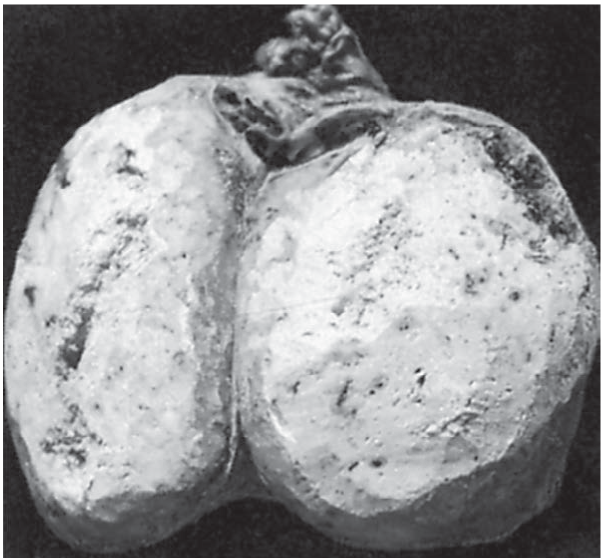
Spot diagnosis
Gross appearance
- Testis is enlarged, usually replaced by a well-circumscribed, homogeneous mass.
- Cut surface is gray-white to pinkish cream, fleshy, and lobulated.
- Tumor commonly compresses the residual testicular tissue.
- Hemorrhage and necrosis are usually absent or minimal. If prominent, consider a non-seminomatous germ-cell tumor, especially embryonal carcinoma.
- Tunica may remain intact in early lesions.
Microscopy
- Sheets or nests of uniform, large polygonal cells.
- Cells have clear glycogen-rich cytoplasm, central nucleus, and prominent nucleolus, giving a “fried-egg” appearance.
- Tumor cells are separated by delicate fibrous septa containing dense lymphocytic infiltrate.
- Occasional syncytiotrophoblastic giant cells may be present.
Clinical and laboratory points
- Peak age: 30-40 years.
- Usually presents as a painless testicular swelling.
- Associated with cryptorchidism and germ-cell neoplasia in situ.
- AFP is not elevated in pure seminoma. Elevated AFP indicates an associated non-seminomatous component until proved otherwise.
- β-hCG can be mildly elevated if syncytiotrophoblastic cells are present.
- Spreads mainly by lymphatics to para-aortic (retroperitoneal) lymph nodes, with hematogenous spread occurring later. Bailey & Love's Short Practice of Surgery, 28th ed., p. 1437-1443.
- It is highly radiosensitive and has an excellent prognosis.
One-line practical presentation
“This is a testis showing a homogeneous gray-white, fleshy tumor compressing the surrounding parenchyma, suggestive of seminoma.”
Important differential: seminoma vs embryonal carcinoma
| Feature | Seminoma | Embryonal carcinoma |
|---|---|---|
| Gross | Homogeneous, gray-white/cream, little hemorrhage or necrosis | Variegated, ill-defined, with prominent hemorrhage and necrosis |
| Cells | Uniform, clear cytoplasm | Pleomorphic, indistinct cell borders |
| Histology | Fibrous septa with lymphocytes | Solid, glandular or papillary patterns; marked atypia |
| Markers | AFP normal in pure tumor | AFP and/or β-hCG may be raised |
| Behavior | Usually more radiosensitive, relatively better prognosis | More aggressive |
Viva questions
-
Why is the cut surface homogeneous?
Because seminoma is composed of relatively uniform tumor cells with little hemorrhage and necrosis. -
What is the route of lymphatic spread?
Para-aortic lymph nodes, because testicular lymphatics follow the gonadal vessels. -
What tumor marker rules out pure seminoma when elevated?
AFP. -
Does it occur in children?
Rarely. It is mainly a tumor of young and middle-aged adults. -
What is the classic microscopic background?
Fibrous septa infiltrated by lymphocytes.
Quiz me on seminoma with rapid-fire viva questions
Seminoma
Other testicular tumor
- Embryonal carcinoma - high-yield differential from seminoma
- Yolk sac tumor - commonest testicular tumor in children
- Choriocarcinoma - hemorrhagic tumor with high β-hCG
- Teratoma - cystic/solid tumor with multiple tissue types
- Leydig cell tumor - sex-cord stromal tumor, may cause gynecomastia
Mature cystic teratomaof ovary
ovarian mature cystic teratoma
ovarian mature cystic teratoma gross pathology dermoid cyst features
Mature cystic teratoma of ovary
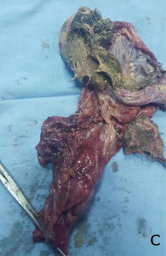
Spot diagnosis
This is an ovarian mature cystic teratoma (dermoid cyst), a benign germ-cell tumor containing mature tissue derived from one or more germ layers, most often ectoderm.
Gross appearance
- Usually unilateral, spherical, and cystic.
- Outer surface is smooth.
- On cutting, it is commonly unilocular.
- Cavity contains thick, greasy pultaceous sebaceous material and a tuft/ball of hair.
- May show teeth, bone, cartilage, or other mature tissues.
- A solid mural projection, the Rokitansky protuberance (dermoid plug), is characteristic. Teeth and bone are often found in it.
Microscopy
- Cyst wall lined by keratinizing stratified squamous epithelium with skin appendages:
- Hair follicles
- Sebaceous glands
- Sweat glands
- Other mature tissues from all three germ layers may occur:
- Ectoderm: skin, neural tissue
- Mesoderm: cartilage, bone, fat, muscle
- Endoderm: respiratory or intestinal epithelium, thyroid tissue
Important complications
- Torsion - most common complication
- Rupture causing chemical peritonitis due to spilled sebaceous material
- Infection
- Rare malignant transformation, most often squamous cell carcinoma, particularly in older patients
- Rarely associated with anti-NMDA receptor encephalitis if neural tissue is present
One-line practical presentation
“This is an ovarian dermoid cyst showing a unilocular cyst with pultaceous sebaceous material, hair, and possibly teeth or bone, consistent with a mature cystic teratoma.”
Differentiate: mature vs immature teratoma
| Feature | Mature cystic teratoma | Immature teratoma |
|---|---|---|
| Nature | Usually benign | Malignant potential |
| Age | Reproductive age | Usually younger patients |
| Gross | Cystic, hair, sebum, teeth, bone | Large, solid and cystic, variegated |
| Histology | Mature adult-type tissues | Immature or embryonal, especially neuroectodermal tissue |
| Prognosis | Excellent | Depends on grade and stage |
Rapid-fire viva
- Most common ovarian germ-cell tumor? Mature cystic teratoma.
- Another name? Dermoid cyst.
- Characteristic contents? Hair and sebaceous material.
- Characteristic mural nodule? Rokitansky protuberance.
- Commonest complication? Torsion.
- Commonest malignant transformation? Squamous cell carcinoma.
- Is it usually unilateral or bilateral? Usually unilateral, but bilateral lesions can occur.
- Why does it contain teeth and hair? It differentiates into mature tissues of multiple germ layers.
Carcinoma penis
penile squamous cell carcinoma
penile squamous cell carcinoma pathology gross appearance risk factors lymph node spread
Carcinoma of penis
Spot diagnosis
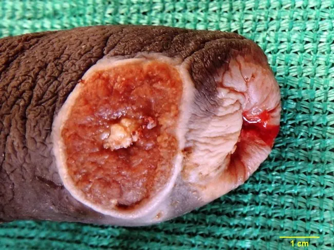
Squamous cell carcinoma of penis, usually arising from the glans penis or prepuce.
Gross appearance
- Commonly affects glans, followed by prepuce and shaft.
- Begins as an ulcerative or papillary/exophytic cauliflower-like growth.
- May form a hard, indurated, non-healing ulcer with everted edges.
- Cut surface is irregular, firm, gray-white or tan-white, sometimes with necrosis.
- Advanced lesions can infiltrate corpora and destroy the glans or shaft.
Etiology and risk factors
- HPV, especially types 16 and 18
- Phimosis and poor genital hygiene
- Chronic balanitis/balanoposthitis
- Lichen sclerosus
- Smoking
- Penile intraepithelial neoplasia (PeIN), including:
- Erythroplasia of Queyrat: carcinoma in situ on glans/prepuce
- Bowen disease: carcinoma in situ on penile shaft
Microscopy
- Invasive nests, cords, or sheets of malignant squamous cells.
- Varying keratinization with keratin pearls in well-differentiated tumors.
- Cellular pleomorphism, hyperchromatic nuclei, abnormal mitoses.
- Infiltration into underlying stroma and possibly corpus spongiosum/cavernosum.
Spread
- First spreads by lymphatics to superficial and deep inguinal lymph nodes.
- Then to external iliac and pelvic nodes.
- Hematogenous spread is late and uncommon, to lung, liver, bone, or brain.
One-line practical presentation
“This is a penectomy specimen showing an ulceroproliferative growth involving the glans penis, suggestive of squamous cell carcinoma of penis.”
Important viva points
- Most common histologic type? Squamous cell carcinoma.
- Most common site? Glans penis.
- Commonest mode of spread? Lymphatic spread to inguinal lymph nodes.
- Important viral association? HPV 16 and 18.
- Precancerous lesion on glans? Erythroplasia of Queyrat.
- Precancerous lesion on shaft? Bowen disease.
- Common clinical presentation? Non-healing ulcer, exophytic growth, bleeding, discharge, or phimosis.
- Best investigation to confirm diagnosis? Biopsy.
- What is the key prognostic factor? Presence and extent of lymph-node metastasis. The current EAU guideline summary describes stepwise inguinal then pelvic nodal spread.
Chronic pyelonephritis
chronic pyelonephritis reflux nephropathy
chronic pyelonephritis pathology gross irregular corticomedullary scars blunted calyces
Chronic pyelonephritis
Causes
- Reflux nephropathy - most common cause
- Chronic urinary obstruction:
- Renal calculi
- Ureteric obstruction
- Posterior urethral valves
- Prostatic enlargement
- Recurrent bacterial urinary infections
Gross pathology
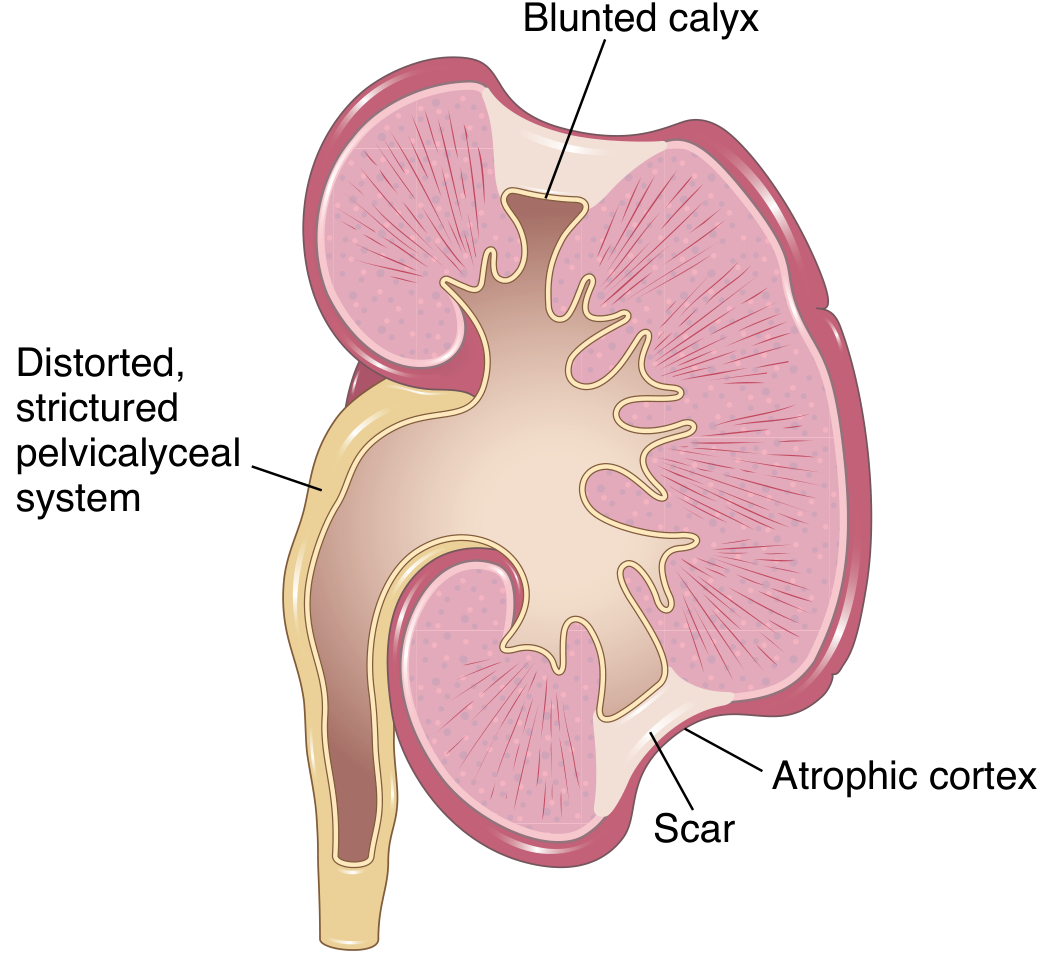
- Kidney is small, contracted, and irregularly scarred.
- Characteristic coarse, irregular corticomedullary scars.
- Scars are usually at the upper and lower poles.
- Underlying calyces are dilated, blunted, clubbed, and deformed.
- Papillae are flattened.
- If both kidneys are involved, scarring is typically asymmetrical.
Microscopy
- Patchy interstitial fibrosis.
- Chronic inflammatory infiltrate: lymphocytes and plasma cells, sometimes neutrophils.
- Tubular atrophy and dilatation.
- Dilated atrophic tubules contain eosinophilic protein casts resembling thyroid colloid: thyroidization of tubules.
- Glomeruli may be normal initially, later showing ischemic sclerosis and periglomerular fibrosis.
One-line practical presentation
“This is a kidney showing irregular coarse corticomedullary scars, with underlying blunted and deformed calyces, suggestive of chronic pyelonephritis.”
Chronic vs acute pyelonephritis
| Feature | Acute pyelonephritis | Chronic pyelonephritis |
|---|---|---|
| Cause | Acute bacterial infection, usually ascending | Recurrent infections due to reflux or obstruction |
| Onset | Sudden fever, flank pain, dysuria | Often insidious, may be asymptomatic |
| Gross kidney | Enlarged, congested; small cortical abscesses or yellow-white streaks | Small, contracted kidney with coarse irregular scars |
| Calyces | Usually not deformed | Blunted, clubbed, deformed calyces |
| Inflammation | Predominantly neutrophils in interstitium and tubules | Lymphocytes, plasma cells, fibrosis |
| Tubules | WBC casts may occur | Tubular atrophy, dilatation, thyroidization |
| Outcome | Usually resolves with treatment, may form abscess/papillary necrosis | Hypertension, CKD, eventually ESRD |
Rapid-fire viva
- Most common cause? Vesicoureteric reflux causing reflux nephropathy.
- Most characteristic gross feature? Coarse corticomedullary scars overlying blunted calyces.
- Why are polar scars common? Intrarenal reflux preferentially affects upper and lower pole papillae.
- What is thyroidization? Atrophic, dilated tubules filled with colloid-like proteinaceous casts.
- How is bilateral chronic pyelonephritis distinguished from chronic glomerulonephritis? Chronic pyelonephritis causes asymmetrical coarse scars and calyceal deformity; chronic glomerulonephritis produces symmetrical fine granular contraction without calyceal deformity.
- Major complications? Hypertension, chronic renal failure, and ESRD.
- Important diagnostic clue on imaging? A small, asymmetrically contracted kidney with polar scars and clubbed calyces.
Fibroadenoma
breast fibroadenoma
fibroadenoma breast pathology gross circumscribed slit like spaces intracanalicular pericanalicular
Fibroadenoma of breast
Typical patient
- Usually age 15-35 years
- Presents as a painless, mobile breast lump
- Called the “breast mouse” because it slips freely under the fingers
Gross pathology
- Single, well-circumscribed, firm, rubbery, ovoid mass.
- Usually not truly encapsulated, but sharply demarcated from surrounding breast tissue.
- Cut surface: homogeneous gray-white to tan, whorled, bulging, with possible slit-like spaces.
- Usually 1-3 cm, but may grow large, especially in pregnancy or adolescence.
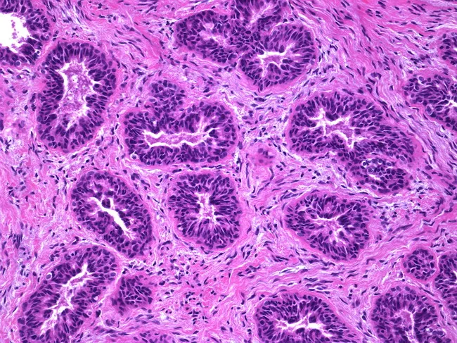
Microscopy
-
Pericanalicular pattern
- Proliferating fibrous stroma surrounds round/open ducts.
- Duct lumina remain patent.
-
Intracanalicular pattern
- Stroma compresses ducts into elongated, slit-like spaces.
- This is often emphasized in viva.
One-line practical presentation
“This is a well-circumscribed, firm, gray-white breast mass with a homogeneous bulging cut surface, suggestive of fibroadenoma.”
Important differential: Fibroadenoma vs phyllodes tumor
| Feature | Fibroadenoma | Phyllodes tumor |
|---|---|---|
| Age | Usually 15-35 years | Usually 40-50 years |
| Growth | Slow | Often rapid |
| Size | Usually small | Often large |
| Gross | Firm, circumscribed, homogeneous | Lobulated, may show cleft-like spaces |
| Histology | Uniform stroma, ducts compressed or surrounded by stroma | Leaf-like architecture, more cellular stroma |
| Behavior | Benign | May be benign, borderline, or malignant |
Rapid-fire viva
- Commonest age group? Young women, usually 15-35 years.
- Is it benign or malignant? Benign.
- Why is it called breast mouse? It is freely mobile.
- Is it encapsulated? No, but it is well circumscribed.
- What are the two microscopic patterns? Pericanalicular and intracanalicular.
- What happens to ducts in intracanalicular fibroadenoma? They are compressed into slit-like spaces by proliferating stroma.
- What happens to ducts in pericanalicular fibroadenoma? They retain rounded, open lumina and are surrounded by stroma.
- What lesion must be differentiated from it? Phyllodes tumor.
- Does it usually increase breast cancer risk? A simple fibroadenoma generally does not materially increase risk.
- What causes rapid enlargement in a young girl? Juvenile fibroadenoma.