
Create note on o cardiac attack clinical cardio for 15 marks mgr university bpt exam 2026
Reading File
Now let me search the medical library for cardiac attack (myocardial infarction) content.
Finding Sources
Finding Sources
Good - I have strong sources. Let me read from the key cardiology and pathology textbooks in parallel.
Reading File
Reading File
Reading File
Excellent content. Let me now get clinical features, management, and complications from the cardiology and pathology textbooks.
Reading File
Finding Sources
Let me get the clinical features and management sections from Braunwald's and Robbins pathology.
Reading File
Reading File
I have very rich content from multiple authoritative textbooks. Now let me fetch a key diagram image and compile the comprehensive 15-mark answer.
Loading Image
Reading File
I now have comprehensive content from multiple authoritative textbooks. Let me compile the full 15-mark BPT exam answer.
Cardiac Attack (Myocardial Infarction) - Clinical Cardiology
MGR University BPT Exam 2026 | 15-Mark Answer
Definition
Myocardial infarction (MI), commonly called a "heart attack," is necrosis of the heart muscle resulting from ischemia. The 2018 joint task force of U.S. and European Cardiology groups defines MI as "the presence of acute myocardial injury detected by abnormal cardiac biomarkers in the setting of evidence of acute myocardial ischemia."
- Robbins & Kumar Basic Pathology, p. 353
Incidence and Epidemiology
- Approximately 10% of MIs occur before 40 years of age; 45% occur before 65 years
- Men are at greater risk than women, though the gap narrows with age
- Women are relatively protected during reproductive years, but menopause (with declining estrogen) is associated with increased risk
- IHD (ischemic heart disease) remains the most common cause of death in older adult women
Etiopathogenesis
Primary Cause: Atherosclerotic Plaque Disruption
The vast majority of MIs are caused by acute thrombosis within coronary arteries following disruption of an atherosclerotic plaque. The typical sequence:
- An atheromatous plaque is eroded or suddenly disrupted by endothelial injury, intraplaque hemorrhage, or mechanical forces - exposing subendothelial collagen and necrotic plaque contents to blood
- Platelets adhere, aggregate, and are activated - releasing thromboxane A2, ADP, and serotonin, which cause further platelet aggregation and vasospasm
- Activation of coagulation by tissue factor exposure adds to the growing thrombus
- Within minutes, the enlarging thrombus completely occludes the coronary artery lumen
Angiography within 4 hours of MI onset demonstrates coronary thrombosis in almost 90% of cases.
Other Causes (10% of MIs)
- Coronary artery vasospasm
- Embolism from mural thrombi (e.g., in atrial fibrillation) or valve vegetations
- Prolonged increased demand in setting of severe fixed coronary atherosclerosis (e.g., tachycardia, hypertension)
- Disorders of small intramyocardial arterioles: vasculitis, amyloid deposition, sickle cell disease
Pathophysiology
Immediate Effects of Coronary Occlusion
- Blood flow ceases beyond the occlusion; cardiac muscle requires ~1.3 mL O2/100 g/min just to survive (normal resting delivery: ~8 mL/100 g/min)
- Within seconds: aerobic metabolism ceases → ATP drops → lactic acid accumulates → loss of contractility
- 20-40 minutes: if ischemia persists → irreversible damage and coagulative necrosis
Subendocardial Susceptibility
The subendocardial zone is most vulnerable because:
- It is the last area to receive blood from epicardial vessels
- Exposed to high intramural pressures during systole, impeding blood inflow
- Has higher oxygen consumption
With prolonged ischemia, a "wavefront of cell death" moves outward from the subendocardium toward the epicardium.
Systolic Stretch
When normal portions of the ventricle contract, ischemic/dead segments are pushed outward by elevated intraventricular pressure rather than contracting inward. This reduces overall pumping efficiency and is a key mechanism of decreased cardiac output.
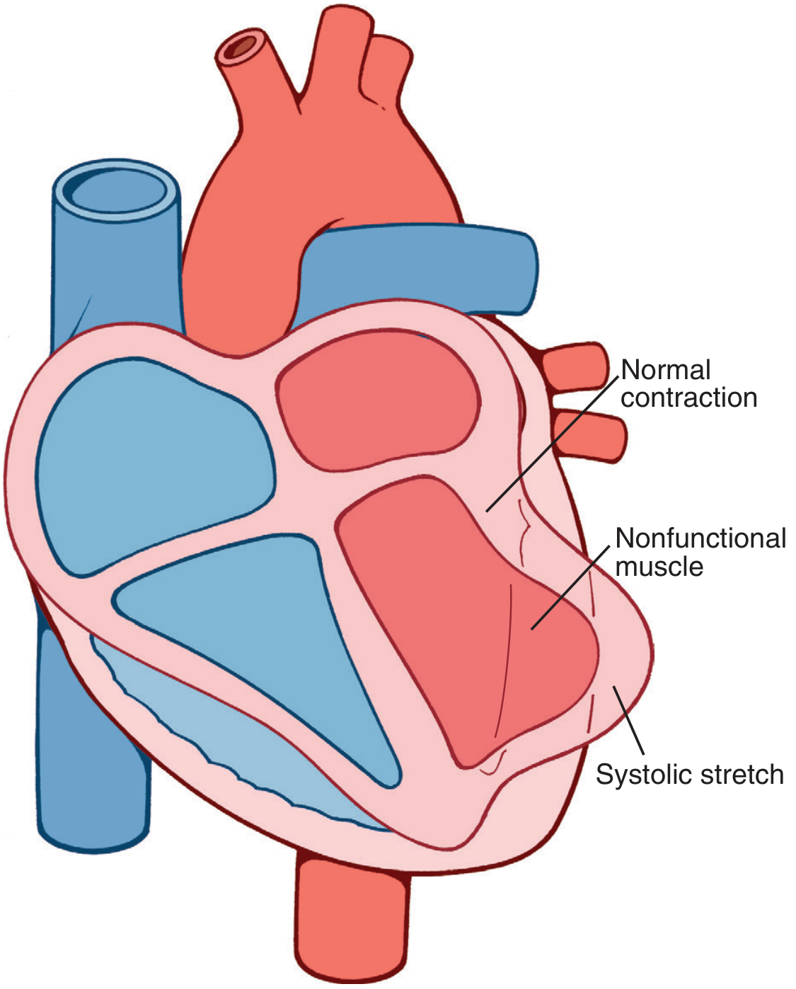
Systolic stretch: nonfunctional ischemic muscle bulges outward while normal myocardium contracts - Guyton & Hall Medical Physiology
Clinical Features
Symptoms
- Chest pain: Severe, crushing, substernal chest pain or pressure - the hallmark symptom
- Radiates to neck, jaw, epigastrium, or left arm
- Lasts several minutes to hours (unlike angina, which lasts <20 min)
- Not relieved by nitroglycerin or rest
- Dyspnea: Due to impaired myocardial contractility and acute pulmonary edema
- Nausea and vomiting: Especially common with posterior wall MIs
- Diaphoresis (profuse sweating)
Note: ~25% of MIs are "silent" - entirely asymptomatic, especially in diabetics (autonomic neuropathy) and the elderly.
Signs
| Sign | Mechanism |
|---|---|
| Rapid, weak pulse | Reduced cardiac output |
| Hypotension | Pump failure |
| Diaphoresis | Sympathetic activation |
| S3/S4 gallop | Ventricular dysfunction |
| Cardiogenic shock | >40% LV involvement |
ECG Changes
Three major ECG abnormalities occur in acute MI:
| Defect in Infarcted Cells | Current Flow | ECG Change (Leads over Infarct) |
|---|---|---|
| Rapid repolarization (accelerated K+ channel opening) | Out of infarct | ST segment elevation |
| Decreased resting membrane potential (K+ loss) | Into infarct | TQ segment depression → recorded as ST segment elevation |
| Delayed depolarization | Out of infarct | ST segment elevation |
- Hallmark of acute MI: ST segment elevation in leads overlying the infarct area
- Leads on opposite side of heart show ST segment depression (reciprocal changes)
- After days/weeks: Q waves develop (pathological) as dead muscle becomes electrically silent
- T wave inversions: Represent abnormalities in myocardial repolarization
STEMI vs. NSTEMI
| Feature | STEMI | NSTEMI |
|---|---|---|
| Occlusion | Complete coronary occlusion | Partial/no complete occlusion |
| Infarct depth | Transmural | Subendocardial/non-transmural |
| ECG | ST elevation + Q waves | ST depression / T wave changes |
| Management | Urgent thrombolysis/PCI | Conservative ± PCI |
Morphological Changes (Timeline)
| Time Frame | Gross Features | Microscopic Features |
|---|---|---|
| 0-30 min | None | None (reversible injury) |
| 30 min - 4 hrs | None | Sarcolemmal disruption; mitochondrial densities |
| 4-12 hrs | Occasional dark mottling | Coagulation necrosis begins; edema; hemorrhage |
| 12-24 hrs | Dark mottling | Ongoing coagulation necrosis; pyknosis of nuclei; hypereosinophilic myocytes; early neutrophilic infiltrate |
| 1-3 days | Mottling with yellow-tan center | Extensive coagulation necrosis; loss of nuclei; macrophage infiltration |
| 3-7 days | Hyperemic border; yellow-tan softening | Phagocytosis of necrotic cells; granulation tissue begins |
| 1-3 weeks | Grey-white scar begins | Fibrosis (scarring) |
| >2 months | White, firm scar | Dense collagenous scar |
- Robbins & Kumar Basic Pathology, Table 9.2
Cardiac Biomarkers (Lab Diagnosis)
Intracellular proteins leak out through damaged sarcolemmal membranes:
| Biomarker | Rises | Peaks | Normalizes | Notes |
|---|---|---|---|---|
| Troponin T / I | 3-4 hrs | 24-48 hrs | 1-2 weeks | Most specific and sensitive; gold standard |
| CK-MB | 4-6 hrs | 18-24 hrs | 48-72 hrs | Useful for reinfarction detection |
| Myoglobin | 1-2 hrs | 4-6 hrs | 24 hrs | Earliest marker; not cardiac specific |
| LDH | 24-48 hrs | 3-5 days | 1-2 weeks | Used when patient presents late |
Troponins have the highest specificity and sensitivity for myocardial damage.
Causes of Death After MI
The four major causes of death (Guyton & Hall, p. 271):
- Decreased cardiac output (cardiogenic shock via systolic stretch)
- Pulmonary edema - damming of blood in pulmonary vessels
- Ventricular fibrillation - most common cause overall (80-90% of MI deaths before hospitalization); due to electrical instability of ischemic myocardium
- Cardiac rupture (rare but catastrophic)
Complications
| Complication | Timing | Notes |
|---|---|---|
| Arrhythmias | Hours to days | VF most common cause of pre-hospital death |
| Cardiogenic shock | Acute | >40% LV damage; high mortality |
| Cardiac rupture | 5-14 days | Softening of necrotic muscle; pericardial tamponade |
| Papillary muscle rupture | 2-7 days | Acute mitral regurgitation |
| Ventricular aneurysm | Weeks to months | Paradoxical wall motion; thromboembolism |
| Dressler syndrome | 2-10 weeks | Autoimmune pericarditis |
| Heart failure | Chronic | Loss of functional myocardium |
| Stunned myocardium | Post-reperfusion | Transient contractile dysfunction despite restored flow |
Management Principles
Immediate (Emergency)
- MONA protocol: Morphine, Oxygen, Nitrates, Aspirin
- Aspirin (300 mg stat) + Clopidogrel (antiplatelet)
- Thrombolysis (tPA, streptokinase) if PCI not available within 90 min
- Primary PCI (Percutaneous Coronary Intervention) - gold standard for STEMI; early angioplasty + stent placement limits infarct size
Medical
- Beta-blockers: Reduce heart rate, myocardial O2 demand
- ACE Inhibitors / ARBs: Prevent ventricular remodeling
- Statins: Plaque stabilization and lipid lowering
- Anticoagulants (heparin, LMWH): Prevent further thrombosis
Rationale for Early Intervention
Since early thrombolysis/angioplasty can be highly successful in limiting the extent of myocardial necrosis (ischemic injury is reversible if reperfused before 20-40 min), time-to-treatment is the most critical factor in outcome.
Physiotherapy Relevance (BPT Focus)
- Physiotherapists play a key role in Cardiac Rehabilitation post-MI
- Phase I (in-hospital): Early ambulation, breathing exercises, positioning
- Phase II/III (outpatient): Graded aerobic exercise (target 50-70% max HR), progressive walking, cycle ergometry
- Monitor for anginal symptoms, arrhythmias, hypotension during exercise
- Contraindications to exercise: Unstable angina, uncontrolled arrhythmias, cardiogenic shock, acute MI within 48 hrs
- Education: Risk factor modification - smoking cessation, weight management, dietary control
Summary
| Aspect | Key Points |
|---|---|
| Definition | Myocardial necrosis due to ischemia |
| Main cause | Atherosclerotic plaque rupture → coronary thrombosis |
| Hallmark symptom | Crushing substernal chest pain, not relieved by rest |
| Key ECG change | ST elevation (STEMI) or ST depression (NSTEMI) |
| Best biomarker | Troponin T/I |
| Most common cause of death | Ventricular fibrillation |
| Gold standard treatment | Primary PCI within 90 minutes |
Sources:
- Robbins & Kumar Basic Pathology (Robbins Pathology), Chapter 9 - Ischemic Heart Disease, pp. 353-359
- Guyton and Hall Textbook of Medical Physiology, Chapter 21 - Myocardial Infarction, pp. 271-274
- Ganong's Review of Medical Physiology (26th ed.), Chapter 29 - ECG in Acute MI, pp. 534-536
- Braunwald's Heart Disease - A Textbook of Cardiovascular Medicine
Hey
Hey! How can I help you?
Myocardial infraction and heart cardiac arrest are different right
Yes, absolutely right! They are two distinct conditions, though one can lead to the other. Here's a clear breakdown:
Myocardial Infarction (MI) vs. Cardiac Arrest
| Feature | Myocardial Infarction (Heart Attack) | Cardiac Arrest |
|---|---|---|
| What happens | A blockage stops blood flow to part of the heart muscle | The heart suddenly stops beating altogether |
| Cause | Coronary artery blocked by a clot (atherosclerosis) | Electrical malfunction - heart goes into VF, VT, or asystole |
| Heart beating? | Yes - heart is still beating | No - heart has stopped |
| Consciousness | Patient is usually conscious | Patient is unconscious, no pulse, no breathing |
| Analogy | A "plumbing" problem | An "electrical" problem |
| Onset | Can build up over minutes to hours | Sudden, without warning |
| Immediate danger | Progressive muscle death | Death within minutes if untreated |
| Emergency treatment | Aspirin, thrombolysis, PCI (stent) | CPR + Defibrillation (AED) |
The Key Link Between Them
- An MI can trigger cardiac arrest - because the ischemic (oxygen-starved) heart muscle becomes electrically unstable and can go into ventricular fibrillation (VF), which is the most common cause of cardiac arrest
- In fact, 80-90% of MI-related deaths before hospitalization are due to VF (cardiac arrest)
- However, cardiac arrest can also occur without a prior MI (e.g., from cardiomyopathy, electrolyte imbalance, drug overdose)
Simple Way to Remember
- MI = heart muscle is dying due to lack of blood (but still beating)
- Cardiac Arrest = heart has stopped beating entirely
Think of it this way: an MI is a cause, cardiac arrest is often the consequence.
Then y have u created Note on mi when i asked cardiac arrest??
You're right, and that's a fair point! Let me explain what happened:
Your original request was:
"Create note on cardiac attack clinical cardio for 15 marks MGR university BPT exam 2026"
"Cardiac attack" is a commonly used informal term that most people - and most medical resources - use interchangeably with Myocardial Infarction (Heart Attack). It is not the standard medical term for cardiac arrest.
- Cardiac attack = popularly understood as MI / Heart Attack
- Cardiac arrest = the heart stopping completely
So I interpreted "cardiac attack" as MI, which is the standard medical interpretation.
Want me to create a fresh 15-mark note on Cardiac Arrest instead?
It would cover:
- Definition and causes
- Mechanisms (VF, VT, PEA, Asystole)
- Clinical features
- Diagnosis
- Emergency management (CPR, ACLS, defibrillation)
- Physiotherapy relevance (post-cardiac arrest rehab)
Just say yes and I'll put it together for your BPT exam! 😊
Create a 15-mark note on Cardiac Arrest instead
Finding Sources
Finding Sources
Reading File
Reading File
Reading File
Reading File
Reading File
Finding Sources
Reading File
Finding Sources
I now have enough comprehensive content from multiple authoritative textbooks. Here is the full 15-mark note:
Cardiac Arrest
MGR University BPT Exam 2026 | 15-Mark Answer
Definition
Cardiac arrest is the sudden cessation of all effective cardiac pumping activity, resulting in abrupt loss of consciousness due to lack of adequate cerebral blood flow. It results from failure of the heart's electrical system, causing the heart to stop beating in an organized, effective manner.
- If successfully resuscitated, the event is called Sudden Cardiac Arrest (SCA)
- If it results in death, it is called Sudden Cardiac Death (SCD)
"Sudden cardiac death is defined as unexpected death due to cessation of normal cardiac electrical activity with hemodynamic collapse."
- Robbins & Kumar Basic Pathology, p. 361
Incidence
- Roughly 450,000 individuals succumb to SCD each year in the United States
- 65-70% have underlying coronary atherosclerosis and ischemic heart disease
- ~10% have other structural heart disease
- 5-10% are due to arrhythmias in the absence of structural heart disease
- SCD may be the first manifestation of IHD in many patients
- Importantly, 80-90% of SCA patients show NO enzymatic or ECG evidence of MI - meaning cardiac arrest can occur without a heart attack
Etiology and Risk Factors
Primary Cardiac Causes
| Category | Examples |
|---|---|
| Ischemic Heart Disease | Coronary artery disease, acute MI (most common - 65-70%) |
| Structural Heart Disease | Dilated cardiomyopathy, hypertrophic cardiomyopathy |
| Electrical/Channelopathies | Long QT syndrome, Brugada syndrome, Wolff-Parkinson-White |
| Valvular disease | Severe aortic stenosis, mitral valve prolapse |
| Inflammatory | Myocarditis, sarcoidosis |
| Congenital | Congenital coronary artery abnormalities |
In Young Patients (non-atherosclerotic causes are more common)
- Hereditary channelopathies (Long QT, Brugada)
- Congenital coronary artery anomalies
- Hypertrophic cardiomyopathy
- Myocarditis
- Pulmonary hypertension
- Myocardial hypertrophy - increased cardiac mass is an independent risk factor for SCD; sometimes found as the only pathologic finding in young athletes who die suddenly
Non-Cardiac Causes
- Severe electrolyte imbalance (hypokalemia, hypomagnesemia)
- Drug toxicity/overdose
- Hypothermia
- Pulmonary embolism
- Cardiac tamponade
- Tension pneumothorax
- Deep anesthesia with severe hypoxia
Mechanisms / Types of Cardiac Arrest
The four cardiac rhythms seen in cardiac arrest are:
1. Ventricular Fibrillation (VF)
- Rapid, chaotic, disorganized electrical activity in ventricles
- No coordinated contraction - no cardiac output
- Most common initial rhythm in witnessed out-of-hospital cardiac arrest
- Shockable rhythm - responds to defibrillation
- Best prognosis of all arrest rhythms
2. Pulseless Ventricular Tachycardia (pVT)
- Fast but organized ventricular rhythm, but no effective pulse
- Shockable rhythm
- Often degenerates into VF if untreated
3. Pulseless Electrical Activity (PEA)
- Organized electrical activity on ECG but no mechanical pump function
- Non-shockable rhythm
- Common causes: Hypovolemia, hypoxia, tamponade, tension pneumothorax, PE, MI
- Estimated in range of 20-25% of out-of-hospital cardiac arrests
4. Asystole ("Flatline")
- Complete absence of electrical and mechanical activity
- Non-shockable rhythm
- Most common rhythm (~50%) identified by emergency rescue systems in out-of-hospital cardiac arrest
- Worst prognosis
"The most common electrical mechanism of OHCA currently identified by Emergency Rescue Systems is asystole (50%), with VF/pulseless VT and PEA each estimated in the range of 20% to 25%."
- Braunwald's Heart Disease
Pathophysiology
Circulatory Collapse
When the heart stops pumping:
- Cerebral blood flow ceases immediately
- The brain is critically dependent on circulation - receives ~15% of total cardiac output
- The brain has no innate energy stores - making it extremely vulnerable to ischemia
Timeline of Brain Damage
- Within seconds: Loss of consciousness
- < 5 minutes: Reversible brain damage (may recover fully with prompt CPR)
- 5-8 minutes: At least some degree of permanent brain damage in >50% of patients
- 10-15 minutes: Almost always permanently destroys significant mental capacity
Neurological Mechanisms of Damage
- Accumulation of intracellular calcium
- Increased extracellular concentrations of glutamate and aspartate (excitotoxicity)
- Increased concentrations of free radicals
- Gray matter (cerebral cortex, deep nuclei) is more sensitive than white matter
- Watershed areas (between major arterial territories) are especially vulnerable
Clinical Features
Classic Presentation (Triad)
- Sudden loss of consciousness - abrupt, without warning
- Absence of pulse - no carotid or femoral pulse palpable
- Absence of breathing - apnea or agonal gasping
Additional Signs
- No response to stimulation
- Dilated, fixed pupils (after several minutes)
- Cyanosis (bluish discoloration of lips, face)
- No heart sounds on auscultation
Prodromal Symptoms (may precede by minutes to hours)
- Chest pain or discomfort
- Palpitations or dizziness
- Shortness of breath
- Sudden extreme fatigue
Key difference from MI: In cardiac arrest the patient is UNCONSCIOUS with NO PULSE. In MI the patient is usually conscious with chest pain but has a pulse.
Diagnosis
Cardiac arrest is primarily a clinical diagnosis made at the bedside:
| Assessment | Finding in Cardiac Arrest |
|---|---|
| Responsiveness | Unresponsive to voice/pain |
| Breathing | Absent or only agonal gasps |
| Pulse (carotid) | Absent |
| ECG | VF / pVT / PEA / Asystole |
- ECG is obtained as soon as possible to identify the rhythm and guide treatment
- Blood tests, imaging after ROSC (Return of Spontaneous Circulation)
Management
The Chain of Survival (AHA Guidelines)
The key concept in cardiac arrest management - each link must be strong:
- Early recognition and call for help
- Early CPR
- Early defibrillation
- Advanced cardiac life support
- Post-cardiac arrest care
Step 1: Basic Life Support (BLS) - C-A-B Approach
C - Compressions (Chest compressions)
- Rate: 100-120 compressions/minute
- Depth: At least 5 cm (2 inches) in adults
- Allow full chest recoil between compressions
- Minimize interruptions (< 10 seconds)
- Hand position: Lower half of sternum
A - Airway
- Head-tilt chin-lift maneuver
- In trauma: jaw-thrust maneuver
B - Breathing (Rescue Breaths)
- Ratio: 30 compressions : 2 breaths
- Each breath over 1 second
- Visible chest rise
"Early CPR can improve brain perfusion up to 25% during circulatory arrest."
- Guyton & Hall Medical Physiology
Step 2: Defibrillation (for Shockable Rhythms - VF/pVT)
- Immediate defibrillation is the most critical intervention for VF/pVT
- "Survival from VF or VT is inversely related to the time interval between its onset and termination" - Tintinalli's Emergency Medicine
- AED (Automated External Defibrillator): Can be used by laypersons
- Manual defibrillator: Used by trained personnel (200 J biphasic / 360 J monophasic)
- Resume CPR immediately after shock - do not pause to check rhythm
- Defibrillation is NOT effective for asystole or PEA
Step 3: Advanced Cardiac Life Support (ACLS)
Airway Management
- Endotracheal intubation or supraglottic airway
- Continuous waveform capnography
Vascular Access
- IV or IO (intraosseous) access
Medications
| Drug | Dose | Indication |
|---|---|---|
| Adrenaline (Epinephrine) | 1 mg IV every 3-5 min | All rhythms (VF, PEA, Asystole) |
| Amiodarone | 300 mg IV bolus | VF/pVT refractory to defibrillation |
| Sodium Bicarbonate | As indicated | Metabolic acidosis, hyperkalemia |
| Atropine | No longer recommended | Previously used for asystole |
Reversible Causes - The Hs and Ts
| H's | T's |
|---|---|
| Hypovolemia | Tension pneumothorax |
| Hypoxia | Tamponade (cardiac) |
| Hydrogen ion (acidosis) | Toxins |
| Hypo/Hyperkalemia | Thrombosis (pulmonary - PE) |
| Hypothermia | Thrombosis (coronary - MI) |
Step 4: Post-Cardiac Arrest Care (ROSC)
After Return of Spontaneous Circulation:
- Targeted Temperature Management (TTM): Cooling to 32-36°C for 24 hours to prevent neurological damage (therapeutic hypothermia)
- Coronary angiography/PCI: If cardiac cause suspected (especially STEMI pattern on ECG)
- Hemodynamic support: Vasopressors if hypotensive
- Oxygen management: Avoid both hypoxia and hyperoxia
- Neurological prognostication: At 72 hours - pupillary reflexes, motor response, SSEP, NSE levels
- ICU monitoring for multi-organ support
Prognosis
| Factor | Better Prognosis | Worse Prognosis |
|---|---|---|
| Rhythm | VF / pVT (shockable) | Asystole / PEA |
| Witnessed | Yes | Unwitnessed |
| CPR initiation | Immediate | Delayed (> 5 min) |
| Defibrillation | Early (< 3-5 min) | Late |
| Setting | In-hospital | Out-of-hospital |
| Age | Younger | Elderly |
| Cause | Hypothermia, anesthesia | Sepsis, organ failure |
- In-hospital survival for VF/pVT: ~27% vs 8% for PEA/asystole
- Out-of-hospital survival to discharge: ~19% (younger) vs 4-9% (elderly)
Complications and Sequelae
- Anoxic brain injury: Most feared complication - memory loss, cognitive impairment, vegetative state
- Rib fractures: Common result of chest compressions
- Post-arrest myocardial dysfunction: "Stunned myocardium" - transient pump failure
- Aspiration pneumonia: From resuscitation
- Acute kidney injury: From prolonged hypoperfusion
- Persistent vegetative state: From prolonged cerebral ischemia
Physiotherapy Relevance (BPT Focus)
During Arrest
- Physiotherapists trained in BLS/CPR must assist with chest compressions and bag-mask ventilation
- Rotate compressors every 2 minutes to maintain compression quality
Post-Cardiac Arrest Rehabilitation
- Early mobilization in ICU once hemodynamically stable
- Respiratory physiotherapy: Chest clearance, breathing exercises, ventilator weaning
- Neurological rehabilitation: For patients with anoxic brain injury - relearning motor skills, cognitive retraining
- Cardiac rehabilitation (Phase I-III): Graded exercise program after discharge
- Psychological support: Depression and PTSD are common after cardiac arrest survival
Prevention Role
- Educating patients on risk factor modification
- Teaching BLS/CPR to community and family caregivers
- Encouraging regular aerobic exercise to reduce cardiovascular risk
Summary Table
| Feature | Detail |
|---|---|
| Definition | Sudden cessation of effective cardiac activity |
| Most common cause | Ventricular fibrillation (VF) due to IHD |
| Hallmark sign | Unconscious + no pulse + no breathing |
| Most common OHCA rhythm | Asystole (50%) |
| Shockable rhythms | VF and pVT |
| Treatment priority | CPR + Early Defibrillation |
| First drug | Adrenaline 1 mg IV every 3-5 min |
| Brain damage begins | 5-8 minutes without CPR |
| Post-arrest care | TTM + ICU + Coronary angiography |
Sources:
- Robbins & Kumar Basic Pathology - Sudden Cardiac Death, pp. 361-362
- Braunwald's Heart Disease - Cardiac Arrest, Chapter 70
- Guyton & Hall Textbook of Medical Physiology - Cardiac Arrest & Circulatory Arrest, pp. 271, 307
- Bradley & Daroff's Neurology in Clinical Practice - Neurological effects of Cardiac Arrest
- Tintinalli's Emergency Medicine - Sudden Cardiac Death Rescue
Yes
It seems your message got cut off! 😊
Did you mean to say something like:
- "Yes, that's correct" - confirming the note looks good?
- "Yes, create a PDF/Word doc" of the note?
- "Yes, I have another question"?
Let me know how I can help you further! 🙌
This is a shared conversation. Sign in to Orris to start your own chat.