
Make a PPT of this topic minimum 20 slides First page topic Name : Talha Khan Group: 610A Subject : Out Patient Polyclinic Therapy Cardiomegaly & Causes of cardiomegaly, types of cardiomegaly, specific clinical symptoms, ECG and radiological changes. Treatment, and specific features of using B-blockers and cardiac glycosides. Not Pptx required
cardiomegaly AND heart failure
cardiomegaly causes radiographic ECG changes treatment beta blockers cardiac glycosides guideline
https://pmc.ncbi.nlm.nih.gov/articles/PMC8743869/
chest radiograph cardiomegaly pulmonary edema enlarged cardiac silhouette
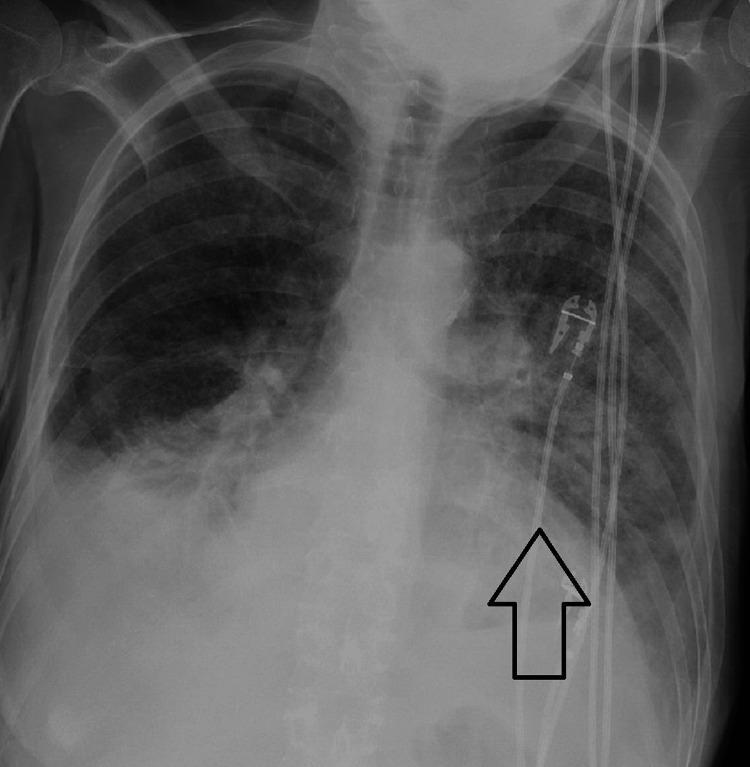
Anteroposterior (AP) chest radiograph demonstrating acute bilateral pulmonary edema. The image shows diffuse, ill-defined, hazy opacities and infiltrates throughout both lung fields, with a notable predominance and increased density in the left lung (indicated by a black arrow). These findings are consistent with alveolar and interstitial fluid accumulation. The cardiac silhouette appears enlarged, suggesting underlying cardiomegaly. Several medical devices are visible: multiple radiopaque lines (likely monitoring leads or catheters) descend through the superior mediastinum toward the cardiac silhouette, and a port-like device or pacemaker component is visible in the upper chest region. The radiograph illustrates a clinical scenario of acute heart failure or hyperbaric-induced pulmonary edema, emphasizing the loss of normal lung lucency and the obscuration of pulmonary vascular markings.
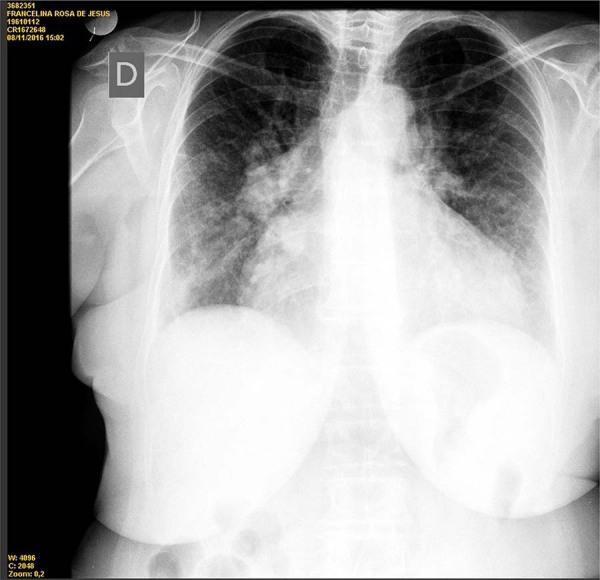
This diagnostic image is a posterior-anterior (PA) chest radiograph demonstrating significant cardiopulmonary pathology. The most prominent finding is marked cardiomegaly, with the cardiac silhouette significantly enlarged, resulting in an increased cardiothoracic ratio. There is evidence of pulmonary venous congestion, characterized by prominent hilar vasculature and diffuse, bilateral reticular opacities most concentrated in the perihilar and central lung fields. The lungs show vascular engorgement, consistent with congestive heart failure. The mediastinal contour appears widened, and the left heart border is shifted laterally due to ventricular enlargement. The diaphragmatic domes are partially obscured by the enlarged cardiac mass and overlying breast tissue shadows. This radiograph is an educational example of how chronic cardiac dysfunction manifests visually as volume overload and interstitial pulmonary edema in a clinical setting.
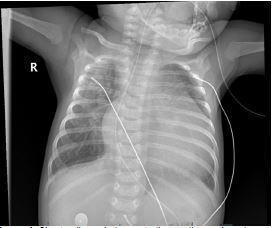
This diagnostic image is an anteroposterior (AP) pediatric chest radiograph demonstrating significant cardiomegaly and signs of pulmonary congestion. The cardiac silhouette is markedly enlarged, occupying a substantial portion of the thoracic cavity, which is indicative of cardiac dysfunction or congenital heart disease. The lung parenchyma exhibits diffuse interstitial prominence and hazy opacities, characteristic of pulmonary interstitial edema. Multiple medical devices are visible, including external monitoring leads and an enteric tube. The radiographic findings are clinically significant for evaluating pediatric patients presenting with respiratory distress or suspected cardiogenic shock, such as that seen in Shone complex or other left-sided obstructive lesions. The image serves as a critical educational tool for identifying neonatal/infant cardiomegaly and secondary pulmonary vascular congestion in a critical care or emergency setting.
electrocardiogram left ventricular hypertrophy strain pattern
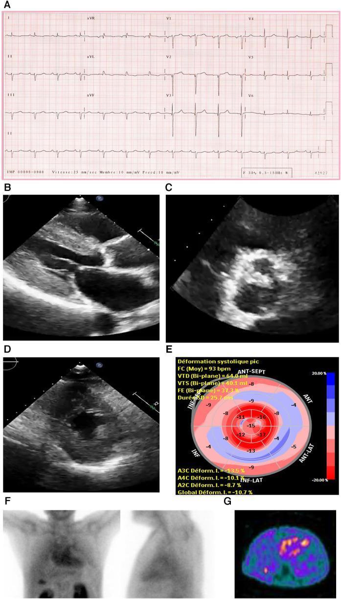
This composite diagnostic image illustrates clinical investigations for cardiac amyloidosis. Panel A shows a 12-lead electrocardiogram (ECG) demonstrating a regular sinus rhythm, prolonged PR interval (first-degree AV block), and low voltage in the limb leads, which contrasts with the ventricular hypertrophy seen in imaging. Panels B and D are 2D echocardiograms showing a small left ventricular (LV) cavity with marked concentric LV hypertrophy. Panel C displays a parasternal short-axis view of a severely calcified trileaflet aortic valve, characteristic of aortic stenosis. Panel E presents a global longitudinal strain (GLS) polar map ('bull's-eye' plot) showing reduced strain in the basal and mid-ventricular segments with relative apical sparing (a classic 'cherry on top' pattern). Panel F consists of anterior and lateral planar bone scintigraphy (99mTc-PYP) showing intense myocardial tracer uptake (Grade 3). Panel G shows a matching SPECT scan confirming strong cardiac uptake, confirming a diagnosis of transthyretin cardiac amyloidosis.
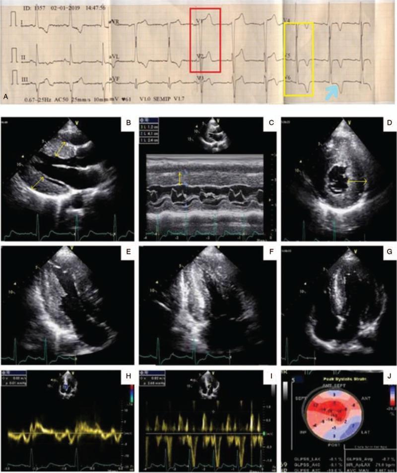
A multi-panel clinical diagnostic figure comprising a surface electrocardiogram (ECG) and transthoracic echocardiography (TTE) images related to cardiac amyloidosis evaluation. Panel A displays a 12-lead ECG showing signs of left ventricular hypertrophy (LVH), with high-amplitude QRS complexes in V2 (red box) and V5-V6 (yellow box) accompanied by a classic ST-segment depression and T-wave inversion indicative of a strain pattern (blue arrow). Panels B-G show 2D and M-mode echocardiography images illustrating significant asymmetric septal hypertrophy, with the interventricular septum measuring 2.4 cm. A distinctive 'speckled' or granular myocardial texture is visible in the short-axis views (D-E). Panels H-I utilize Tissue Doppler imaging to evaluate diastolic dysfunction, showing an E/e' ratio suggestive of grade 2 dysfunction. Panel J presents a bull's-eye map from speckle-tracking strain echocardiography, demonstrating a characteristic 'apical sparing' pattern (marked diminution of longitudinal strain in the basal and mid-segments with preserved apical function), which is highly suggestive of cardiac amyloidosis.
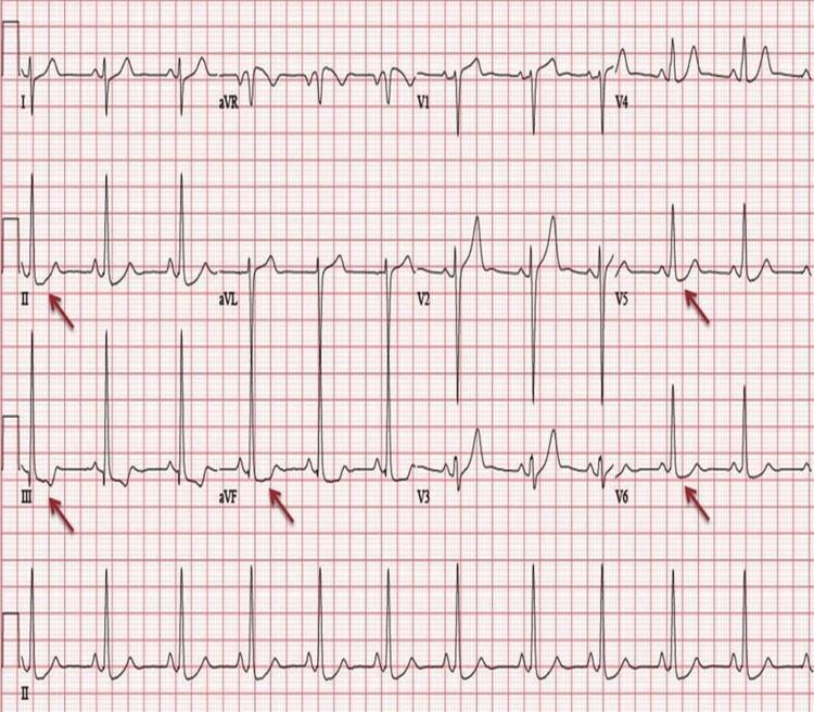
This diagnostic image is a 12-lead electrocardiogram (ECG) demonstrating classic features of Left Ventricular Hypertrophy (LVH) with associated strain patterns. The tracing shows a sinus rhythm with high QRS voltage, characterized by tall R-waves in the lateral leads (V5, V6) and deep S-waves in the right precordial leads (V1, V2), satisfying Sokolow-Lyon criteria for LVH. Red arrows highlight significant ST-segment depression and T-wave inversions in the inferolateral leads (II, III, aVF, V5, and V6). This repolarization abnormality is characteristic of a 'strain pattern' often seen in severe chronic pressure or volume overload of the left ventricle. Clinical relevance includes the assessment of hypertensive heart disease and cardiomyopathies, such as left ventricular non-compaction (LVNC). The ECG serves as a primary screening tool for identifying structural heart changes and associated ischemic or metabolic disturbances.
Slide 1 - Title Page
Group: 610A
Subject: Out Patient Polyclinic Therapy
Slide 2 - Definition
- Cardiomegaly means enlargement of the heart.
- It may involve enlargement of one or more cardiac chambers.
- It is a clinical/radiological finding, not a diagnosis by itself.
- It may result from cardiac disease, hypertension, valve disease, congenital disease, or systemic disorders.
- A chest X-ray may suggest cardiomegaly, but echocardiography is needed to assess chambers, valves, and ventricular function.
Slide 3 - Normal Heart Size
- On a posteroanterior (PA) chest X-ray, heart size is assessed using the cardiothoracic ratio (CTR).
- CTR = maximum cardiac width / maximum internal thoracic width.
- Normal adult PA-film CTR: less than 50%.
- A CTR greater than 50% suggests cardiomegaly.
- An AP portable chest X-ray can falsely magnify the cardiac shadow.
Slide 4 - Basic Mechanisms of Cardiomegaly
-
Pressure overload
- Hypertension
- Aortic stenosis
- Pulmonary hypertension
-
Volume overload
- Valve regurgitation
- Congenital left-to-right shunts
- Chronic kidney disease with fluid overload
-
Primary myocardial disease
- Dilated cardiomyopathy
- Hypertrophic cardiomyopathy
- Myocarditis
-
Pericardial causes
- Large pericardial effusion can mimic an enlarged heart shadow.
Slide 5 - Types of Cardiomegaly
- Left ventricular cardiomegaly
- Right ventricular cardiomegaly
- Left atrial enlargement
- Right atrial enlargement
- Biventricular cardiomegaly
- Generalized cardiomegaly
- True cardiomegaly
- Pseudo-cardiomegaly, for example due to pericardial effusion, obesity, AP X-ray projection, or poor inspiration.
Slide 6 - Left Ventricular Hypertrophy
- Long-standing systemic hypertension
- Aortic stenosis
- Hypertrophic cardiomyopathy
- Aortic regurgitation
- Chronic kidney disease
- Increased left ventricular wall thickness
- Initially preserved ejection fraction may occur
- Later, diastolic dysfunction and heart failure may develop
- ECG may show high-voltage QRS complexes and strain pattern.
Slide 7 - Right-Sided Cardiac Enlargement
- Pulmonary hypertension
- Chronic obstructive pulmonary disease
- Pulmonary embolic disease
- Tricuspid valve disease
- Congenital heart disease
- Left-sided heart failure causing secondary pulmonary hypertension
- May progress to right-sided heart failure.
- Produces systemic venous congestion and peripheral edema.
Slide 8 - Causes of Cardiomegaly: Cardiac Causes
- Hypertensive heart disease
- Ischemic heart disease and previous myocardial infarction
- Dilated cardiomyopathy
- Hypertrophic cardiomyopathy
- Myocarditis
- Valvular heart disease:
- Aortic stenosis
- Aortic regurgitation
- Mitral regurgitation
- Mitral stenosis
- Congenital heart disease
- Arrhythmia-induced cardiomyopathy, especially persistent tachyarrhythmia.
Slide 9 - Causes of Cardiomegaly: Non-Cardiac/Systemic Causes
- Severe anemia
- Thyroid disease, especially hyperthyroidism
- Chronic kidney disease and fluid overload
- Pregnancy-related cardiomyopathy
- Alcohol misuse
- Cocaine and cardiotoxic drugs
- Hemochromatosis
- Amyloidosis
- Acromegaly
- Nutritional deficiency, for example thiamine deficiency
- Pericardial effusion
Slide 10 - Specific Clinical Symptoms
- Exertional dyspnea
- Fatigue and reduced exercise tolerance
- Orthopnea
- Paroxysmal nocturnal dyspnea
- Palpitations
- Chest pain
- Dizziness or syncope
- Nocturia
- Ankle swelling
- Abdominal distension due to ascites
- Rapid weight gain from fluid retention
Slide 11 - Physical Examination Findings
- Tachycardia or irregular pulse
- Raised jugular venous pressure
- Displaced and diffuse apical impulse
- Parasternal heave in right ventricular enlargement
- S3 gallop in systolic heart failure
- S4 sound in a stiff hypertrophied ventricle
- Cardiac murmurs due to valvular disease
- Bibasal lung crackles
- Peripheral pitting edema
- Hepatomegaly and ascites in right-sided failure
Slide 12 - Important Red-Flag Symptoms
- Severe breathlessness at rest
- Acute pulmonary edema
- Cyanosis
- Chest pain suggestive of acute coronary syndrome
- Syncope
- Severe hypotension
- New rapid atrial fibrillation
- Marked bradycardia
- Confusion or reduced urine output
- Severe leg edema with rapidly increasing weight
Slide 13 - Diagnostic Evaluation
- History and physical examination
- Blood pressure measurement
- ECG
- Chest radiograph
- Echocardiography
- Blood tests:
- Complete blood count
- Renal function
- Electrolytes
- Thyroid function tests
- BNP or NT-proBNP
- Troponin where indicated
- Cardiac MRI for cardiomyopathy or infiltrative disease
- Coronary assessment if ischemic heart disease is suspected
Slide 14 - ECG Changes in Cardiomegaly
- Left ventricular hypertrophy voltage criteria
- Right ventricular hypertrophy pattern
- Left or right atrial enlargement
- Left-axis or right-axis deviation
- Bundle branch block
- ST-segment depression and T-wave inversion
- Atrial fibrillation
- Other tachyarrhythmias or bradyarrhythmias
- Pathological Q waves in previous myocardial infarction
- Low voltage in pericardial effusion or infiltrative cardiomyopathy
Slide 15 - ECG Features of Left Ventricular Hypertrophy
- Tall R waves in left precordial leads, especially V5 and V6
- Deep S waves in V1 and V2
- Increased QRS voltage
- Left-axis deviation may occur
- ST depression and T-wave inversion in lateral leads, called the LV strain pattern
- Left atrial enlargement may coexist
Slide 16 - Radiological Changes
- Increased cardiothoracic ratio, usually greater than 50% on PA view
- Enlarged left ventricular contour
- Enlarged right heart border in right atrial enlargement
- Pulmonary venous congestion
- Upper lobe venous diversion
- Kerley B lines due to interstitial edema
- Perihilar “bat-wing” pulmonary edema
- Pleural effusions
- Pulmonary artery enlargement in pulmonary hypertension
Slide 17 - Role of Echocardiography
- Chamber size
- Left ventricular ejection fraction
- Wall thickness
- Regional wall motion abnormalities
- Valve stenosis or regurgitation
- Intracardiac shunts
- Pulmonary artery pressure estimation
- Pericardial effusion
- Diastolic function
Slide 18 - General Principles of Treatment
- Control hypertension
- Treat ischemic heart disease
- Correct valvular disease when indicated
- Treat arrhythmias, especially atrial fibrillation
- Treat anemia, thyroid disease, and renal dysfunction
- Limit salt intake in congestive symptoms
- Use diuretics for fluid overload
- Encourage weight monitoring, smoking cessation, and alcohol avoidance
- Refer for cardiology assessment when structural heart disease is suspected
Slide 19 - Drug Treatment in Heart Failure With Reduced Ejection Fraction
- ARNI, ACE inhibitor, or ARB
- Evidence-based beta-blocker
- Mineralocorticoid receptor antagonist
- SGLT2 inhibitor
- Loop diuretic for congestion and edema
- Hydralazine plus nitrate in selected patients
- Ivabradine in selected patients with sinus rhythm and high heart rate
- Digoxin in selected symptomatic patients or for rate control in atrial fibrillation
Slide 20 - Specific Features of Beta-Blockers
- Bisoprolol
- Carvedilol
- Metoprolol succinate
- Reduce sympathetic overactivity
- Slow heart rate
- Improve ventricular filling time
- Reduce arrhythmia risk
- Improve symptoms and survival when appropriately used
- Reduce hospitalization
- Start with a low dose.
- Titrate gradually every 2 to 4 weeks if tolerated.
- Use in stable, euvolemic patients.
- Do not initiate or rapidly increase during acute decompensated heart failure.
- Monitor pulse, blood pressure, weight, congestion, and fatigue.
- Severe bradycardia
- High-grade AV block without a pacemaker
- Cardiogenic shock
- Severe acute decompensated heart failure
- Severe active bronchospasm
Slide 21 - Specific Features of Cardiac Glycosides: Digoxin
- Inhibits Na+/K+-ATPase.
- Increases intracellular calcium.
- Produces positive inotropic effect.
- Increases vagal tone and slows AV-node conduction.
- Selected patients with symptomatic HFrEF despite standard therapy
- Ventricular rate control in atrial fibrillation, especially when heart failure is present
- May improve symptoms.
- May reduce heart-failure hospital admissions.
- Does not clearly improve overall survival.
- Nausea, vomiting, anorexia
- Confusion
- Yellow or blurred vision
- Bradycardia
- AV block
- Atrial or ventricular arrhythmias
- Monitor renal function, potassium, magnesium, heart rate, and digoxin concentration where appropriate.
- Risk of toxicity increases with renal impairment, hypokalemia, hypomagnesemia, and interacting drugs.
- The medical literature notes that digoxin lowers heart-failure admissions but does not reduce mortality, so it should be used selectively.
Slide 22 - Conclusion
- Cardiomegaly is a sign of underlying structural or functional heart disease.
- Common causes include hypertension, ischemic heart disease, valvular disease, cardiomyopathy, and congenital heart disease.
- ECG and chest X-ray provide helpful clues, but echocardiography is essential for confirmation and cause assessment.
- Treatment depends on the underlying cause and the presence of heart failure.
- Beta-blockers are central in stable HFrEF.
- Digoxin has a limited, selective role and requires careful toxicity monitoring.
Make a ppt of it
~/
2022 AHA ACC HFSA heart failure guideline beta blockers digoxin recommendations
~/
mkdir -p "/tmp/workspace/cardiomegaly_presentation/assets" "/tmp/workspace/cardiomegaly_presentation/src" "/tmp/workspace/cardiomegaly_presentation/output" && python /tmp/skills/shared/scripts/fetch_images.py "https://cdn.orris.care/cdss_images/pmc_clinical_VQA_0fde282d454a822fe884ea088f5c3318cbd3c01c11b5160049da91feb632c520.jpg" "https://cdn.orris.care/cdss_images/pmc_clinical_VQA_03152b722b1cb86ef1d57f4d755aa687989488df7da9defa36d7d35f9ab80e28.jpg" > "/tmp/workspace/cardiomegaly_presentation/assets/image_data.json"
~/cardiomegaly_presentation/src/create_ppt.py
import os, json, base64
from io import BytesIO
from pptx import Presentation
from pptx.util import Inches, Pt
from pptx.enum.shapes import MSO_SHAPE
from pptx.enum.text import PP_ALIGN, MSO_ANCHOR
from pptx.dml.color import RGBColor

OUT='/tmp/workspace/cardiomegaly_presentation/output/Cardiomegaly_Talha_Khan_610A.pptx'
ASSET='/tmp/workspace/cardiomegaly_presentation/assets/image_data.json'
prs=Presentation(); prs.slide_width=Inches(13.333); prs.slide_height=Inches(7.5)
blank=prs.slide_layouts[6]
NAVY=RGBColor(12,38,66); BLUE=RGBColor(20,92,150); TEAL=RGBColor(0,151,167); PALE=RGBColor(239,247,250); INK=RGBColor(25,42,58); MUTED=RGBColor(88,105,120); WHITE=RGBColor(255,255,255); RED=RGBColor(190,55,62); GOLD=RGBColor(241,179,54)

def rect(s,x,y,w,h,color, radius=False, line=None):
    sh=s.shapes.add_shape(MSO_SHAPE.ROUNDED_RECTANGLE if radius else MSO_SHAPE.RECTANGLE, Inches(x), Inches(y), Inches(w), Inches(h))
    sh.fill.solid(); sh.fill.fore_color.rgb=color
    sh.line.color.rgb = color if line is None else line
    return sh

def text(s, txt, x,y,w,h, size=20, color=INK, bold=False, align=PP_ALIGN.LEFT, font='Aptos', valign=MSO_ANCHOR.TOP):
    tb=s.shapes.add_textbox(Inches(x), Inches(y), Inches(w), Inches(h)); tf=tb.text_frame
    tf.clear(); tf.word_wrap=True; tf.margin_left=Pt(2); tf.margin_right=Pt(2); tf.margin_top=Pt(1); tf.margin_bottom=Pt(1); tf.vertical_anchor=valign
    lines=txt.split('\n')
    for i,line in enumerate(lines):
        p=tf.paragraphs[0] if i==0 else tf.add_paragraph(); p.text=line; p.alignment=align; p.space_after=Pt(5)
        for r in p.runs: r.font.name=font; r.font.size=Pt(size); r.font.bold=bold; r.font.color.rgb=color
    return tb

def bullets(s, items, x,y,w,h, size=19):
    tb=s.shapes.add_textbox(Inches(x), Inches(y), Inches(w), Inches(h)); tf=tb.text_frame; tf.clear(); tf.word_wrap=True
    tf.margin_left=Pt(3); tf.margin_right=Pt(2); tf.margin_top=Pt(2); tf.margin_bottom=Pt(2)
    for i,item in enumerate(items):
        p=tf.paragraphs[0] if i==0 else tf.add_paragraph(); p.text=item; p.level=0; p.font.size=Pt(size); p.font.name='Aptos'; p.font.color.rgb=INK; p.space_after=Pt(9); p.bullet=True
    return tb

def footer(s,n):
    rect(s,0,7.18,13.333,.32,NAVY); text(s,'Out Patient Polyclinic Therapy',.45,7.205,4,.18,9,WHITE); text(s,f'{n:02d}',12.25,7.195,.55,.18,10,WHITE,True,PP_ALIGN.RIGHT)

def header(s,title,sub='',n=1):
    rect(s,0,0,13.333,.18,TEAL); text(s,title,.55,.4,12.1,.52,27,NAVY,True)
    if sub: text(s,sub,.57,.93,11.8,.25,11,MUTED)
    rect(s,.55,1.24,1.0,.05,TEAL); footer(s,n)

def add_image(s, stream,x,y,w,h):
    s.shapes.add_picture(stream, Inches(x), Inches(y), width=Inches(w), height=Inches(h))

def diagram_heart(s,x,y,scale=1):
    # stylized heart icon made with circles and triangle
    c1=s.shapes.add_shape(MSO_SHAPE.OVAL,Inches(x),Inches(y),Inches(1.05*scale),Inches(1.05*scale)); c1.fill.solid(); c1.fill.fore_color.rgb=RED; c1.line.color.rgb=RED
    c2=s.shapes.add_shape(MSO_SHAPE.OVAL,Inches(x+.8*scale),Inches(y),Inches(1.05*scale),Inches(1.05*scale)); c2.fill.solid(); c2.fill.fore_color.rgb=RED; c2.line.color.rgb=RED
    tri=s.shapes.add_shape(MSO_SHAPE.ISOSCELES_TRIANGLE,Inches(x+.28*scale),Inches(y+.53*scale),Inches(1.35*scale),Inches(1.35*scale)); tri.rotation=180; tri.fill.solid(); tri.fill.fore_color.rgb=RED; tri.line.color.rgb=RED

def content_slide(title, items, n, sub='', side_label=None):
    s=prs.slides.add_slide(blank); header(s,title,sub,n)
    rect(s,.6,1.55,8.05,5.18,WHITE,True,RGBColor(220,230,236)); bullets(s,items,.92,1.85,7.45,4.7,20)
    rect(s,9.05,1.55,3.65,5.18,PALE,True,RGBColor(215,232,236))
    diagram_heart(s,10.1,2.05,1.25)
    label=side_label or title
    text(s,label,9.42,4.1,2.9,1.25,20,NAVY,True,PP_ALIGN.CENTER,valign=MSO_ANCHOR.MIDDLE)
    return s

with open(ASSET) as f: im=json.load(f)
imgs=[]
for d in im:
    imgs.append(BytesIO(base64.b64decode(d['base64'].split(',',1)[1])))

# 1 title
s=prs.slides.add_slide(blank); rect(s,0,0,13.333,7.5,NAVY); rect(s,0,0,13.333,.22,TEAL); diagram_heart(s,9.5,1.35,1.7)
text(s,'CARDIOMEGALY',.75,1.25,8.4,.75,34,WHITE,True); text(s,'Causes, Types, Clinical Symptoms, ECG and Radiological Changes, Treatment, Beta-Blockers and Cardiac Glycosides',.78,2.12,7.7,1.25,19,RGBColor(215,235,242))
rect(s,.78,4.45,5.8,1.15,BLUE,True); text(s,'Talha Khan\nGroup: 610A',1.05,4.65,3.2,.65,20,WHITE,True); text(s,'Subject: Out Patient Polyclinic Therapy',.8,6.68,6,.25,12,RGBColor(195,218,227))

slides=[
('Definition of Cardiomegaly',['Cardiomegaly means enlargement of the heart or one or more cardiac chambers.','It is a radiological and clinical finding, not a diagnosis by itself.','It may result from pressure overload, volume overload, or primary myocardial disease.','Echocardiography identifies chamber size, valve disease, and ventricular function.'],'What it means'),
('Normal Heart Size',['On a PA chest radiograph, the cardiothoracic ratio (CTR) is used.','CTR = maximum cardiac width divided by maximum internal thoracic width.','A normal adult PA-film CTR is less than 50%.','AP portable films may magnify the heart shadow and give a false impression of enlargement.'],'How to assess'),
('Mechanisms of Enlargement',['Pressure overload: hypertension, aortic stenosis, pulmonary hypertension.','Volume overload: valve regurgitation, shunts, renal fluid overload.','Myocardial disease: dilated cardiomyopathy, myocarditis, ischemic damage.','Pericardial fluid can create pseudo-cardiomegaly.'],'Why it enlarges'),
('Types of Cardiomegaly',['Left ventricular enlargement or hypertrophy.','Right ventricular enlargement or hypertrophy.','Left or right atrial enlargement.','Biventricular or generalized cardiomegaly.','True cardiomegaly versus pseudo-cardiomegaly.'],'Classification'),
('Left Ventricular Hypertrophy',['Common causes: long-standing hypertension and aortic stenosis.','Also occurs in hypertrophic cardiomyopathy and chronic volume overload.','Initially, ejection fraction may be preserved.','Progression can cause diastolic dysfunction, myocardial ischemia, and heart failure.'],'Pressure-loaded LV'),
('Right-Sided Enlargement',['Causes include pulmonary hypertension, COPD, pulmonary embolic disease, and tricuspid valve disease.','Congenital heart disease may cause chronic right-sided volume overload.','May progress to systemic venous congestion and right-sided heart failure.'],'Right heart'),
('Cardiac Causes',['Hypertensive heart disease and ischemic heart disease.','Dilated or hypertrophic cardiomyopathy.','Myocarditis.','Valvular disease: aortic stenosis/regurgitation and mitral disease.','Congenital heart disease and persistent tachyarrhythmia.'],'Common causes'),
('Systemic and Non-Cardiac Causes',['Severe anemia and thyroid disease.','Chronic kidney disease with fluid overload.','Pregnancy-related cardiomyopathy.','Alcohol, cocaine, and cardiotoxic medications.','Hemochromatosis, amyloidosis, acromegaly, and nutritional deficiency.'],'Think beyond heart'),
('Specific Clinical Symptoms',['Exertional dyspnea and reduced exercise tolerance.','Orthopnea and paroxysmal nocturnal dyspnea.','Fatigue, palpitations, chest pain, dizziness, or syncope.','Ankle swelling, rapid weight gain, abdominal distension, and nocturia.'],'Patient presentation'),
('Physical Examination',['Tachycardia or irregular pulse.','Raised jugular venous pressure and peripheral pitting edema.','Displaced diffuse apical impulse; parasternal heave in RV enlargement.','S3 or S4 gallop, cardiac murmurs, basal crackles, hepatomegaly, or ascites.'],'Clinical signs'),
('Red Flags',['Severe breathlessness at rest or acute pulmonary edema.','Cyanosis, chest pain, syncope, marked hypotension.','New rapid atrial fibrillation or marked bradycardia.','Confusion, reduced urine output, or rapidly worsening edema.'],'Urgent referral'),
('Diagnostic Evaluation',['Focused history, examination, blood pressure, and medication review.','ECG and chest radiograph.','Echocardiography is central for structure, ejection fraction, valves, and pericardial fluid.','CBC, renal function, electrolytes, thyroid tests, BNP/NT-proBNP, and troponin when appropriate.'],'Work-up'),
('ECG Changes',['Changes reflect the underlying cause and affected chamber.','LVH or RVH voltage criteria; atrial enlargement and axis deviation.','Bundle branch block, ST-T abnormalities, atrial fibrillation, tachyarrhythmias, or prior infarct Q waves.','Low voltage can occur with pericardial effusion or infiltrative cardiomyopathy.'],'Electrical clues'),
]
for i,(t,items,label) in enumerate(slides,2): content_slide(t,items,i,side_label=label)

# 15 ECG image slide
s=prs.slides.add_slide(blank); header(s,'ECG Features of Left Ventricular Hypertrophy','High voltage and secondary repolarization abnormalities',15)
rect(s,.55,1.5,5.0,5.3,PALE,True); bullets(s,['Tall R waves in V5-V6 and deep S waves in V1-V2.','Increased QRS voltage and possible left-axis deviation.','ST depression and T-wave inversion in lateral leads: LV strain pattern.','Left atrial enlargement can coexist.'],.85,1.85,4.4,4.5,18)
add_image(s,imgs[1],6.0,1.55,6.75,4.65); text(s,'Example ECG: LVH with strain pattern',6.05,6.3,6.55,.3,13,MUTED,False,PP_ALIGN.CENTER)

# 16 chest image
s=prs.slides.add_slide(blank); header(s,'Radiological Changes','Chest radiography supports the diagnosis but does not establish the cause',16)
add_image(s,imgs[0],.58,1.48,6.05,4.9); rect(s,6.95,1.5,5.75,4.9,PALE,True); bullets(s,['Increased CTR on a PA film.','Enlarged ventricular or atrial contours.','Pulmonary venous congestion and upper lobe diversion.','Kerley B lines, interstitial edema, bat-wing edema, or pleural effusions.','Pulmonary artery enlargement in pulmonary hypertension.'],7.28,1.85,5.1,4.35,18); text(s,'Example: enlarged cardiac silhouette with pulmonary congestion',.65,6.45,5.9,.3,12,MUTED,False,PP_ALIGN.CENTER)

# 17-22
more=[
('Role of Echocardiography',['Measures chamber dimensions and ventricular wall thickness.','Assesses left ventricular ejection fraction and regional wall motion.','Identifies valvular stenosis or regurgitation.','Estimates pulmonary pressures and detects intracardiac shunts or pericardial effusion.','It clarifies the cause suggested by X-ray or ECG.'],'Key investigation'),
('General Treatment Principles',['Treat the underlying cause rather than the enlarged cardiac shadow alone.','Control blood pressure and treat ischemic, valvular, thyroid, renal, or arrhythmic disease.','Limit dietary salt when congested; encourage daily weight monitoring.','Use diuretics for fluid overload and refer for cardiology review when structural disease is suspected.'],'Individualize care'),
('HFrEF Drug Treatment',['For suitable patients: ARNI, ACE inhibitor, or ARB.','Evidence-based beta-blocker.','Mineralocorticoid receptor antagonist and SGLT2 inhibitor.','Loop diuretic for congestion.','Selected cases: ivabradine, hydralazine-nitrate, or digoxin.'],'Guideline-directed therapy'),
('Beta-Blockers: Role and Benefits',['Examples: bisoprolol, carvedilol, and metoprolol succinate.','Reduce sympathetic drive, slow heart rate, and reduce arrhythmia risk.','Improve symptoms and survival in stable HFrEF when used appropriately.','Start low and titrate gradually in stable, euvolemic patients.'],'Beta-blockers'),
('Beta-Blockers: Safety Features',['Do not start or rapidly increase dose during acute decompensated heart failure or cardiogenic shock.','Monitor heart rate, blood pressure, congestion, fatigue, and weight.','Use caution with severe bradycardia, high-grade AV block without pacemaker, or active severe bronchospasm.','Avoid abrupt withdrawal unless medically necessary.'],'Safe use'),
('Cardiac Glycosides: Digoxin',['Digoxin inhibits Na+/K+-ATPase, increasing intracellular calcium and contractility.','It increases vagal tone and slows AV nodal conduction.','Consider in selected symptomatic HFrEF despite standard therapy or for rate control in atrial fibrillation.','It may reduce heart-failure admissions but does not clearly improve overall survival.'],'Digoxin'),
('Digoxin: Toxicity and Monitoring',['Possible toxicity: nausea, vomiting, anorexia, confusion, visual disturbance, bradycardia, AV block, and arrhythmias.','Risk increases in renal impairment, hypokalemia, hypomagnesemia, and drug interactions.','Monitor renal function, electrolytes, pulse, symptoms, and serum level when clinically indicated.','Use the lowest effective dose and review medications carefully.'],'Use cautiously'),
('Key Take-Home Messages',['Cardiomegaly is a sign that requires cause-directed evaluation.','Hypertension, ischemic disease, valve disease, and cardiomyopathy are major causes.','ECG and chest X-ray provide clues; echocardiography confirms anatomy and function.','Beta-blockers are essential in stable HFrEF; digoxin has a selective, monitored role.'],'Summary'),
('References',['Fuster and Hurst’s The Heart, 15th ed: Digoxin section.','Goldman-Cecil Medicine: Heart failure and cardiac glycosides.','2022 AHA/ACC/HFSA Guideline for the Management of Heart Failure.','Images used for teaching: medical image library examples of cardiomegaly and LVH ECG.'],'Selected sources')]
for idx,(t,items,label) in enumerate(more,17): content_slide(t,items,idx,side_label=label)

prs.save(OUT)
print(OUT, len(prs.slides))
python "/tmp/workspace/cardiomegaly_presentation/src/create_ppt.py"
python -c "from pptx import Presentation; p=Presentation('/tmp/workspace/cardiomegaly_presentation/output/Cardiomegaly_Talha_Khan_610A.pptx'); print('slides=',len(p.slides)); [print(i+1, ' | '.join(sh.text.replace(chr(10),' / ')[:110] for sh in s.shapes if getattr(sh,'has_text_frame',False) and sh.text.strip())[:260]) for i,s in enumerate(p.slides)]"
~/cardiomegaly_presentation/output
Cardiomegaly Talha Khan 610A
Presentation · PPTX