
Glanzmann thrombasthenia
Glanzmann thrombasthenia
Glanzmann thrombasthenia platelet aggregation blood smear
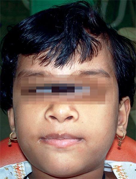
This clinical photograph shows the frontal facial view of a 6-year-old girl diagnosed with Glanzmann's thrombasthenia, a rare autosomal recessive bleeding disorder characterized by a defect in platelet aggregation. The patient exhibits a medium skin tone and short dark hair. While the eyes are obscured for patient anonymity, the visible facial features demonstrate a subtle, generalized puffiness. Clinically, the image serves to document the baseline physical appearance of a pediatric patient who presents with a significant medical history of spontaneous ecchymoses, easy bruising, and recurrent epistaxis. These manifestations are hallmark signs of the underlying qualitative platelet disorder involving the glycoprotein IIb/IIIa complex. In a dental context, identifying such patients is critical for pre-operative planning to prevent uncontrolled hemorrhage during procedures like extractions. The photograph highlights the importance of recognizing systemic hematological conditions in pediatric dentistry to ensure appropriate hematological consultation and hemostatic management.

This composite diagnostic image features eight light microscopy frames (45x magnification) of blood smears, labeled (a) through (h). The primary educational focus is the qualitative assessment of platelet aggregation and blood compatibility following exposure to Berberine (BBR) in various formulations: naked nanoparticles (BBR-NNPs), surface-modified nanoparticles (BBR-SMNPs), pure BBR, and placebo controls at concentrations of 10 mcg/mL and 100 mcg/mL. Each panel shows a field of view containing evenly distributed erythrocytes (red blood cells) against a clear background. Red circular annotations are overlaid to highlight individual platelets or small clusters. Across all treatment groups and concentrations, there is a notable absence of large-scale platelet clumping or pathological aggregation, suggesting the drug delivery systems maintain hemocompatibility. The erythrocytes maintain a characteristic biconcave appearance without significant morphological alteration. This comparison chart is used in pharmacological research to demonstrate that nanocarrier surface modifications, such as Vitamin E TPGS, do not adversely affect blood cellular components.
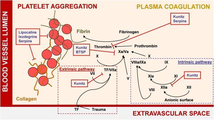
A pathophysiology diagram illustrating the mechanisms of hemostasis and the inhibitory effects of tick salivary proteins on platelet aggregation and plasma coagulation. The diagram is divided into the 'Blood Vessel Lumen' and the 'Extravascular Space.' On the left, 'Platelet Aggregation' is shown as a cluster of red spheres representing platelets cross-linked by fibrin strands; this process is inhibited by Lipocalins, Ixodegrins, and Serpins. On the right, the 'Plasma Coagulation' cascade is detailed, featuring both the Extrinsic pathway (initiated by Tissue Factor/TF due to trauma) and the Intrinsic pathway (activated via anionic surfaces). The diagram highlights key enzymatic conversions, such as Factor X to Xa/Va, Prothrombin to Thrombin, and Fibrinogen to Fibrin. Red annotation boxes identify specific tick protein families—including Kunitz-type inhibitors, BTSPs, and Serpins—that target and inhibit various coagulation factors like Thrombin, Factor Xa, and the XIIa/XIa complex. This visual resource effectively demonstrates the interference of exogenous biological agents with human blood clotting mechanisms.
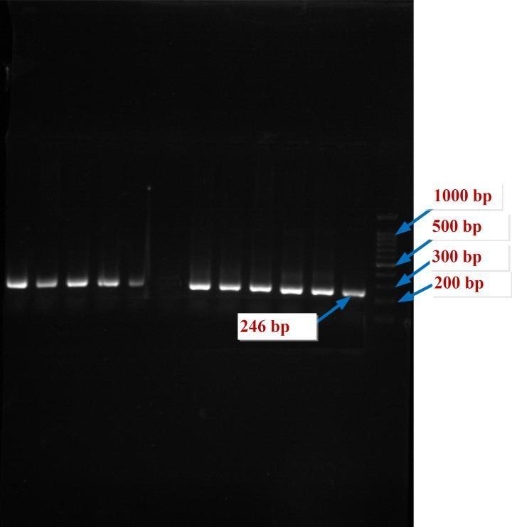
This diagnostic image shows an agarose gel electrophoresis of PCR products related to the HPA-5 gene in a study of human platelet antigens. The gel features multiple vertical lanes displaying bright DNA bands against a dark background, typical of ethidium bromide or similar staining. The rightmost lane contains a DNA ladder with marked reference points at 1000 bp, 500 bp, 300 bp, and 200 bp. The experimental lanes display highly homogeneous, singular bands indicating successful target amplification. A specific label points to a band at 246 bp, representing the expected size of the PCR product for the HPA-5 allele. This molecular diagnostic technique is used in hematology to determine genotype frequencies in patients, such as those with Glanzmann Thrombasthenia, to assess genetic susceptibility to treatment resistance or platelet alloimmunization. The clear, sharp bands suggest high specificity and concentration of the amplified DNA fragments.
Glanzmann Thrombasthenia
Definition and Genetics
- Goldman-Cecil Medicine, p. 1668
- Henry's Clinical Diagnosis and Management by Laboratory Methods, p. 971
Pathophysiology
- Binding fibrinogen (and vitronectin, fibronectin, vWF) after platelet activation
- Cross-linking platelets to form the platelet plug (aggregation)
- Clot retraction via interaction with the platelet cytoskeleton
Classification
| Type | GPIIb/IIIa Level | Clot Retraction |
|---|---|---|
| Type 1 | <5% of normal (complete absence) | Absent |
| Type 2 | 10-20% of normal | Reduced |
| Variant | Near-normal quantity but dysfunctional | Variable |
Clinical Features
- Onset: Bleeding typically presents from childhood, often severe
- Pattern: Mucocutaneous bleeding - epistaxis, gingival bleeding, easy bruising, menorrhagia (often severe at menarche), gastrointestinal bleeding
- Platelet count: Normal
- Platelet morphology: Normal size (not macrothrombocytopenic, unlike Bernard-Soulier)
- Bleeding time: Markedly prolonged
- PT/aPTT: Normal
- Clot retraction: Abnormal/absent
Laboratory Diagnosis
Platelet Aggregometry (Key Finding)
- Shape change (initial upward deflection) is preserved - platelets can still respond to activation signals
- Both primary AND secondary waves of aggregation are absent (unlike storage pool defects where only the secondary wave is lost)
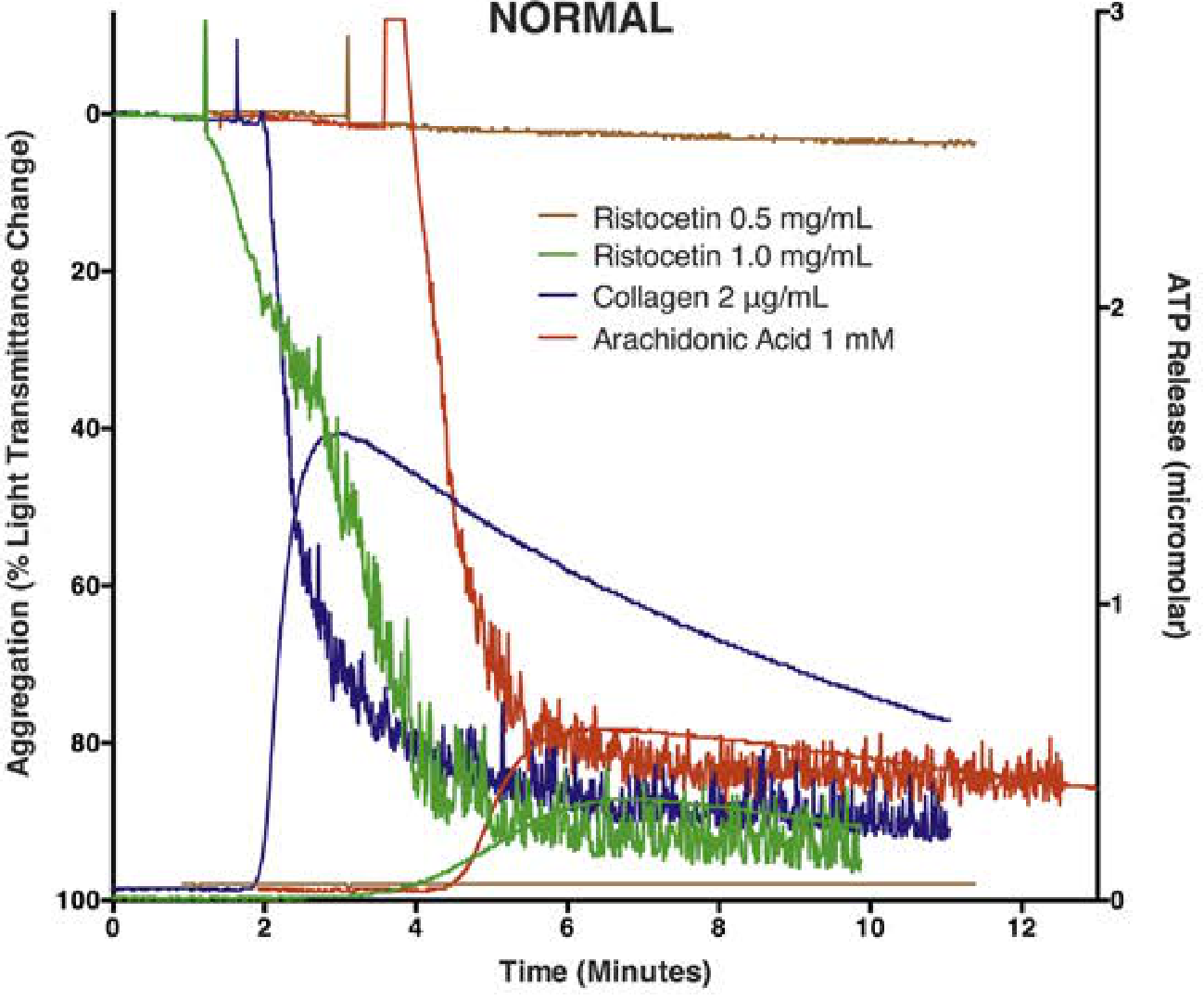
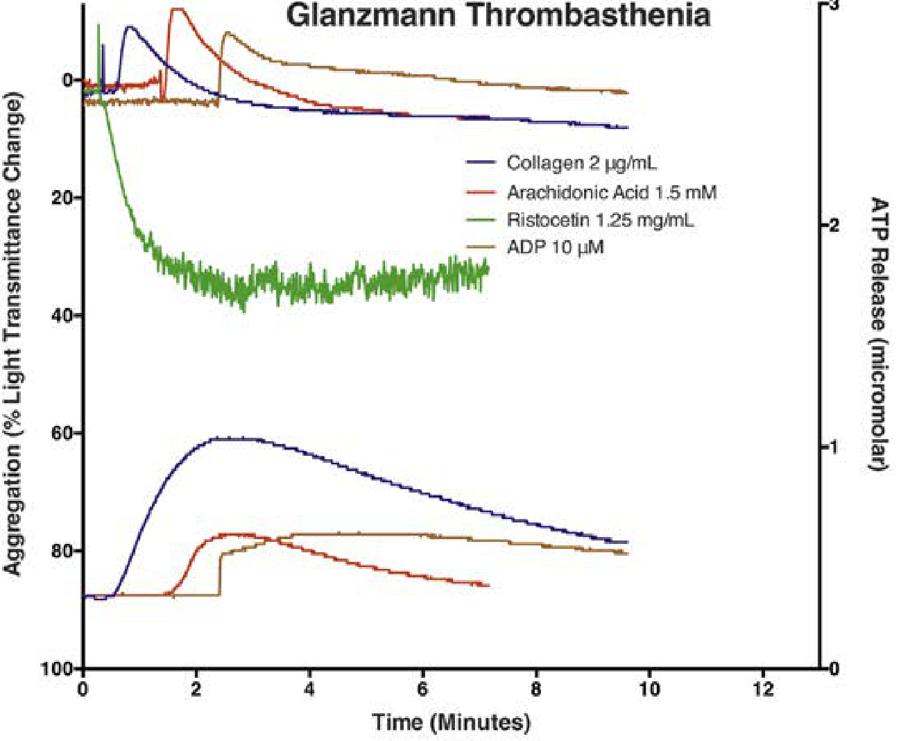
Comparison with Bernard-Soulier Syndrome
| Feature | Glanzmann Thrombasthenia | Bernard-Soulier Syndrome |
|---|---|---|
| Defect | GPIIb/IIIa (αIIbβ3) | GPIb/IX/V complex |
| Platelet size | Normal | Macrothrombocytopenia |
| Platelet count | Normal | Low-normal to decreased |
| ADP/collagen/epinephrine aggregation | Absent | Normal |
| Ristocetin aggregation | Normal | Absent |
| Gene mutations | ITGA2B, ITGB3 | GPIBA, GPIBB, GP9 |
| Clot retraction | Abnormal | Normal |
Confirmatory Tests
- Flow cytometry - confirms absent/reduced surface GPIIb/IIIa (CD41/CD61)
- Genetic testing - ITGA2B and ITGB3 mutation analysis (database: https://glanzmann.mcw.edu/)
- Heterozygous carriers have ~50% of normal GPIIb/IIIa levels but no bleeding disorder and normal aggregation
Treatment
Acute Bleeding
- Platelet transfusion - first-line for significant bleeds; HLA-matched single-donor platelets preferred to limit alloimmunization
- Repeated transfusions can induce anti-GPIIb/IIIa and anti-HLA antibodies, causing refractoriness
- Recombinant Factor VIIa (rFVIIa) - FDA-approved specifically for GT; bypasses the platelet aggregation defect by generating thrombin at the injury site; particularly useful when alloimmunization has made platelets ineffective
Adjunctive
- Antifibrinolytics (tranexamic acid, epsilon-aminocaproic acid) - especially effective for mucosal bleeding (epistaxis, menorrhagia, dental procedures); often used first-line for minor bleeds
- DDAVP - may provide some benefit, especially for milder bleeding; acts by releasing vWF and increasing platelet adhesion
Special Situations
-
Pregnancy/delivery: Platelet transfusion is the primary treatment intrapartum; postpartum hemorrhage occurs in ~50% of pregnancies despite prophylaxis. Women who have previously received transfusions should be evaluated for HLA- and GP-specific antibodies before delivery.
-
Dental procedures: Pre-procedure antifibrinolytics or platelet support are important
-
Emerging therapies: Gene therapy and emicizumab (off-label) are being investigated per recent literature
-
Harrison's Principles of Internal Medicine 22E, p. 1802
-
Goldman-Cecil Medicine, p. 1668
Recent Literature (2023-2026)
- A 2025 review by Nurden & Nurden in Semin Thromb Hemost (PMID 38499192) reviewed 10 years of progress, highlighting advances in gene therapy, novel hemostatic agents, and molecular understanding of ITGA2B/ITGB3 mutations.
- A 2024 review in Br J Haematol (PMID 38744450) addressed pregnancy management specifically.
- A 2025 review on molecular pathogenesis (PMID 41301446) covers the full landscape of inherited platelet dysfunction including GT.
Key High-Yield Summary
| Feature | Detail |
|---|---|
| Inheritance | Autosomal recessive |
| Defect | GPIIb/IIIa (αIIbβ3) absent or dysfunctional |
| Genes | ITGA2B, ITGB3 (chromosome 17) |
| Platelet count | Normal |
| Platelet size | Normal |
| Bleeding time | Prolonged |
| PT/aPTT | Normal |
| Clot retraction | Absent |
| Aggregation defect | Absent with all agonists except ristocetin |
| PIA1 antigen | Negative (on GPIIIa) |
| Confirm by | Flow cytometry (CD41/CD61), genetic testing |
| Rx mild bleed | Tranexamic acid, DDAVP |
| Rx severe bleed | HLA-matched platelets, rFVIIa |
Refer Pediatrics textbook and literature and give answer
Glanzmann thrombasthenia children pediatric
PMID: 39604156
PMID: 37386449
PMID: 40928240
Glanzmann thrombasthenia management bleeding treatment
Glanzmann thrombasthenia gene therapy diagnosis
PMID: 38499192
PMID: 41301446
Glanzmann thrombasthenia stem cell transplantation emicizumab
Glanzmann Thrombasthenia - Pediatric Perspective
Library note: The textbook library contains two pediatrics references: The Harriet Lane Handbook (23rd ed.) and Red Book 2021. Harriet Lane lists Glanzmann thrombasthenia as a key inherited cause of platelet dysfunction (Table 14.10), but neither pediatric text has a dedicated chapter. The following answer integrates Harriet Lane's pediatric framework with content from Harrison's 22E, Goldman-Cecil, and Henry's Clinical Diagnosis, supplemented by current peer-reviewed literature (2021-2026).
1. Overview and Epidemiology in Children
- Harriet Lane Handbook, 23rd ed., Table 14.10 (platelet dysfunction causes in children)
- Nurden & Nurden, Semin Thromb Hemost 2025 (PMID 38499192)
2. Molecular Basis
| Gene | Protein encoded | Chromosome |
|---|---|---|
| ITGA2B | GPIIb (αIIb subunit) | 17q21.31 |
| ITGB3 | GPIIIa (β3 subunit) | 17q21.32 |
3. Classification
| Type | GPIIb/IIIa Surface Level | Clot Retraction | Mechanism |
|---|---|---|---|
| Type 1 | <5% of normal | Absent | Null mutations (nonsense, frameshift) |
| Type 2 | 10-20% of normal | Reduced | Reduced expression |
| Variant | Near-normal or normal level | Variable | Qualitative/dysfunctional complex |
4. Clinical Features in Children
Bleeding pattern
- Onset: Often at birth or in infancy (purpura neonatorum, umbilical stump bleeding, circumcision bleeding)
- Mucocutaneous dominant: Epistaxis (most common - often severe and recurrent), gingival bleeding, easy bruising, petechiae
- Dental eruption bleeding - characteristic in young children
- GI bleeding (less common)
- Intracranial hemorrhage - rare but life-threatening
- Menorrhagia - major problem at menarche; often the presenting symptom in adolescent girls
Important negatives
- Normal platelet count - this is a qualitative, not quantitative, defect
- Normal platelet size - distinguishes GT from Bernard-Soulier syndrome (which has macrothrombocytopenia)
- No joint bleeds - distinguishes from hemophilia
5. Diagnosis
Blood indices
| Test | Result |
|---|---|
| Platelet count | Normal |
| Platelet morphology | Normal size |
| Bleeding time | Markedly prolonged |
| PT / aPTT / fibrinogen | Normal |
| Clot retraction | Absent or severely reduced |
Platelet aggregometry (gold standard functional test)
- Initial shape change is preserved (the tracing shows an upward deflection) but no subsequent aggregation follows
- This is the opposite pattern to Bernard-Soulier syndrome
| Agonist | GT Result | Bernard-Soulier Result |
|---|---|---|
| ADP | Absent | Normal |
| Collagen | Absent | Normal |
| Epinephrine | Absent | Normal |
| Arachidonic acid | Absent | Normal |
| Ristocetin | Normal | Absent |
| Clot retraction | Absent | Normal |
- Henry's Clinical Diagnosis and Management by Laboratory Methods, p. 971
Confirmatory tests
- Flow cytometry - absent or markedly reduced CD41 (GPIIb) and CD61 (GPIIIa) on platelet surface; this is particularly useful in neonates and infants where adequate PRP for aggregometry is hard to obtain
- Genetic testing (ITGA2B, ITGB3) - confirms diagnosis; important for genetic counseling; mutations catalogued at https://glanzmann.mcw.edu/
- Anti-GPIIb/IIIa antibody testing - critical before platelet transfusion; detects alloimmunization
- Rodriguez-Alen et al., Biomolecules 2025 (PMID 41301446)
6. Management in Children
A. First-line for minor/mucosal bleeding
- Antifibrinolytics - tranexamic acid (TXA) or epsilon-aminocaproic acid (EACA) are the first-line for epistaxis, gingival bleeding, and dental procedures in children
- TXA: 15-25 mg/kg orally (or IV) every 8 hours; topical application in epistaxis
- Local measures - nasal packing, pressure, cauterization for nosebleeds
B. Desmopressin (DDAVP)
- 0.3 mcg/kg IV or intranasal; may augment platelet adhesion via vWF release; useful adjunct in moderate bleeding, though efficacy is variable in GT
C. Platelet transfusion
- First-line for significant or surgical bleeding
- HLA-matched, single-donor apheresis platelets preferred to minimize alloimmunization
- Risk: repeated transfusions cause anti-HLA and anti-GPIIb/IIIa (PIA1) antibody formation, which leads to platelet transfusion refractoriness - a major clinical challenge in children who will need lifelong management
- In children, limiting early platelet transfusions (by using rFVIIa when possible) is especially important to preserve transfusion efficacy for later life
D. Recombinant Factor VIIa (rFVIIa, NovoSeven)
- FDA-approved specifically for GT (and Bernard-Soulier) in patients with anti-GPIIb/IIIa antibodies or refractoriness
- Standard dose: 90 mcg/kg IV every 2-3 hours until hemostasis
- A 2025 systematic review of 100 subjects showed rFVIIa was effective in 93% of non-surgical bleeds, 91-92% of surgical procedures, with only 4 (2.7%) serious adverse events. The authors recommend considering rFVIIa as frontline even without antibodies, to prevent alloimmunization - especially relevant in children (Saultier et al., Haemophilia 2025, PMID 39604156)
- Mechanism: generates sufficient thrombin on the platelet surface even without GPIIb/IIIa-fibrinogen bridging
E. Emergency management consensus (2023)
- Minor bleeds: antifibrinolytics ± DDAVP
- Moderate bleeds with no refractoriness: platelets OR rFVIIa
- Severe/refractory bleeds: rFVIIa (90 mcg/kg) + antifibrinolytics
- Avoid unnecessary platelet exposure in children to preserve future options
F. Hematopoietic stem cell transplantation (HSCT)
- The only potentially curative treatment for severe GT
- Indicated in children with: life-threatening bleeding, refractoriness to platelets and rFVIIa, or severely impaired quality of life
- A 2025 pediatric report from J Pediatr Hematol Oncol (Al-Antary et al., PMID 40928240) documented successful allo-HSCT in 2 siblings with GT, supported by CIBMTR registry data (2008-2022), confirming HSCT prevents life-threatening bleeding
- Key post-HSCT monitoring: flow cytometric detection of GPIIb/GPIIIa on platelets to identify late graft rejection - particularly important when pre-transplant anti-GPIIb/IIIa antibodies are present
G. Emerging/future therapies
- Gene therapy using lentiviral vectors: preclinical stage; CRISPR/Cas9 approaches being explored
- Bispecific antibodies that link platelets to coagulation factors (e.g., emicizumab analogs) - under investigation
- Monoclonal antibodies to accelerate fibrin formation at injury sites - promising prophylactic approach
- Nurden & Nurden, Semin Thromb Hemost 2025 (PMID 38499192)
7. Perioperative and School/Activity Considerations
- Pre-procedural planning is critical - dental cleaning, tonsillectomy, and even minor procedures require hematology consultation
- Tranexamic acid mouthwash for dental procedures
- Children should wear medical alert identification
- Avoid aspirin, NSAIDs, and SSRIs (all impair residual platelet function)
- Contact sports restrictions based on severity
8. Differential Diagnosis in a Child with Mucocutaneous Bleeding + Normal Platelet Count
| Condition | Key Distinguishing Feature |
|---|---|
| Glanzmann thrombasthenia | No aggregation with all agonists except ristocetin; absent clot retraction |
| Bernard-Soulier syndrome | Macrothrombocytopenia; aggregates with all agonists except ristocetin |
| von Willebrand disease | Reduced ristocetin aggregation; corrected by normal plasma; vWF antigen low |
| Storage pool defects (HPS, CHS) | Secondary wave only absent; abnormal dense granules on EM |
| Platelet release defects | Absent secondary wave; normal first wave |
Summary Table
| Feature | Details |
|---|---|
| Inheritance | Autosomal recessive |
| Genes | ITGA2B (GPIIb), ITGB3 (GPIIIa) |
| Defect | αIIbβ3 integrin (fibrinogen receptor) absent/dysfunctional |
| Platelet count | Normal |
| Platelet size | Normal |
| Bleeding time | Markedly prolonged |
| Clot retraction | Absent |
| Aggregation | Absent with all agonists except ristocetin |
| Confirm | Flow cytometry (CD41/CD61), genetics |
| Mild bleeding Rx | Tranexamic acid, DDAVP |
| Severe bleeding Rx | HLA-matched platelets, rFVIIa (90 mcg/kg) |
| Curative | Allogeneic HSCT |
| Future | Gene therapy, bispecific antibodies |