
Anemia
Reading File
The topic "Anemia" is broad. Let me clarify what would be most useful before diving deep.
Asking for Preferences
What aspect of anemia are you looking for? · What is this for?
Searching the medical library for sickle cell disease and thalassemia now.
Finding Sources
Finding Sources
Good sources found. Let me read from Robbins Pathology (best for exam) for both topics simultaneously.
Finding Sources
Reading File
Excellent content from Robbins Basic Pathology. Now let me get the sickle cell section from Robbins Pathologic Basis of Disease and also read more of the thalassemia content.
Reading File
Reading File
Excellent data from both books. Let me also fetch the thalassemia pathophysiology diagram and read more sickle cell content (clinical features/complications).
Loading Image
Reading File
Now let me get the clinical features and management sections for sickle cell disease.
Reading File
Finding Sources
Let me read the rest of the clinical features and management sections, and also fetch the sickle cell pathophysiology diagram.
Reading File
Loading Image
I now have all the key content I need from Robbins. Let me compile the comprehensive exam-focused study guide.
Sickle Cell Disease & Thalassemia - Exam Study Guide
Sources: Robbins, Cotran & Kumar Pathologic Basis of Disease; Robbins & Kumar Basic Pathology
SICKLE CELL DISEASE
Genetics & Molecular Basis
- Mutation: Single missense point mutation in the β-globin gene (chromosome 11) - glutamate replaced by valine at position 6
- This converts HbA (α₂β₂) to HbS (α₂β^S₂)
- Inheritance: Autosomal recessive
- Sickle cell trait (HbAS): ~8-10% of African Americans; ~40% HbS, rest HbA - largely asymptomatic; HbA interferes with HbS polymerization
- Sickle cell disease: Homozygous HbSS; ~70,000-100,000 affected in the US
- Malaria protection: HbS mutation arose in West Africa; provides protection against P. falciparum malaria by promoting clearance of parasitized cells and impairing PfEMP-1 membrane knob formation
Pathophysiology
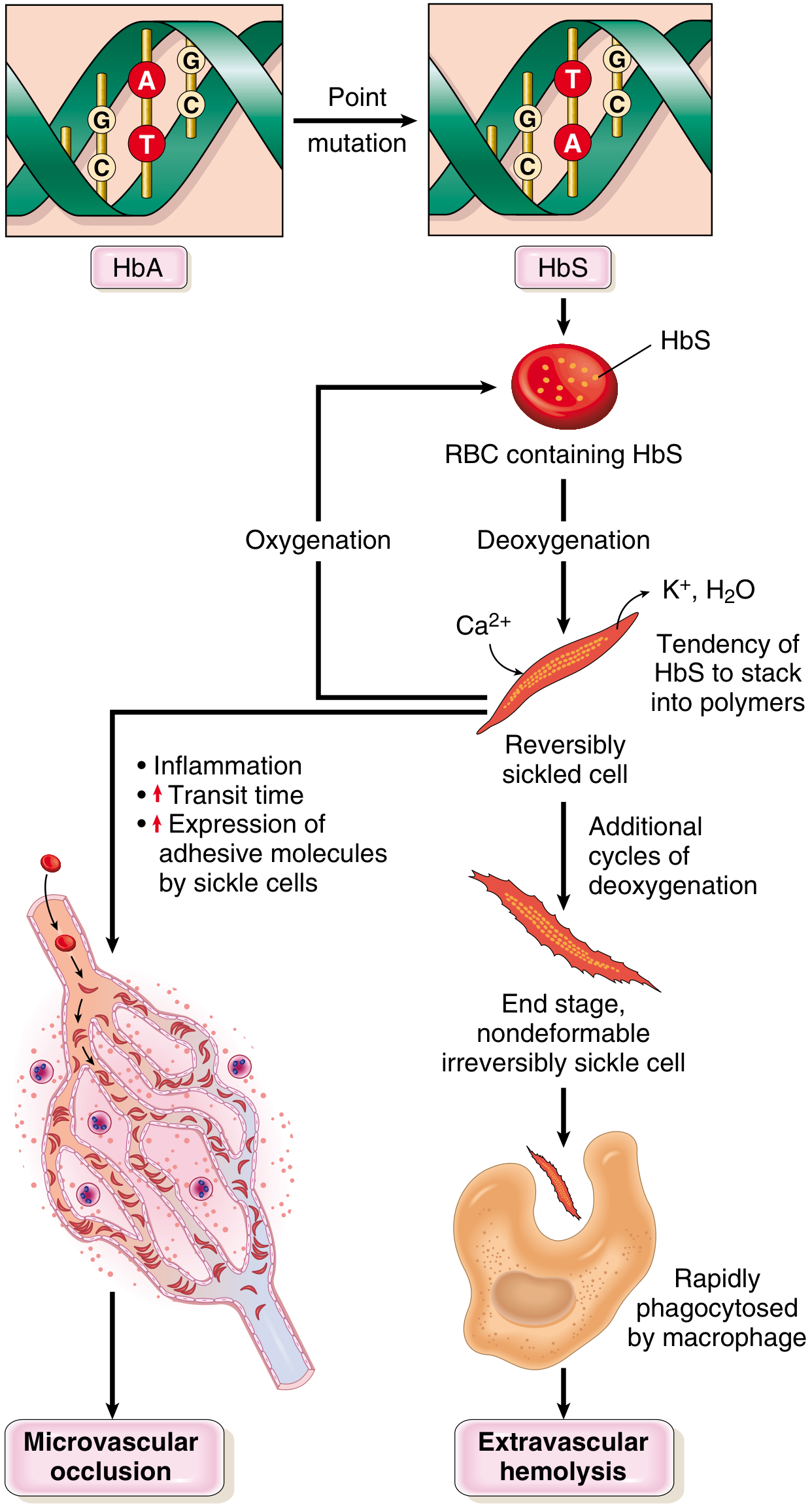
Key mechanism: Deoxygenated HbS molecules polymerize into long needlelike fibers → red cell distortion into sickle/holly-leaf shape
Factors that increase sickling:
| Factor | Effect |
|---|---|
| Low O₂ tension | Core trigger - deoxygenated HbS polymerizes |
| High HbS concentration / dehydration (high MCHC) | More aggregation |
| Low pH / acidosis | Reduces O₂ affinity → more deoxyHbS |
| Slow microvascular transit | Prolonged deoxygenation (spleen, bone marrow) |
| Inflammation | Up-regulates endothelial adhesion molecules |
| HbC co-inheritance | More sickling than HbAS |
Factors that decrease sickling:
- HbF (fetal hemoglobin) - interferes with polymerization
- Co-existing α-thalassemia (reduces MCHC)
- HbA (in heterozygotes)
Downstream cascade:
- Repeated sickling → Ca²⁺ influx → K⁺ & H₂O efflux → dehydrated, dense, rigid cells
- Irreversibly sickled cells (ISCs) form - retain sickle shape even when oxygenated
- ISCs cleared by extravascular hemolysis (spleen/liver macrophages)
- Intravascular hemolysis releases free Hb → binds/inactivates NO → vasoconstriction + platelet aggregation → vascular occlusion
- Sickle cells express increased adhesion molecules → adhere to endothelium → microvascular occlusion
Morphology
- Peripheral blood smear: Irreversibly sickled cells, target cells, reticulocytosis, Howell-Jolly bodies (nuclear remnants, due to functional asplenia)
- Bone marrow: Hyperplastic (compensatory erythroid hyperplasia)
- Spleen in children: Enlarged (up to 500g), red pulp congested with trapped sickled cells
- Spleen in adults: Small fibrotic nubbin = autosplenectomy (repeated infarctions)
Clinical Features
Baseline anemia: Hematocrit 18-30%; moderate hemolytic anemia, reticulocytosis, hyperbilirubinemia
Crises (high-yield exam topics):
| Crisis Type | Mechanism | Key Features |
|---|---|---|
| Vaso-occlusive (pain) crisis | Ischemic infarction from sickling in microvasculature | Most common; fever, severe pain; bones, lungs, liver, brain, spleen, penis |
| Acute chest syndrome | Vaso-occlusion in pulmonary vessels | Fever, cough, chest pain, pulmonary infiltrates; potentially fatal; requires exchange transfusion |
| Sequestration crisis | Massive splenic trapping of sickled RBCs | Rapid splenomegaly, hypovolemia, shock; children with intact spleens |
| Aplastic crisis | Parvovirus B19 infects erythroid progenitors | Sudden severe anemia; transient cessation of erythropoiesis |
Organ complications:
- Dactylitis / hand-foot syndrome: Painful bone crises in small bones of hands/feet in infants/children - often first manifestation
- Stroke: Due to adhesion of sickled RBCs to arterial endothelium + NO depletion causing vasoconstriction
- Priapism: Up to 45% of males after puberty; can cause erectile dysfunction
- Retinopathy: Vascular occlusion; may cause blindness
- Leg ulcers: Adults; subcutaneous vascular stagnation
- Hyposthenuria: Renal medullary damage (sickling promoted by hypertonicity) → inability to concentrate urine → dehydration risk
- Infections: Encapsulated organisms (especially Streptococcus pneumoniae, H. influenzae) due to functional asplenia + complement pathway defects
Diagnosis
- Peripheral smear: sickled cells
- Hemoglobin electrophoresis: Confirms HbS; differentiates trait vs. disease
- Sickling tests (metabisulfite test)
- Newborn screening: routine in all 50 US states (heel stick)
- Prenatal diagnosis: fetal DNA from amniocentesis or chorionic biopsy
Treatment
- Hydroxyurea (mainstay): Inhibits DNA synthesis; benefits include:
- Increases HbF synthesis (HbF inhibits sickling)
- Decreases adhesion molecule expression on RBCs
- Reduces WBC count (anti-inflammatory)
- Reduces frequency of painful crises and acute chest syndrome; improves survival
- Exchange transfusion: For acute chest syndrome, stroke
- Bone marrow / stem cell transplant: Only curative option
- Supportive: Hydration, analgesics, vaccinations, prophylactic penicillin in children
- Gene therapy: Emerging curative approach
THALASSEMIA
Overview
Inherited disorders with decreased synthesis of α- or β-globin chains. Double problem:
- Insufficient HbA → microcytic, hypochromic anemia
- Excess unpaired globin chains → precipitates → RBC membrane damage → hemolysis
Particularly common in Mediterranean, African, and Asian populations (malaria-endemic regions).
β-Thalassemia
Genetics
- Gene: β-globin gene, chromosome 11 (single gene per haploid)
- Mutations: >100 mutations, mostly point mutations (not deletions) → affect transcription, mRNA splicing, or translation
- β⁰: No β-globin produced
- β⁺: Reduced β-globin produced
Classification
| Syndrome | Genotype | Clinical Features |
|---|---|---|
| β-Thalassemia major (Cooley's anemia) | β⁰/β⁰ or β⁰/β⁺ (homozygous) | Severe anemia; requires regular transfusions |
| β-Thalassemia intermedia | Variable β⁺/β⁺ or mild β⁰/β⁺ | Moderate anemia; transfusions NOT regularly required |
| β-Thalassemia minor (trait) | Heterozygous β/β⁰ or β/β⁺ | Asymptomatic or mild; red cell abnormalities on smear |
Pathophysiology
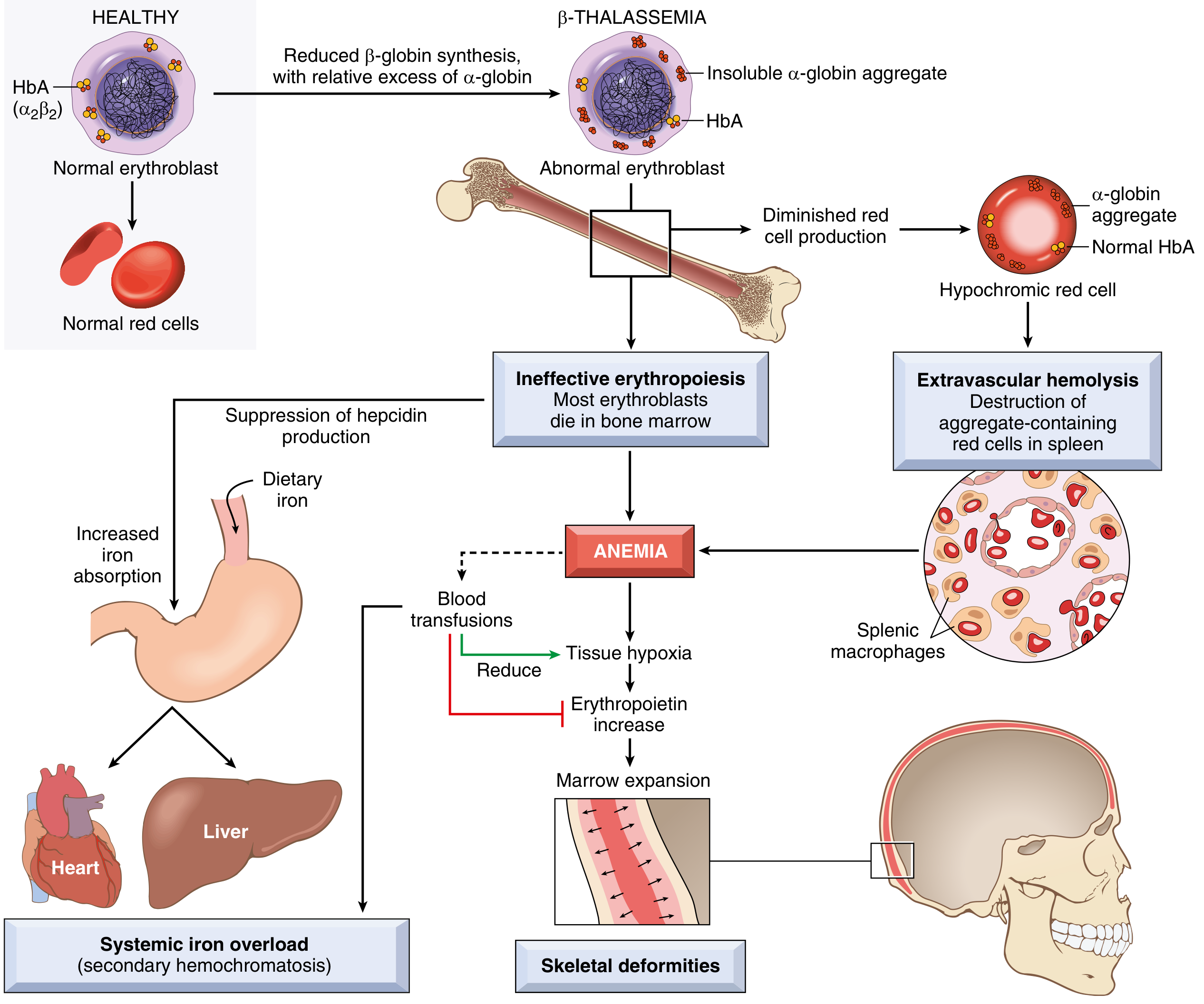
- Reduced β-globin → relative excess of α-globin chains
- Excess α-chains form toxic insoluble precipitates that damage erythroid precursor membranes → apoptosis in bone marrow = ineffective erythropoiesis (most important mechanism)
- Few surviving RBCs have shortened lifespan → extravascular hemolysis in spleen
- Anemia → ↑ erythropoietin → massive erythroid marrow expansion → bone deformities
- Low hepcidin levels → increased dietary iron absorption → secondary hemochromatosis (even without transfusions)
- Transfusions worsen iron overload
Clinical Features of β-Thalassemia Major
- Presents 6-12 months after birth (when HbF→HbA switch occurs)
- Severe anemia: pallor, jaundice, hepatosplenomegaly
- Skeletal deformities: Erythroid marrow expansion causes:
- "Crew-cut" skull X-ray appearance (hair-on-end pattern)
- Frontal bossing, maxillary hypertrophy ("chipmunk facies")
- Thinning of cortical bone → pathological fractures
- Growth retardation, failure to thrive
- Iron overload (secondary hemochromatosis):
- Cardiac failure (leading cause of death)
- Cirrhosis
- Endocrine failure (diabetes, hypogonadism)
- Increased susceptibility to infections
Treatment of β-Thalassemia Major
- Regular blood transfusions (maintain Hb >9-10 g/dL)
- Iron chelation therapy: Deferoxamine (IV/SC) or deferasirox (oral) - mandatory to prevent iron overload
- Splenectomy: If hypersplenism causing excessive transfusion needs
- Bone marrow / stem cell transplant: Curative; best results in young patients
- Hydroxyurea: Can increase HbF in some patients
α-Thalassemia
Genetics
- Gene: α-globin genes - TWO genes per chromosome 16 (total 4 α-globin genes)
- Mutations: Mostly gene deletions (contrast with β-thalassemia which is mostly point mutations)
Classification (by number of deleted α genes)
| # Genes Deleted | Syndrome | Genotype | Clinical Features |
|---|---|---|---|
| 1 | Silent carrier | -α/αα | Asymptomatic; normal CBC |
| 2 | α-Thalassemia trait | --/αα (Asian) or -α/-α (African/Asian) | Asymptomatic; mild microcytic anemia (like β-thal minor) |
| 3 | HbH disease | --/-α | Moderately severe hemolytic anemia; HbH = β₄ tetramers (unstable); resembles β-thal intermedia |
| 4 | Hydrops fetalis | --/-- | Lethal in utero; Hb Barts (γ₄ tetramers); fetus cannot deliver O₂; stillbirth or death shortly after birth |
Key exam point: With loss of α-globin genes, excess β-globin forms HbH (β₄), and excess γ-globin in fetal life forms Hb Barts (γ₄) - both have very high O₂ affinity and cannot deliver O₂ to tissues.
HIGH-YIELD COMPARISON TABLE
| Feature | Sickle Cell Disease | β-Thalassemia Major | α-Thalassemia Trait |
|---|---|---|---|
| Gene | β-globin (chr 11) | β-globin (chr 11) | α-globin (chr 16) |
| Mutation type | Point mutation (missense) | Point mutations | Gene deletions |
| RBC morphology | Sickle cells, target cells, Howell-Jolly | Target cells, nucleated RBCs, anisopoikilocytosis | Mild microcytosis |
| MCV/MCH | Usually normal/high (with reticulocytosis) | Low (microcytic, hypochromic) | Low (microcytic) |
| Reticulocytes | High | High (but ineffective) | Normal/slightly high |
| Spleen | Autosplenectomy in adults | Massive splenomegaly | Normal |
| Iron stores | Normal to low | Overloaded (even without transfusions) | Normal |
| HbF effect | Protective - decreases sickling | Partially compensatory | N/A |
| Diagnosis | Hb electrophoresis | Hb electrophoresis + elevated HbA2 | Hb electrophoresis + gene testing |
| Definitive cure | Stem cell transplant / gene therapy | Stem cell transplant | Usually none needed |
QUICK MNEMONICS
Sickle cell crises - "PASA":
- Pain (vaso-occlusive) - most common
- Acute chest syndrome - most dangerous
- Sequestration - spleen trapping in children
- Aplastic - parvovirus B19
Hydroxyurea mechanism - "4F's":
- Increases Fetal Hb (HbF)
- Reduces adhesion molecule Forces
- Fewer WBCs (anti-inFlammatory)
- Fewer painful crises (fewer Flare-ups)
α-thalassemia severity: 1-2-3-4 deleted genes = Silent-Trait-HbH-Hydrops (lethal)
Sources: Robbins, Cotran & Kumar Pathologic Basis of Disease, pp. 599-601; Robbins & Kumar Basic Pathology, pp. 387-389
This is a shared conversation. Sign in to Orris to start your own chat.