
Treaent of pneumonia for 50 year old
"community-acquired pneumonia" AND treatment
community acquired pneumonia chest x-ray infiltrate consolidation
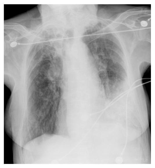
This diagnostic image is an anteroposterior (AP) chest X-ray. The primary clinical finding is a dense, focal opacification in the left lower lobe, consistent with an alveolar infiltrate or consolidation. The left costophrenic angle is partially obscured by the increased density, which borders the cardiac silhouette. In contrast, the right lung field appears relatively clear of acute consolidation, though some mild interstitial markings are visible. Multiple radiopaque medical artifacts are present, including electrocardiogram (ECG) leads and wires superimposed over the thoracic cavity, indicating acute care monitoring. The mediastinal contour appears stable, and the trachea is midline. This imaging is characteristic of community-acquired pneumonia, specifically localized to the left basal region, often associated with pathogens like Moraxella catarrhalis in appropriate clinical contexts.
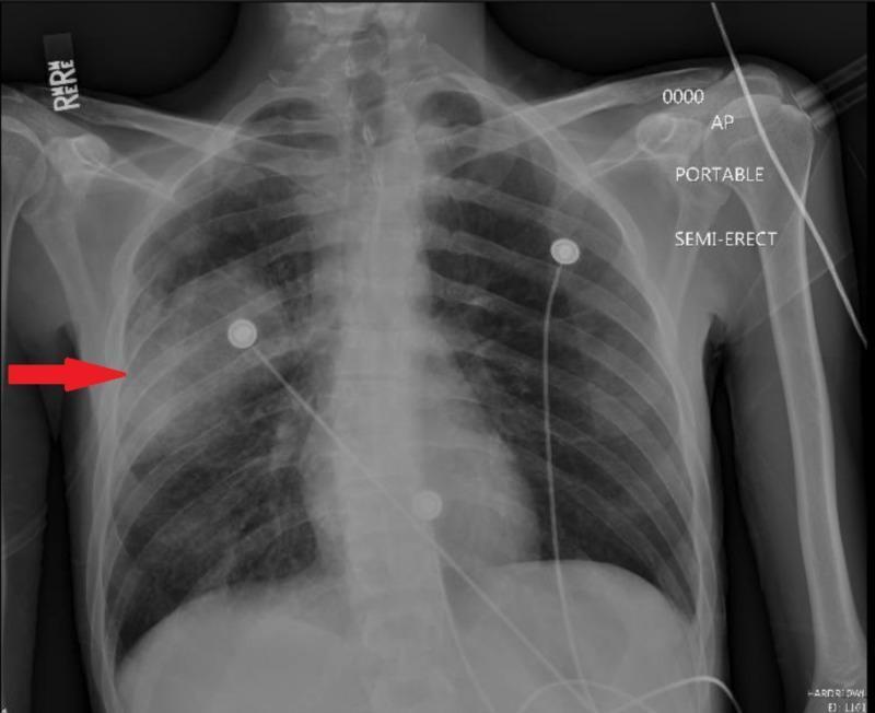
This diagnostic image is a portable anteroposterior (AP) chest X-ray of a human male in a semi-erect position. The primary finding is a focal area of increased opacity in the right mid-lung zone, indicated by a red arrow, which is characteristic of consolidation in the right middle lobe. This infiltrate suggests an infectious process such as community-acquired pneumonia. The left lung field remains relatively clear. Several non-anatomical features are present, including radiopaque monitoring electrodes (ECG leads) with associated wiring across the chest and a 'RIGHT' (R) marker in the upper left corner. The image labels 'PORTABLE' and 'SEMI-ERECT' provide clinical context regarding the acquisition technique in an emergency or acute care setting. This visual is used for teaching the radiological presentation of lobar pneumonia and the identification of anatomical versus non-anatomical structures on a portable chest radiograph.
Treatment of Community-Acquired Pneumonia (CAP) in a 50-Year-Old
Step 1: Risk Stratification Before Treatment
- Confusion (new onset)
- Urea > 7 mmol/L (BUN > 19 mg/dL)
- Respiratory rate >= 30/min
- Blood pressure < 90 systolic or <= 60 diastolic
- Age >= 65 years
| Score | Mortality | Recommendation |
|---|---|---|
| 0-1 | Low | Outpatient treatment |
| 2 | Moderate | Consider short admission or close follow-up |
| 3-5 | High | Hospitalize; ICU if 4-5 |
Step 2: Antibiotic Therapy
Outpatient Treatment (no comorbidities, not severely ill)
| Scenario | Drug(s) | Notes |
|---|---|---|
| Otherwise healthy, no recent antibiotics | Amoxicillin 1g TID OR Azithromycin 500mg day 1, then 250mg x 4 days | Cover typical + atypical organisms |
| Comorbidities (COPD, diabetes, heart/liver/renal disease, malignancy, asplenia, alcohol use) | Respiratory fluoroquinolone - Levofloxacin 750mg OD or Moxifloxacin 400mg OD | Single-agent broad coverage |
| Comorbidities (alternative) | Beta-lactam + macrolide - Amoxicillin-clavulanate 875/125mg BD + Azithromycin | Dual coverage |
Inpatient Treatment (non-ICU)
- Beta-lactam + macrolide: e.g., Ampicillin-sulbactam IV + Azithromycin IV/PO
- OR Respiratory fluoroquinolone monotherapy: Levofloxacin 750mg IV/PO once daily
ICU-level / Severe CAP
- Beta-lactam IV (Ceftriaxone or Ampicillin-sulbactam) + Azithromycin IV
- OR Beta-lactam IV + Respiratory fluoroquinolone IV
- If Pseudomonas risk (structural lung disease, recent hospitalization, steroids, broad-spectrum AB use): add anti-pseudomonal coverage (Piperacillin-tazobactam or Cefepime)
- If MRSA risk: add Vancomycin or Linezolid
Step 3: Duration of Treatment
- Minimum 5 days, and continue at least 48-72 hours after the patient's last fever or signs of clinical instability
- Most uncomplicated CAP: 5-7 days total is adequate per recent systematic reviews (PMID 36948555)
- Atypical pneumonia (Mycoplasma, Chlamydia): some guidelines suggest 7-10 days
- Legionella: 10-14 days
Step 4: Supportive Care
- Oxygen - target SpO2 >= 94%
- IV fluids if dehydrated or hemodynamically compromised
- Antipyretics/analgesics - Paracetamol (Acetaminophen) for fever/pleuritic pain
- Chest physiotherapy in select cases
- Bronchodilators if wheeze or underlying COPD
- Avoid corticosteroids routinely (not recommended in non-severe CAP)
Step 5: Common Causative Organisms
| Type | Organisms |
|---|---|
| Typical | Streptococcus pneumoniae (most common), H. influenzae, Staphylococcus aureus |
| Atypical | Mycoplasma pneumoniae, Chlamydophila pneumoniae, Legionella pneumophila |
| Viral | Influenza A/B, SARS-CoV-2, RSV |
| Aspiration risk | Anaerobes (alcoholics, aspiration) |
Step 6: Monitoring and Follow-Up
- Repeat CXR at 4-6 weeks to confirm resolution (rule out underlying malignancy in a 50-year-old smoker)
- Check for clinical improvement within 48-72 hours - if no improvement, broaden coverage or investigate for resistant organisms, empyema, or wrong diagnosis
- Blood cultures before antibiotics if hospitalized (though yield is low ~5-10%)
Step 7: Prevention - Important for age 50
- Influenza vaccine annually (especially recommended for patients > 50 years) - Textbook of Family Medicine 9e, p.321
- Pneumococcal vaccine (PPSV23 and/or PCV13/15/20 depending on comorbidities) - see indications based on risk factors above
Chest X-Ray Appearances