
Submandibular gland
submandibular gland anatomy diagram
![This three-panel composite image illustrates an experimental in vivo microscopy setup for analyzing the microvasculature of the submandibular gland. Panel A is a schematic diagram showing the vertical orientation of the imaging system: a dipping objective [1] is positioned above a glass imaging window [2], which rests directly on the submandibular gland [4], all supported by a mounting stage [3]. Panel B provides a macroscopic photograph of the custom-built microscopy stage, highlighting the physical components including the glass window [2], the adjustable mounting arm [3], and a circular fixation ring [5] designed to secure skin sutures around the surgical site. Panel C is a clinical photograph of the surgical field (operation situs) in the anterior neck region. It demonstrates the exposed anatomy after dissection, specifically highlighting the submandibular gland [4] as a pinkish, lobulated, and vascularized glandular structure. The setup is designed to stabilize living tissue for high-resolution imaging of leukocyte trafficking and microvascular permeability in real-time.](https://cdn.orris.care/cdss_images/pmc_clinical_VQA_09808406a59f7094aa58e1eed8c5258bac44c68053e0bb8cc63cad9ed1abd195.jpg)
This three-panel composite image illustrates an experimental in vivo microscopy setup for analyzing the microvasculature of the submandibular gland. Panel A is a schematic diagram showing the vertical orientation of the imaging system: a dipping objective [1] is positioned above a glass imaging window [2], which rests directly on the submandibular gland [4], all supported by a mounting stage [3]. Panel B provides a macroscopic photograph of the custom-built microscopy stage, highlighting the physical components including the glass window [2], the adjustable mounting arm [3], and a circular fixation ring [5] designed to secure skin sutures around the surgical site. Panel C is a clinical photograph of the surgical field (operation situs) in the anterior neck region. It demonstrates the exposed anatomy after dissection, specifically highlighting the submandibular gland [4] as a pinkish, lobulated, and vascularized glandular structure. The setup is designed to stabilize living tissue for high-resolution imaging of leukocyte trafficking and microvascular permeability in real-time.
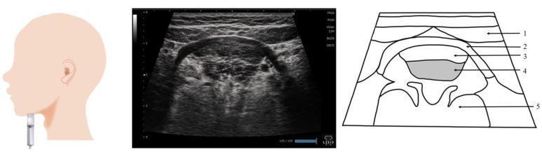
This composite educational graphic details the ultrasound anatomy of the submandibular region. It consists of three panels: a probe placement guide, a representative ultrasound image, and a labeled anatomical diagram. The leftmost panel shows a linear probe positioned vertically on the submandibular surface, perpendicular to the Frankfurt plane. The central ultrasound image displays a coronal cross-section of the suprahyoid musculature. The rightmost diagram provides structural identification: 1) Digastric muscle (superficial), 2) Mylohyoid muscle, 3) Geniohyoid muscle, 4) Genioglossus muscle (deep, shaded grey), and 5) The body of the tongue. The muscles are visualized in a layered, hierarchical arrangement from superficial to deep. Key visual markers include the hyperechoic fascia lines separating the muscle groups. This imaging technique is clinically relevant for assessing tongue thickness, swallowing disorders (dysphagia), and the cross-sectional area of floor-of-mouth musculature in geriatric or rehabilitation medicine.
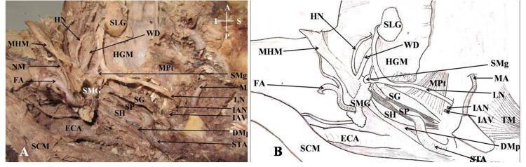
This composite educational resource comprises a clinical photograph of a cadaveric dissection (A) and a matching schematic diagram (B), providing a lateral view of the human infratemporal fossa and submandibular region. The visual focus is the spatial relationship between the salivary glands, neurovascular structures, and musculature. Key anatomical landmarks include the submandibular gland (SMG) and sublingual gland (SLG), with the Wharton duct (WD) shown originating from the SMG. The dissection reveals the mylohyoid muscle (MHM) partially reflected to expose the deeper hyoglossus muscle (HGM). Neural structures are clearly labeled, including the lingual nerve (LN), hypoglossal nerve (HN), inferior alveolar nerve (IAN), and the submandibular ganglion (SMg). The styloid apparatus is demonstrated through the styloglossus (SG), stylohyoid (SH), and stylopharyngeus (SP) muscles. Vascular components include the external carotid artery (ECA), maxillary artery (MA), and facial artery (FA). This image serves as an advanced anatomical reference for medical students and surgical trainees focusing on head and neck anatomy, particularly regarding the complex arrangement of the floor of the mouth and the parapharyngeal space.
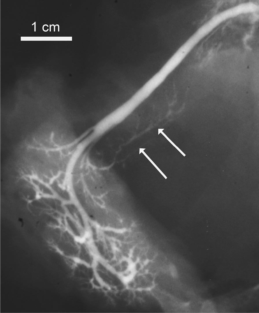
This diagnostic sialogram captures the ductal anatomy of a submandibular gland. The image features a prominent, radiopaque main submandibular duct (Wharton's duct) characterized by a thick, curvilinear morphology. Distally, the duct branches into an intricate, dendritic network of smaller intraglandular ducts that decrease in caliber as they penetrate the glandular parenchyma. Two white arrows indicate an accessory salivary lobe, visible as a cluster of small, rounded contrast-filled structures joining the lateral aspect of the main duct. This accessory tissue exhibits slightly lower contrast intensity compared to the primary duct. A 1 cm scale bar is provided in the upper left corner for morphometric reference. This image is used in clinical education to demonstrate normal anatomical variations, specifically the presence of accessory submandibular lobes, and to illustrate standard salivary ductal architecture.
submandibular gland histology mixed serous mucous acini
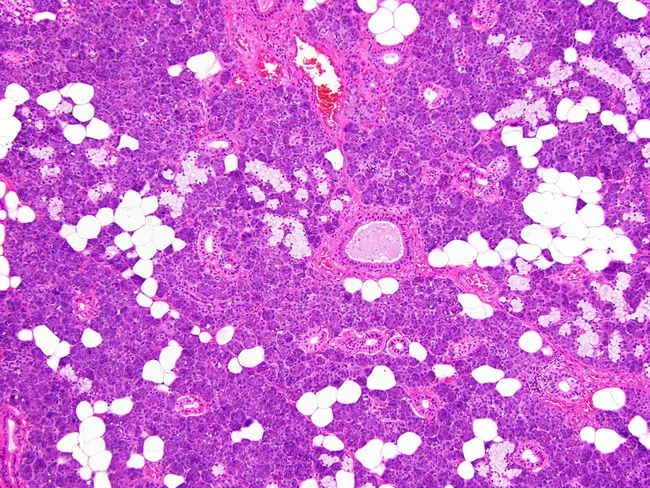
This histology image shows a hematoxylin and eosin stained paraffin-embedded section of human submandibular gland tissue, examined by light microscopy at high power. The gland demonstrates mixed seromucinous acini in which mucous cells form the central portions of the acini and serous cells lie at the periphery, with serous demilunes sometimes evident at mucous borders. Excretory and intercalated ducts are readily visible within the lobular architecture, appearing as columnar to cuboidal ductal elements with clear lumens. The acini are small to medium in size, and the surrounding stroma contains adipose tissue along with delicate connective tissue septa that partition the lobules. The overall pattern reflects normal salivary gland histology without overt cytologic atypia, notable inflammatory infiltrate, or neoplastic change in this field. Clinically, this image is pertinent for education on glandular microanatomy, including acinar polarity (serous periphery vs mucous center), ductal morphology, and the relationship between acini and excretory ducts. It supports differential considerations such as normal variant anatomy, sialadenitis, and salivary gland neoplasms in comparative studies. The image is a valuable teaching tool for pathologists, residents, and students to recognize typical histologic landmarks and enhance reproducibility in diagnostic reporting.
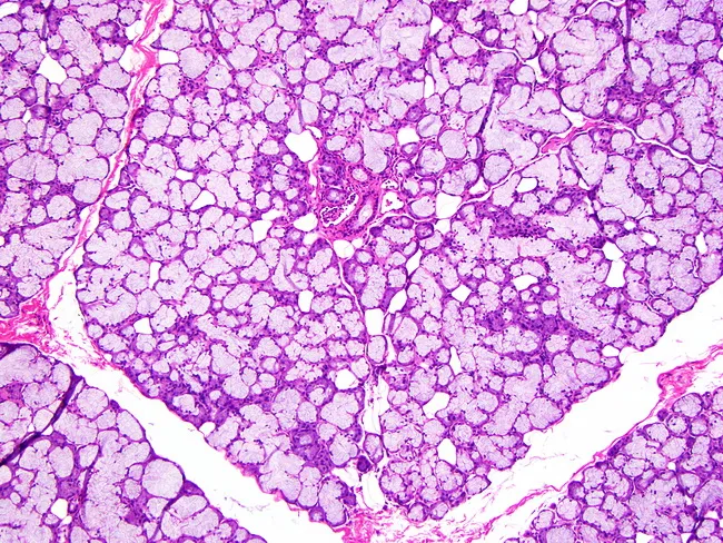
This histology image depicts sublingual salivary gland tissue examined by light microscopy after routine hematoxylin and eosin staining. The mucous acini predominate, appearing as large, pale-staining units with mucus-filled cytoplasm and relatively flattened basal nuclei; serous demilunes are minimal or absent in this field. Acini are arranged in a lobular architecture embedded in a connective tissue stroma that contains scattered capillaries and occasional adipose elements at the periphery. The excretory duct system is relatively inconspicuous compared with serous-rich glands such as the parotid or mixed glands like the submandibular; ducts are small, inconspicuous channels coursing between acini and encased in slender septa. The overall pattern reflects the mucous-dominant phenotype characteristic of the sublingual gland, with larger mucous acini than those observed in the submandibular counterpart. The image emphasizes glandular polarity with secretory cells facing a lumen and myoepithelial cells surrounding acini and ducts. Clinically, this histology supports identification of sublingual gland tissue in surgical specimens and serves as a reference for comparisons in glandular pathology, including evaluation for chronic sialadenitis, mucous retention phenomena, or rare mucous-secreting neoplasms. Overall, the slide demonstrates classic duct-acinar organization, mucous-rich cytoplasm, and a subtle ductal network within a loose connective tissue capsule.
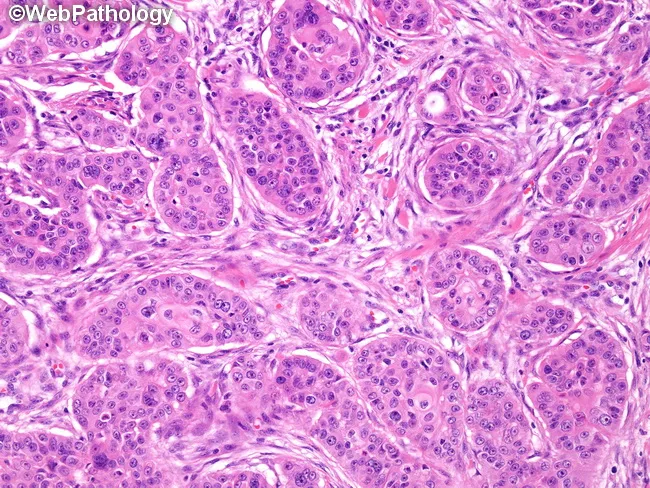
Imaging modality: light microscopy of an H&E–stained histology section from a recurrent submandibular gland mass. Primary subject: high‑grade mucoepidermoid carcinoma with scant cyst formation. Precise anatomical location: submandibular region (salivary gland), head and neck. Visual features: predominantly solid sheets and clusters of epidermoid (squamoid) and intermediate cells displaying cytologic atypia, marked nuclear pleomorphism, prominent nucleoli, and brisk mitotic activity. Keratinization is limited; extensive keratin pearls are not present; occasional small keratin pearls or individual cell keratinization may be seen. Mucicarmine-positive mucous cells are scattered throughout the tumor, indicating mixed differentiation with mucous-secreting components. The stroma is variable with embedded tumor nests and occasional duct-like structures; no prominent cystic spaces are described. Imaging perspective: micrographic cross-sections at high magnification highlight cellular morphology and intercellular bridges. Diagnostic interpretation: high-grade MEC with aggressive features, potential to mimic moderately differentiated squamous cell carcinoma but mucous cell component and mucicarmine positivity support MEC; absence of large keratin pearls argues against classic keratinizing SCC. Clinical relevance: biopsy morphology is essential for grading, prognosis, and therapeutic planning, as high‑grade MEC carries higher risk of recurrence and regional/metastatic spread; findings justify surgical management with consideration of adjuvant therapy and close follow-up.
Submandibular Gland
Overview
Embryology
Anatomy
Location and Shape
- Situated in the submandibular triangle (Level Ib) - bounded superiorly by the mandible, inferiorly by the anterior and posterior bellies of the digastric muscle.
- Consists of a larger superficial lobe (in the digastric triangle) and a smaller deep lobe, which are continuous around the posterior border of the mylohyoid muscle.
Relations
| Surface | Relations |
|---|---|
| Inferior | Skin, platysma, deep cervical fascia; crossed by the facial vein and cervical branch of facial nerve |
| Lateral | Submandibular fossa on medial surface of mandible; medial pterygoid muscle attachment |
| Medial (superficial part) | Mylohyoid muscle anteriorly; styloglossus posteriorly |
| Medial (intermediate) | Hyoglossus muscle; separated by lingual nerve, submandibular ganglion, hypoglossal nerve, deep lingual vein |
Capsule
Wharton's Duct (Submandibular Duct)
- Approximately 5 cm long.
- Emerges from the medial surface of the superficial lobe, passes through the deep lobe, then runs between the sublingual gland and genioglossus muscle.
- Opens in the floor of the mouth at the sublingual papilla (summit), at the side of the frenulum of the tongue.
- On the hyoglossus muscle it is crossed laterally by the lingual nerve - an important surgical landmark (the nerve "hooks" under and then over the duct: "twice bitten by the lingual nerve").
Vascular Supply
| Vessel | Course |
|---|---|
| Facial artery | Enters deep to the posterior belly of digastric and stylohyoid; courses through or superficial to the gland to reach the anterior border of the mandible. Glandular branches must be ligated during excision. Commonly used as recipient artery in free tissue transfer. |
| Facial vein | Lies superficial (lateral) to the gland |
Nerve Supply
Secretomotor (Parasympathetic)
- Preganglionic fibres travel via: Facial nerve (CN VII) → chorda tympani → lingual nerve → submandibular ganglion.
- The submandibular ganglion is a small, fusiform body on the upper part of hyoglossus, suspended from the lingual nerve. Postganglionic fibres supply both the submandibular and sublingual glands.
- These ganglion connections must be divided to free the gland while preserving the lingual nerve during excision.
Other nerves at surgical risk
- Lingual nerve (CN V3): sensory; attached to the deep surface of the gland via the ganglion - loops around Wharton's duct.
- Hypoglossal nerve (CN XII): motor to tongue; lies medial to the digastric and the gland.
- Marginal mandibular branch of facial nerve (CN VII): lies in the superficial fascia, traversing over the facial vessels - at risk during neck incision. Martin's technique (ligation and superior retraction of facial vessels) helps protect it.
Histology
Clinical Relevance
1. Sialolithiasis (Salivary Calculi)
- Wharton's duct is longer, wider, more tortuous, and runs against gravity around the mylohyoid - all promoting salivary stasis.
- Saliva from the gland is more viscous with higher calcium/phosphate concentration.
- Stones are composed of calcium phosphate and carbonate with an organic glycoprotein/mucopolysaccharide matrix.
- In the submandibular gland, stone formation is the primary event (causing stasis and retrograde bacterial sialadenitis), whereas in the parotid, chronic inflammation is the inciting event.
- Ultrasound: detects >90% of stones >2 mm; cost-effective, no radiation, can be used intraoperatively.
- CT (thin cuts, 1-2 mm, non-contrast): very accurate; contrast avoided to prevent confusion with opacified vessels.
- Plain radiographs historically used but now avoided (miss radiolucent stones, ~30% are radiolucent).
2. Submandibular Gland Tumours
- Bimanual palpation (intraoral + external) is essential to assess extent and fixation to mandible or skin.
- Warning signs of malignancy:
- Numbness of tongue → lingual nerve involvement
- Weakness of tongue → hypoglossal nerve involvement
- Weakness of lower lip → marginal mandibular nerve involvement
- 25-28% of submandibular malignancies have regional lymph node metastases at presentation.
- MRI is the preferred imaging modality for suspected neoplasms.
3. Submandibular Gland Excision (Sialadenectomy)
- Incision placed >2 cm below the mandible to protect the marginal mandibular nerve.
- Facial vessels identified and ligated; Martin's manoeuvre (flipping ligated vessels superiorly) protects the marginal mandibular nerve.
- Submandibular ganglion connections divided to free the gland without damaging the lingual nerve.
- Hypoglossal nerve identified and preserved on the hyoglossus muscle.
- Wharton's duct ligated/divided at its anterior extent.
Key Summary Points
| Feature | Detail |
|---|---|
| Size | Second largest major salivary gland |
| Output | ~60-65% of resting saliva |
| Secretion type | Mixed, predominantly serous |
| Duct | Wharton's duct, ~5 cm, opens at sublingual papilla |
| Secretomotor supply | Chorda tympani → lingual nerve → submandibular ganglion |
| Key surgical nerve risks | Marginal mandibular, lingual, hypoglossal |
| Most common pathology | Sialolithiasis (>80% of all salivary calculi) |
| Lymph nodes | Periglandular only (no intraglandular nodes) |