
How to correct pulled elbow
pulled elbow radial head subluxation reduction technique
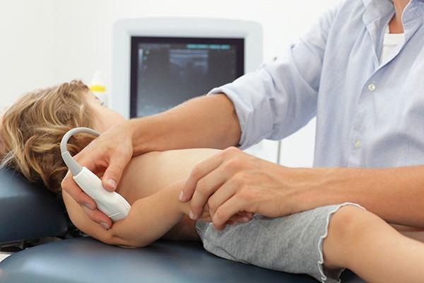
This clinical procedural photograph demonstrates the point-of-care ultrasound (POCUS) technique for diagnosing a 'pulled elbow' (radial head subluxation) in a pediatric patient. The child is positioned supine on a medical examination table with the head turned laterally. A medical professional is seen applying a high-frequency linear ultrasound transducer to the posterior-lateral aspect of the patient's elbow joint. The probe is oriented caudally to visualize the radiohumeral joint and the annular ligament. In the background, an ultrasound monitor displays a real-time grayscale musculoskeletal image. This procedure is used in pediatric emergency medicine to identify signs such as an increased distance between the radial head and the humerus capitate or the interposition of the annular ligament, which confirms subluxation. The image illustrates proper hand placement for stabilizing the young patient's limb while manipulating the transducer to achieve diagnostic views.
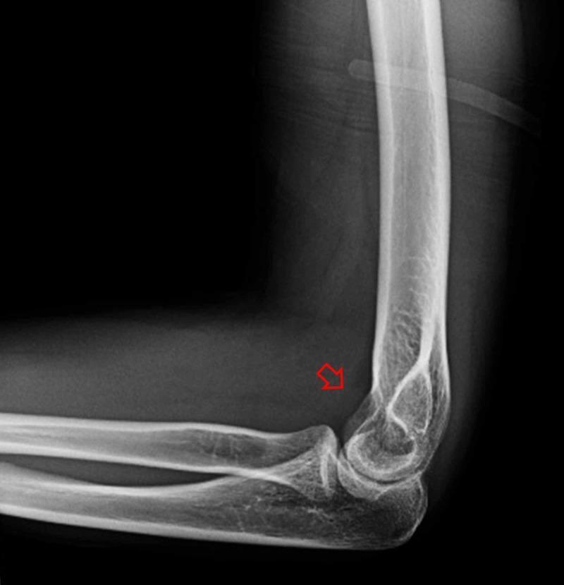
This diagnostic image is a lateral X-ray of a human elbow joint following procedural reduction of a radial head dislocation and elbow subluxation. The radiograph demonstrates restored anatomical alignment: the radial head is congruent with the humeral capitellum, and the ulnohumeral articulation appears well-positioned. No obvious cortical fractures or avulsion fragments are visible in the distal humerus, proximal radius, or ulna. A red arrow points to a subtle, localized area of increased radiopacity within the anterior joint space, suggestive of joint effusion or hemarthrosis. This finding is characterized by a density that is higher than the surrounding soft tissue but significantly lower than the adjacent cortical bone. The image serves as an educational example of post-reduction radiological assessment in emergency orthopedics, highlighting successful joint realignment and secondary signs of intra-articular trauma such as hemarthrosis.
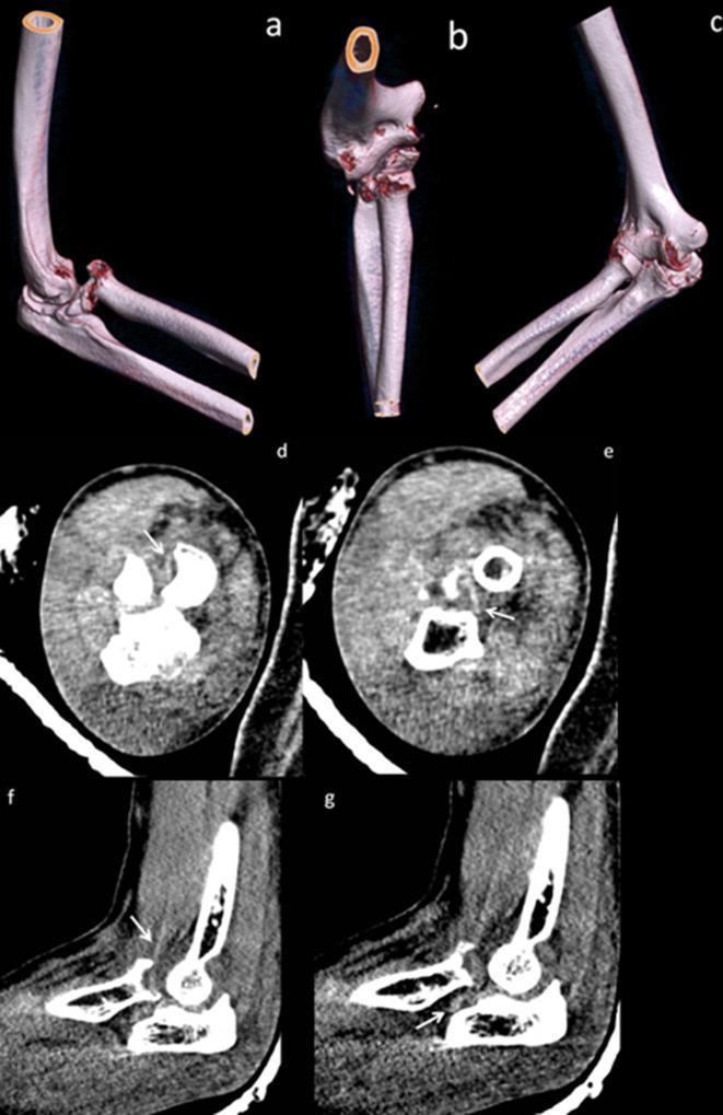
Diagnostic imaging series of the right elbow demonstrating an anteromedial fracture-dislocation of the radial head. Panels (a-c) present volume-rendered 3D CT reconstructions in anterior oblique and anteroposterior views, highlighting a comminuted radial head fracture involving approximately 25% of the articular surface. Small osseous fragments are visible wedged posterior to the radial head and within the radial notch of the ulna. Rotatory subluxation is indicated by the malalignment of the ulnar trochlear notch relative to the humeral trochlea. Panels (d-e) show axial CT slices, and (f-g) show sagittal slices in soft tissue windows. These cross-sectional images identify the brachialis tendon (white arrows) as a linear hyperdensity traversing posterolateral to the radial head fracture site. This anatomical relationship is clinically significant as it illustrates a soft tissue 'sling' effect that can impede manual reduction of the dislocation. The content is suitable for orthopedic and radiology education focusing on complex elbow trauma and the identification of mechanical blocks to reduction.
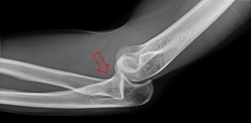
This diagnostic image is a lateral X-ray radiograph of the elbow joint. It demonstrates significant musculoskeletal pathology involving the proximal forearm and distal humerus. The primary finding is a radial head dislocation, where the radial head is displaced anteriorly and is no longer articulating with the capitellum of the humerus. This misalignment is indicated by a red arrow pointing toward the displaced radial head. Additionally, the image shows signs of elbow subluxation, characterized by an incongruous relationship between the trochlear notch of the ulna and the trochlea of the humerus, suggesting partial loss of contact between the joint surfaces. Key anatomical landmarks visible include the distal humeral epicondyles, the olecranon process, and the coronoid process of the ulna. There is no evidence of an acute fracture in this projection. This radiographic study is clinically significant for diagnosing complex elbow injuries and assessing the need for orthopedic reduction. It serves as an educational example of traumatic joint disruption for students and clinicians in emergency medicine and orthopedics.
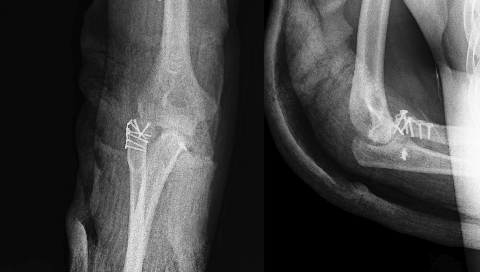
Postoperative X-ray of the right elbow in anteroposterior (AP) and lateral views. The images demonstrate the status after internal fixation of a radial head fracture. Open reduction and internal fixation (ORIF) hardware is clearly visible, consisting of a low-profile mini plate and multiple cortical screws spanning the proximal radius. In the AP view, the hardware is positioned along the lateral aspect of the radial head and neck, maintaining stable reduction of the fracture fragments. The ulnohumeral and radiocapitellar joints appear congruent, with no evidence of subluxation or dislocation. The coronoid process and olecranon are intact. There is a single screw or suture anchor visible in the region of the coronoid/proximal ulna on the lateral view, suggesting possible repair of a concomitant coronoid fracture. Soft tissue swelling is present, consistent with the immediate postoperative period. The radiographic findings confirm appropriate alignment and stabilization of the complex elbow injury, typically classified as a Mason type 4 radial head fracture with associated instability.
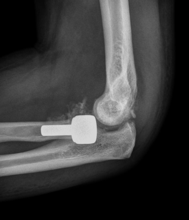
This lateral view X-ray of the elbow shows a radial head prosthesis following a complex injury, likely a 'terrible triad' injury. The imaging reveals a metallic, radiopaque radial head prosthesis consisting of a rounded articular head and a cylindrical intramedullary stem. The primary finding is a posterior subluxation of the radial head prosthesis; the prosthetic head is displaced posteriorly and is no longer articulating correctly with the capitellum of the distal humerus. While the ulnohumeral joint appears to maintain a degree of congruence, the malalignment of the radial component signifies joint instability. Additional soft tissue shadowing or heterotopic ossification may be present anterior to the joint space. This image demonstrates a significant postoperative complication, specifically prosthetic instability and subluxation, which is critical for medical students and orthopedic residents to recognize as a potential failure of stabilization following radial head arthroplasty.
pulled elbow radial head subluxation reduction technique hyperpronation supination flexion
Pulled Elbow (Radial Head Subluxation) - Reduction
Background
Before Attempting Reduction
- Take radiographs first (AP, lateral, and oblique views) to rule out fracture, growth plate injury, or fat pad sign (joint effusion)
- Do NOT manipulate if there is edema, ecchymosis, obvious deformity, fracture on x-ray, or distal neurovascular compromise
- No analgesia or sedation is typically needed
- The child can sit in a parent's lap or lie on the examination table
Two Reduction Techniques
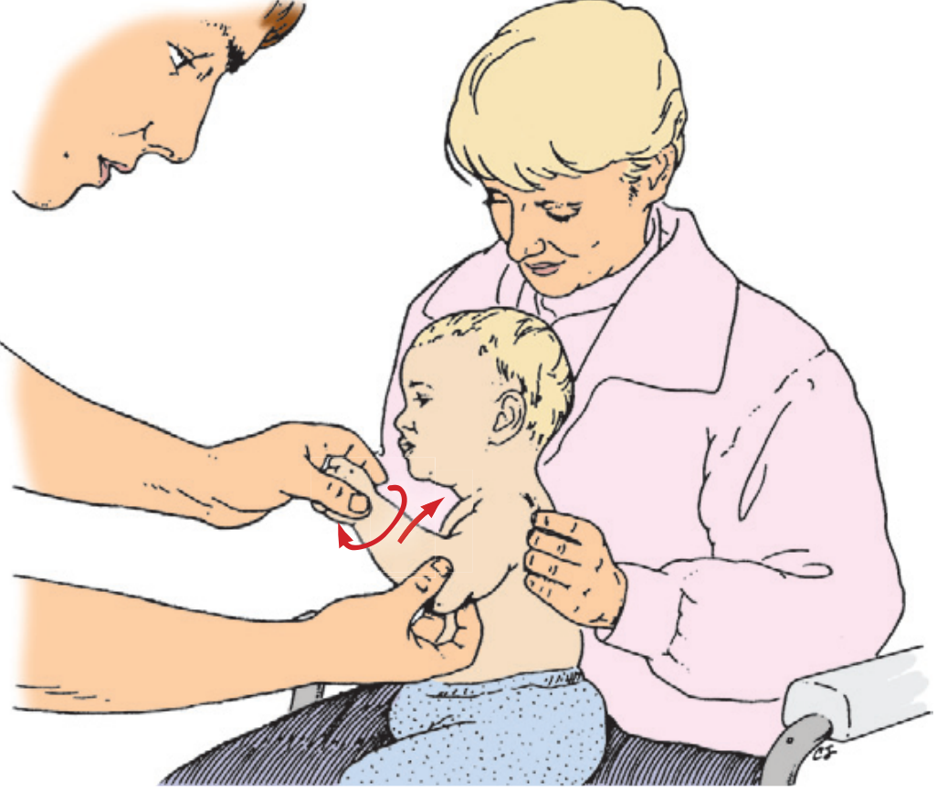
1. Hyperpronation Method (Preferred - First Line)
- Support the child's elbow in your nondominant hand, placing your thumb firmly over the radial head (lateral aspect of the elbow) with gentle medial pressure.
- With your other hand holding the child's wrist, apply slight distal traction.
- Hyperpronated the forearm (turn the palm firmly downward/inward) in one smooth, firm motion.
- Immediately follow with rapid flexion of the elbow.
- Extend back to 90° and release.
- A palpable or audible click/clunk over the radial head signals successful reduction.
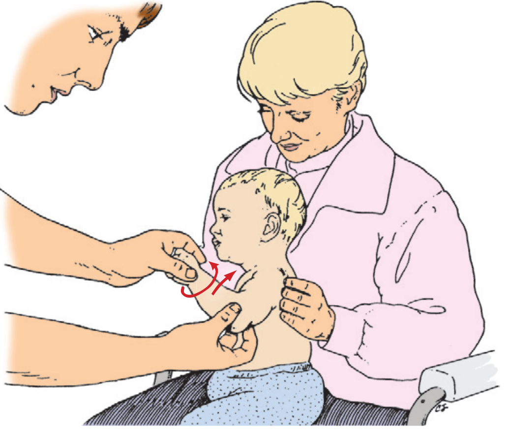
2. Supination-Flexion Method (Second Line / Alternative)
- Support the child's elbow in your nondominant hand with the thumb over the radial head, applying constant gentle medial pressure.
- Apply slight distal traction at the wrist with your other hand.
- Supinate the forearm (palm rotating upward).
- Rapidly flex the elbow fully (past 90°, toward the shoulder) in one smooth arc.
- Hold at 90° flexion, then release.
- Feel for the click/clunk confirming reduction.
Confirming Successful Reduction
- A click or clunk is often felt (not always present)
- Offer the child a toy or sweet on the affected side - if they reach out with that arm, reduction is confirmed
- The child should be using the arm normally within 30 minutes
- No post-reduction immobilization is needed in uncomplicated cases
If First Attempt Fails
- Wait 15-30 minutes - spontaneous reduction can occur
- Attempt the alternative technique (e.g., if hyperpronation failed, try supination-flexion, or vice versa)
- A "combination" approach (hyperpronation immediately followed by supination-flexion) is used by some practitioners with good anecdotal success
- If both fail after 2-3 attempts, obtain orthopedic consultation and repeat imaging
After Reduction
- No splint or sling required in straightforward cases
- Counsel parents to avoid picking the child up by the hands/wrists or swinging by the arms - the same mechanism will re-sublux the joint
- Recurrence is common (up to 27% of cases) until the annular ligament matures (usually by age 6-7)