
Somites 5m embryology short note answer
somites embryology development diagram
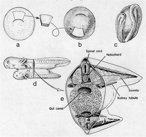
This medical illustration depicts the Spemann-Mangold organizer experiment, a fundamental study in developmental biology and embryology. The series of diagrams (a-e) visualizes the process of neural induction and embryonic axis duplication.
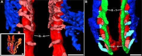
This comparative pathophysiology diagram utilizes 3D surface-rendered imaging to demonstrate the development and remodeling of occipital intersomitic vessels in an embryo. Panel A (8-somite stage) illustrates the early vascular network where the dorsal aorta (DA, red) gives rise to intersegmental arteries (ISA, pink). A transient arteriovenous shunt (AVS), indicated by yellow arrowheads, directly connects the DA to the early cardinal vein (ECV, blue). A white arrowhead marks a regressing ISA at the level of somite S-4. Panel B (15-somite stage) shows the progression after the AVS has regressed (yellow arrowheads). The persistent vasculature includes the vertebral artery (VTA, green), which connects the dorsal ISAs. These are linked via light blue intersomitic vessels (ISVs) to the anterior cardinal vein (ACV) or common cardinal vein (CCV, blue). The somites S-1 and S-4 serve as anatomical landmarks. This visualization highlights the complex transition from transient embryonic shunts to a stable circulatory pattern in the occipital region, providing an educational model for vascular embryology and morphogenesis.
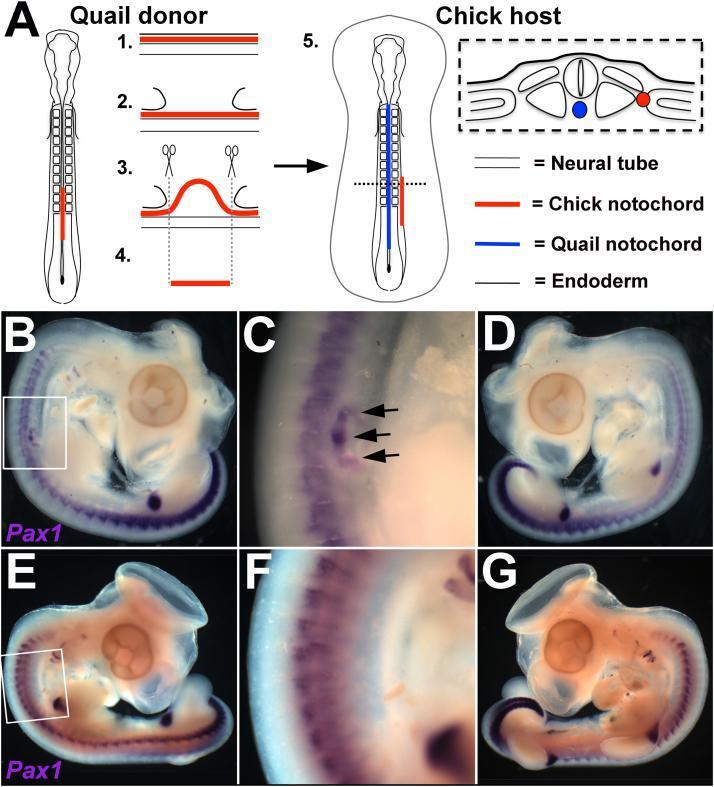
This composite educational material illustrates embryonic development and experimental embryology focusing on sclerotome patterning.
Somites - Short Note (5 marks)
Definition
Origin
Timing and Number
| Stage | Detail |
|---|---|
| First somite pair appears | End of 3rd week (~day 20) |
| Somite period | ~Day 26-32 (38-39 pairs form) |
| Total by end of 5th week | 42-44 pairs |
| Rate (humans) | 2 somites on each side every 4-5 hours |
Mechanism of Formation (Somitogenesis)
- The wavefront involves opposing gradients of FGF8 (high posteriorly) and retinoic acid/RA (high anteriorly) - when cells reach a critical concentration threshold, segmentation is triggered.
- The segmentation clock involves oscillating gene expression in the Notch, WNT, SHH, and FGF signaling pathways. The Notch pathway gene HES7 is a key downstream oscillator.
- Tbx6 (T-box family), FoxC1/FoxC2 (forkhead transcription factors), and HOX genes govern the craniocaudal segmental pattern.
Mutations in HES7 cause spondylocostal dysostosis (severe axial skeletal malformations).
Differentiation of Somites
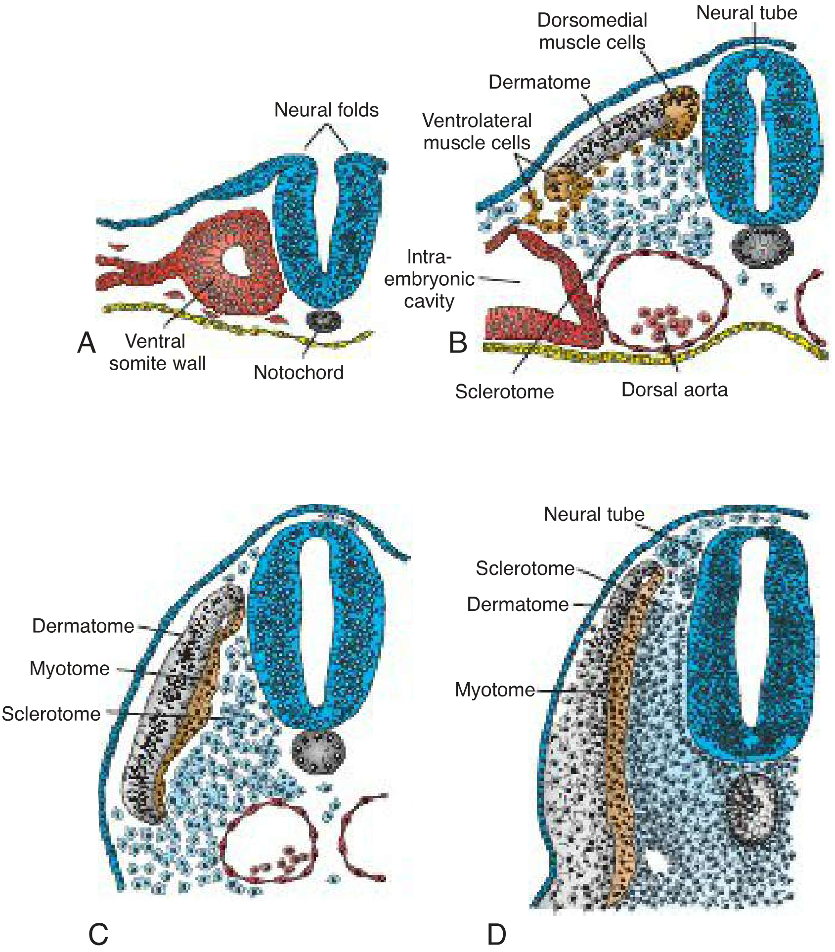
1. Sclerotome (ventromedial)
- Cells lose epithelial characteristics, become mesenchymal again, and migrate to surround the notochord and neural tube.
- Derivatives: vertebrae, ribs, and intervertebral discs (via endochondral ossification).
- Induced by SHH from the notochord and floor plate, and NOGGIN gene products.
2. Dermomyotome (dorsolateral)
| Region | Cells | Derivatives |
|---|---|---|
| Dermatome | Dorsolateral dermomyotome cells | Dermis of the back (fibroblasts) |
| Myotome | Dorsomedial (DML) + ventrolateral (VLL) edges | Striated skeletal muscles |
- Dorsomedial myotome cells → muscles of the back
- Ventrolateral myotome cells migrate → muscles of the body wall (external/internal oblique, transversus abdominis) and limb muscles
- Induced by WNT proteins from the dorsal neural tube and overlying ectoderm, and Neurotrophin-3 (NT-3).
Key Principle of Segmental Innervation
- Each sclerotome forms its own skeletal segment
- Each myotome has its own segmental muscle
- Each dermatome has its own cutaneous nerve supply
Clinical Relevance
| Anomaly | Mechanism |
|---|---|
| Hemivertebrae / fused vertebrae | Mutations in somitogenesis or HOX patterning genes |
| Spondylocostal dysostosis | HES7 mutation |
| Caudal regression syndrome | Disruption of paraxial mesoderm formation from the tail bud |
| Spina bifida | Failure of somite-derived neural arch sclerotome to fuse |