
Pathology of adrenal cortical adenoma 10 marks md pathology exam question
adrenal cortical adenoma gross cut surface yellow
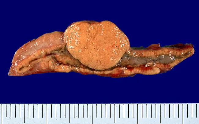
Gross pathology photograph of an excised human adrenal gland displaying a solitary, sharply circumscribed, yellow cortical adenoma arising from the adrenal cortex. The tumor presents as a discrete, well-demarcated nodule with a smooth capsule, contrasting against a relatively thickened surrounding cortex. The cut surface is homogeneous, lipid-rich, and tan-yellow, characteristic of adrenal cortical adenomas, reflecting abundant intracellular lipids. The mass measures approximately a few centimeters in diameter. The rest of the adrenal cortex shows mild concentric thickening or hyperplasia adjacent to the lesion. The adrenal medulla appears gray and well preserved. No hemorrhage or necrosis identified. The specimen is consistent with a benign cortical lesion associated with Conn syndrome, a form of primary hyperaldosteronism. This macroscopic appearance supports aldosterone-secreting adenoma; differentiation from cortisol-producing adenomas, pheochromocytoma, or adrenal cortical carcinoma requires histology and immunohistochemistry. Clinically, such adenomas present with hypertension, hypokalemia, metabolic alkalosis, and suppressed renin. The gross features are crucial for surgical pathology documentation and correlate with preoperative biochemical hyperaldosteronism. This image is useful for educational illustration of adrenal adenoma morphology, gross differential diagnosis, and surgical consideration in endocrine hypertension.
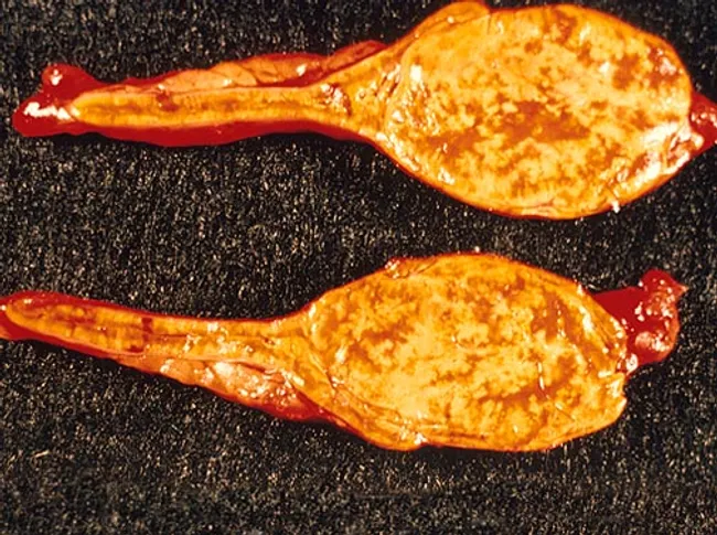
Gross pathology photograph of an adrenal gland cortical lesion from a 35-year-old male with Cushing syndrome, showing a yellow, lipid-rich cortical surface consistent with zona fasciculata. The resections reveal an encapsulated, well-circumscribed adrenal cortical adenoma projecting from the adrenal cortex. The tumor surface is smooth to slightly lobulated with uniform golden-yellow hue, reflecting abundant intracellular lipid in clear/polyhedral cells. On gross examination, the lesion appears discrete and distinct from surrounding yellow-brown medulla and normal cortex. The cut surface (if available) would likely be homogeneous, pale to bright yellow, without necrosis or hemorrhage, aligning with benign adenomatous growth. These features correlate with cortisol hypersecretion and the clinical phenotype of Cushing’s syndrome. Histologically, adenomas originate from adrenal cortex and show benign architecture; cytoplasmic lipid leads to the characteristic color. The image supports a diagnosis of cortisol-producing adrenal cortical adenoma, a common cause of endogenous Cushing’s, typically cured by distal excision. Differential considerations include adrenocortical carcinoma (larger, irregular, invasive) and bilateral micronodular hyperplasia though this specimen demonstrates a solitary, well-defined lesion. Clinically, the correlation with endocrine evaluation—elevated serum cortisol, suppressed ACTH in primary adrenal disease—guides management and prognosis. Overall, this specimen provides crucial gross-pathology correlation for endocrinologic syndromes and surgical pathology education contexts.
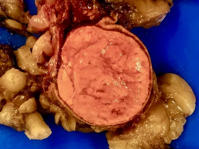
Comprehensive Description: This gross pathology photograph depicts an adrenal gland tumor, a 5.2 × 4.6 cm, round, well-circumscribed adrenal cortical adenoma, excised from a young adult male. Modality: macroscopic gross specimen photography of a fresh surgical specimen with standard lighting. Anterior-midline transsection reveals a golden yellow cut surface, characteristic of lipid-rich adrenal cortical tissue, surrounded by a thin rim of preserved yellow adrenal cortex. The tumor appears homogeneous, with minimal fibrous septation and no evident necrosis or hemorrhage. The color and texture reflect lipid-rich steroidogenic cells typical of aldosterone-producing adenomas. The lesion is solitary and encapsulated within the adrenal cortex, without overt invasion into periadrenal fat. Size corresponds to 5.2 cm in the greatest dimension, consistent with clinically significant adenoma in young patients. This pathology supports primary hyperaldosteronism (Conn syndrome) if functionally active, commonly presenting with hypertension, hypokalemia, and metabolic alkalosis; however, operation and histology would be definitive. Differential considerations include adrenocortical adenoma versus carcinoma; size, circumscription, and lipid-rich greenish-yellow appearance favor adenoma. This specimen may be used for teaching gross anatomy, endocrinology, urology, and surgical pathology, with correlation to immunohistochemistry if available (CYP11B2/aldosterone synthase). Correlation with hormonal assays and preoperative imaging is essential for definitive diagnosis and surgical planning.
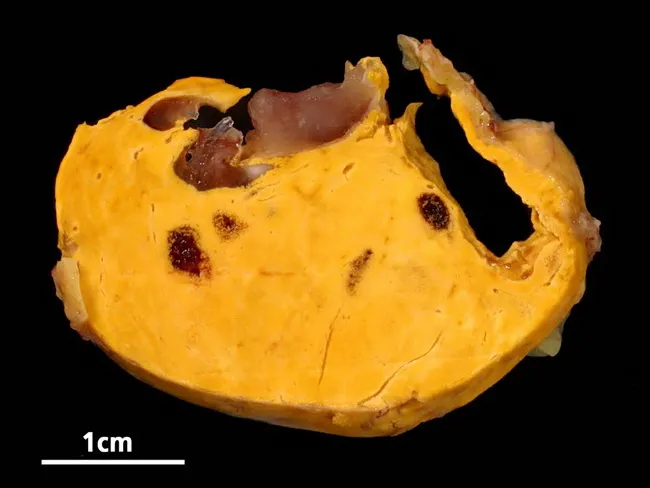
Gross pathology photograph of a suprarenal gland showing an adrenal cortical adenoma with focal myelolipomatous change. The specimen presents a well‑circumscribed, lobulated, bright yellow cortical mass with scattered dark brown to burgundy gelatinous foci representing myelolipoma components. The yellow adipocytic tumor contrasts with the darker, nonlipomatous areas; a cross section reveals a heterogeneous cut surface with preserved capsule and occasional fibrous septa. The architecture is consistent with a benign adrenal cortical neoplasm containing adipose tissue and hematopoietic elements within myelolipomatous foci. Myelolipomas are typically nonfunctional and incidental but may accompany adenomas; their presence does not imply malignancy. Pathologic correlation suggests a nonfunctioning or minimally functioning adrenal cortical adenoma with incidental myelolipomatous change. Clinically, this image is relevant for educational references on adrenal incidentalomas, differential diagnosis (myelolipoma versus liposarcoma or cortical carcinoma), and gross confirmation prior to histology. Potential diagnostic uses include teaching radiologic–pathologic correlation, gross morphology description, and surgical pathology workflow. This specimen demonstrates how macroscopic features guide sampling for histology and how adipose tissue with hematopoietic elements appears within an adrenal cortical tumor, aiding recognition of myelolipomatous change. The 1 cm scale bar in the accompanying image provides size context for tumor dimensions and pathologic assessment and educational value.
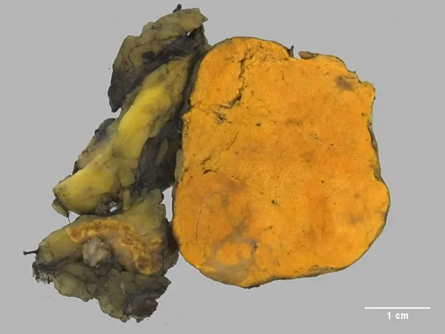
Gross pathology image of a unilateral adrenal gland resection specimen showing a well-circumscribed, golden-yellow cortical adenoma measuring approximately 3 cm in greatest dimension. The tumor is encapsulated and demonstrates a homogeneous, lipid-rich appearance typical of adrenal cortical adenomas; the surrounding adrenal cortex is partially compressed. Coloration is characteristic of lipid-rich adenomatous tissue with a bright yellow/orange hue. The surface contour appears smooth with minimal necrosis or hemorrhage. This macroscopic phenotype aligns with an aldosterone-producing adenoma, a common cause of primary hyperaldosteronism (Conn syndrome) accounting for about 70% of cases; some adenomas are small to moderate in size and may exhibit a lipid-dense cut surface that contrasts with non-neoplastic cortex. The image’s scale bar indicates 1 cm, supporting a lesion size near 3 cm. Clinically, excess aldosterone leads to potassium loss with hypokalemia, sodium retention, hypertension, and suppression of plasma renin activity; a thorough evaluation of the renin-angiotensin-aldosterone axis and radiologic assessment of both adrenals is essential. In cases of benign adenoma, surgical resection is often curative. Differential considerations include adrenal cortical hyperplasia and, less commonly, adrenal cortical carcinoma or pheochromocytoma, though macroscopic appearance and size favor adenoma. Correlation with hormonal studies and postoperative improvement supports the diagnostic conclusion.
Pathology of Adrenal Cortical Adenoma — Model Answer (10 Marks)
1. Definition
2. Etiology and Associations
- Most are sporadic.
- May occur in the setting of hereditary syndromes affecting the adrenal cortex (e.g., MEN1, Carney complex), though the strong familial associations (Li-Fraumeni via germline TP53, Beckwith-Wiedemann via IGF-2 imprinting) are more classically linked to adrenocortical carcinoma rather than adenoma.
- Somatic mutations in genes of the cAMP/PKA pathway (PRKACA) and Wnt/β-catenin pathway (CTNNB1) have been implicated in cortisol- and aldosterone-producing adenomas respectively.
3. Functional Classification
- Non-functional (majority) — clinically silent, incidental finding.
- Functional:
- Aldosterone-producing adenoma (aldosteronoma) → Conn syndrome (primary hyperaldosteronism): hypertension, hypokalemia, metabolic alkalosis, suppressed plasma renin.
- Cortisol-producing adenoma → Cushing syndrome: central obesity, striae, hypertension, hyperglycemia, low ACTH (feedback suppression).
- Virilizing/androgen-producing adenoma is rare; when androgen excess is present, a carcinoma should be strongly suspected instead.
- Functionality cannot be predicted from gross or routine microscopic appearance alone; it is established biochemically/clinically.
4. Gross Morphology
- Solitary, well-circumscribed, encapsulated nodule, typically up to 2.5 cm in diameter (larger functional adenomas are described but this is the classic teaching cut-off).
- Expands the adrenal gland.
- Cut surface is yellow to yellow-brown due to abundant intracytoplasmic lipid (cholesterol, precursor steroids).
- Functional adenomas cause atrophy of the adjacent, non-neoplastic cortex (due to suppression of ACTH/renin-angiotensin drive); non-functional adenomas have a normal adjacent cortex.
- No necrosis or hemorrhage in the typical benign lesion.
5. Microscopic Morphology
- Composed of cells resembling normal adrenocortical cells, arranged in cords, nests, or a trabecular pattern.
- Nuclei: generally small and uniform; mild pleomorphism ("random endocrine atypia") can occur even in benign lesions and does not by itself indicate malignancy.
- Cytoplasm: ranges from eosinophilic (compact, lipid-poor) to vacuolated/clear (lipid-rich), depending on intracellular lipid content.
- Mitotic activity: inconspicuous; Ki-67 labeling index typically <5% (helps distinguish from carcinoma).
- No capsular, vascular, or sinusoidal invasion; no necrosis.
6. Immunohistochemistry
- Steroidogenic factor-1 (SF-1)
- Inhibin-alpha
- Also typically Melan-A (MART-1) and synaptophysin (variable), which help distinguish adrenocortical tumors from metastatic carcinoma or pheochromocytoma. IHC does not reliably separate adenoma from carcinoma — that requires architectural/behavioral criteria (see below).
7. Differentiating Adenoma from Carcinoma — Weiss Criteria
- High nuclear grade
- Mitotic rate >5/50 high-power fields
- Presence of atypical mitoses
- Low proportion of clear (lipid-rich) cells (≤25%)
- Diffuse architecture (loss of normal nesting pattern)
- Necrosis
- Venous invasion
- Sinusoidal invasion
- Capsular invasion
- Adenoma - Weiss score 0-2, well-organized trabecular pattern, uniform cells, minimal atypia, no invasion.
- Carcinoma - Weiss score ≥3, high-grade atypia, necrosis, invasion; carcinomas are also generally larger (often >5-6 cm, many >20 cm), variegated with hemorrhage/necrosis on cut surface, and have a strong tendency to invade the adrenal vein, vena cava, and lymphatics.
- Ultimate proof of malignancy is local invasion or metastasis; the Weiss system approximates this when neither is evident.
8. Clinical Correlation / Diagnostic Workup
- Most adenomas are picked up as an incidentaloma on CT/MRI.
- Imaging features favoring adenoma: small size, homogeneous, low Hounsfield units (<10 HU) on unenhanced CT due to high lipid content, and rapid contrast washout.
- Biochemical screening (plasma aldosterone/renin ratio, overnight dexamethasone suppression test, urinary catecholamines) is done to exclude functionality and rule out pheochromocytoma before biopsy/surgery.
- Management: functional or enlarging/indeterminate lesions are resected (adrenalectomy); small, non-functional, imaging-benign lesions may be observed with serial imaging.
- Prognosis after excision of a true adenoma is excellent (curative), in contrast to adrenocortical carcinoma, which carries a median survival of about 2 years.
| Feature | Adenoma | Carcinoma |
|---|---|---|
| Size | Usually <2.5-5 cm | Often >5-20 cm |
| Circumscription | Well-circumscribed, encapsulated | Poorly demarcated, invasive |
| Cut surface | Homogeneous yellow | Variegated, necrosis/hemorrhage |
| Nuclear atypia | Minimal | Marked |
| Mitoses/Ki-67 | Low (<5%) | High |
| Invasion | Absent | Vascular/capsular/sinusoidal |
| Weiss score | 0-2 | ≥3 |
| Behavior | Benign | Metastasizing, poor prognosis |