
Nephritic bs nephrotic
Reading File
Now let me search the library for nephritic and nephrotic syndrome content in parallel.
Finding Sources
Finding Sources
Finding Sources
Finding Sources
Reading File
Reading File
Reading File
Excellent. I have rich content. Now let me get the Robbins nephrotic syndrome section and the Washington Manual comparison, plus one image.
Reading File
Reading File
Loading Image
I now have everything needed for a thorough comparison. Here is the full breakdown:
Nephritic vs. Nephrotic Syndrome
These are two distinct glomerular syndromes with different mechanisms, features, and causes - though overlap exists.
Core Mechanism
| Nephritic | Nephrotic | |
|---|---|---|
| Pathology | Glomerular inflammation - proliferation of cells + leukocyte infiltration damage the capillary wall | Disruption of the filtration barrier - structural/physicochemical changes increase permeability to protein |
| Histology | Mesangial cell proliferation, leukocyte infiltration; crescents in severe disease | GBM thickening or podocyte foot process fusion/effacement (podocytopathy) |
| Key insult | Inflammatory injury → capillary wall disrupts → blood leaks into urine | Protein leaks across damaged filtration barrier → massive proteinuria |
- Comprehensive Clinical Nephrology, 7th Ed. - Robbins & Cotran Pathologic Basis of Disease
Clinical Features Side-by-Side
| Feature | Nephrotic | Nephritic |
|---|---|---|
| Onset | Insidious | Abrupt |
| Edema | ++++ (massive, periorbital, dependent) | ++ (mild-moderate) |
| Blood pressure | Normal | Raised |
| JVP | Normal / low | Raised |
| Proteinuria | ++++ (>3.5 g/day) | ++ (subnephrotic range) |
| Hematuria | May or may not occur | +++ (brown/tea-colored urine) |
| RBC casts | Absent | Present (pathognomonic) |
| Serum albumin | Low (<3 g/dL) | Normal / slightly reduced |
| GFR / azotemia | Usually preserved early | Reduced - oliguria, azotemia |
| Lipids | Hyperlipidemia + lipiduria | Usually normal |
- Comprehensive Clinical Nephrology, 7th Ed., Table 16.4
Pathophysiology Details
Nephrotic
The cascade follows a logical sequence:
- Glomerular barrier damaged → massive proteinuria (>3.5 g/day)
- Liver cannot compensate → hypoalbuminemia (<3 g/dL)
- Reduced plasma oncotic pressure → generalized edema (periorbital, dependent, ascites, pleural effusion)
- Hypovolemia → aldosterone + sympathetic activation → sodium/water retention (worsens edema)
- Liver upregulates lipoprotein synthesis (compensatory) → hyperlipidemia and lipiduria
Key complications:
- Thromboembolism (10-40% of cases) - urinary loss of antithrombotics (antithrombin III, protein C/S) + increased clotting factor synthesis. Deep vein thrombosis and renal vein thrombosis are classic. Prophylactic anticoagulation is considered when albumin <2.0-2.5 g/dL with additional risk factors.
- Increased infection risk (loss of immunoglobulins)
- Cardiovascular disease from hyperlipidemia
Nephritic
The inflammatory glomerular injury leads to:
- Capillary wall damage → hematuria with RBC casts
- Reduced GFR → oliguria, azotemia, fluid retention
- Fluid retention + renin from ischemic kidneys → hypertension
- Some protein leak → mild proteinuria (rarely nephrotic range, so albumin stays near normal)
- Robbins & Cotran Pathologic Basis of Disease
Classic Causes
Nephrotic Syndrome (primary causes = podocytopathies)
| Disease | Key feature | Age group |
|---|---|---|
| Minimal Change Disease (MCD) | Foot process effacement on EM only; responds to steroids | Children (most common) |
| Focal Segmental Glomerulosclerosis (FSGS) | Focal/segmental scarring; often steroid-resistant | Adults, HIV |
| Membranous Nephropathy | Subepithelial deposits, GBM thickening; anti-PLA2R Ab | Adults (most common primary cause in adults) |
| Diabetic nephropathy | Nodular glomerulosclerosis (Kimmelstiel-Wilson) | Adults with DM |
| Amyloidosis | Congo red positive deposits | Older adults |
| Lupus nephritis | Can cause either syndrome | Young women |
Nephritic Syndrome (immune complex/inflammatory GN)
| Disease | Key feature |
|---|---|
| Poststreptococcal GN | 1-4 weeks post Group A strep pharyngitis or impetigo; subepithelial "humps"; low C3; children 6-10 yrs |
| IgA Nephropathy | Most common worldwide; IgA mesangial deposits; normal complement |
| Lupus Nephritis | Low C3/C4; ANA+, anti-dsDNA+ |
| ANCA-associated vasculitis | Pauci-immune, ANCA+; normal complement |
| Anti-GBM disease (Goodpasture) | Linear IgG on IF; pulmonary hemorrhage |
| MPGN | Can present as either syndrome; low C3 |
| RPGN (crescentic GN) | Severe nephritic picture with rapid loss of kidney function over days-weeks |
Workup Tips
Suspected nephrotic:
- ANA, C3/C4, cryoglobulins, viral serologies (HIV, HBV, HCV, CMV, EBV)
- Review medications; rule out malignancy
- SPEP + urine immunofixation (for monoclonal gammopathy if large protein-albumin gap)
- Renal biopsy in all adults
Suspected nephritic:
-
Anti-GBM antibodies, ANCA, ASO titer
-
ANA, C3/C4, cryoglobulins, viral serologies
-
Complement levels key:
- Low C3: postinfectious GN, lupus, MPGN, C3 glomerulopathy, SBE
- Normal complement: IgA nephropathy, ANCA vasculitis, anti-GBM disease
-
Washington Manual of Medical Therapeutics
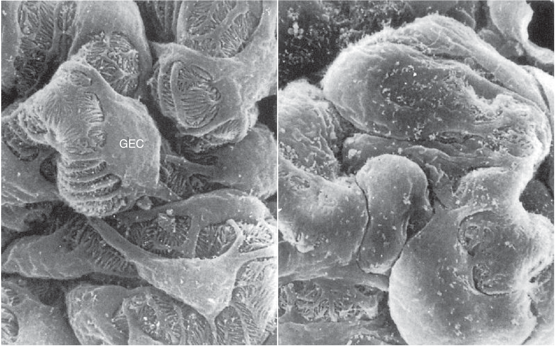
Glomerular Histology - Nephrotic Foot Process Effacement
Below is a scanning electron micrograph comparing normal podocytes (left) with the extensive foot process loss seen in nephrotic syndrome (right - puromycin aminonucleoside model):
General Management Principles
Both syndromes:
- ACEi/ARBs to reduce intraglomerular pressure and proteinuria
- Diuretics + sodium restriction for edema
- Aggressive BP control
Nephrotic-specific:
- Statins for hyperlipidemia (HMG-CoA reductase inhibitors)
- Anticoagulation for thromboembolic complications (heparin → oral anticoagulant)
- Prophylactic anticoagulation if albumin <2.0-2.5 g/dL + risk factors
- Treat underlying cause (steroids for MCD, etc.)
Nephritic-specific:
- Treat underlying cause (immunosuppression for lupus, ANCA; antibiotics for postinfectious)
- RPGN is a uremic emergency - requires urgent evaluation and usually pulse steroids ± cyclophosphamide ± plasmapheresis
One important caveat: the two syndromes are not always mutually exclusive. Diseases like MPGN and lupus nephritis can present with a mixed picture, and the same disease can shift from one presentation to the other. - Comprehensive Clinical Nephrology, 7th Ed.
This is a shared conversation. Sign in to Orris to start your own chat.