Blood supply of brain
Reading File
Finding Sources
Finding Sources
Reading File
Reading File
Reading File
Reading File
Reading File
Loading Image
Loading Image
Reading File
Loading Image
Reading File
Loading Image
I now have comprehensive content from three major textbooks plus excellent diagrams. Let me compile the full answer.
Blood Supply of the Brain
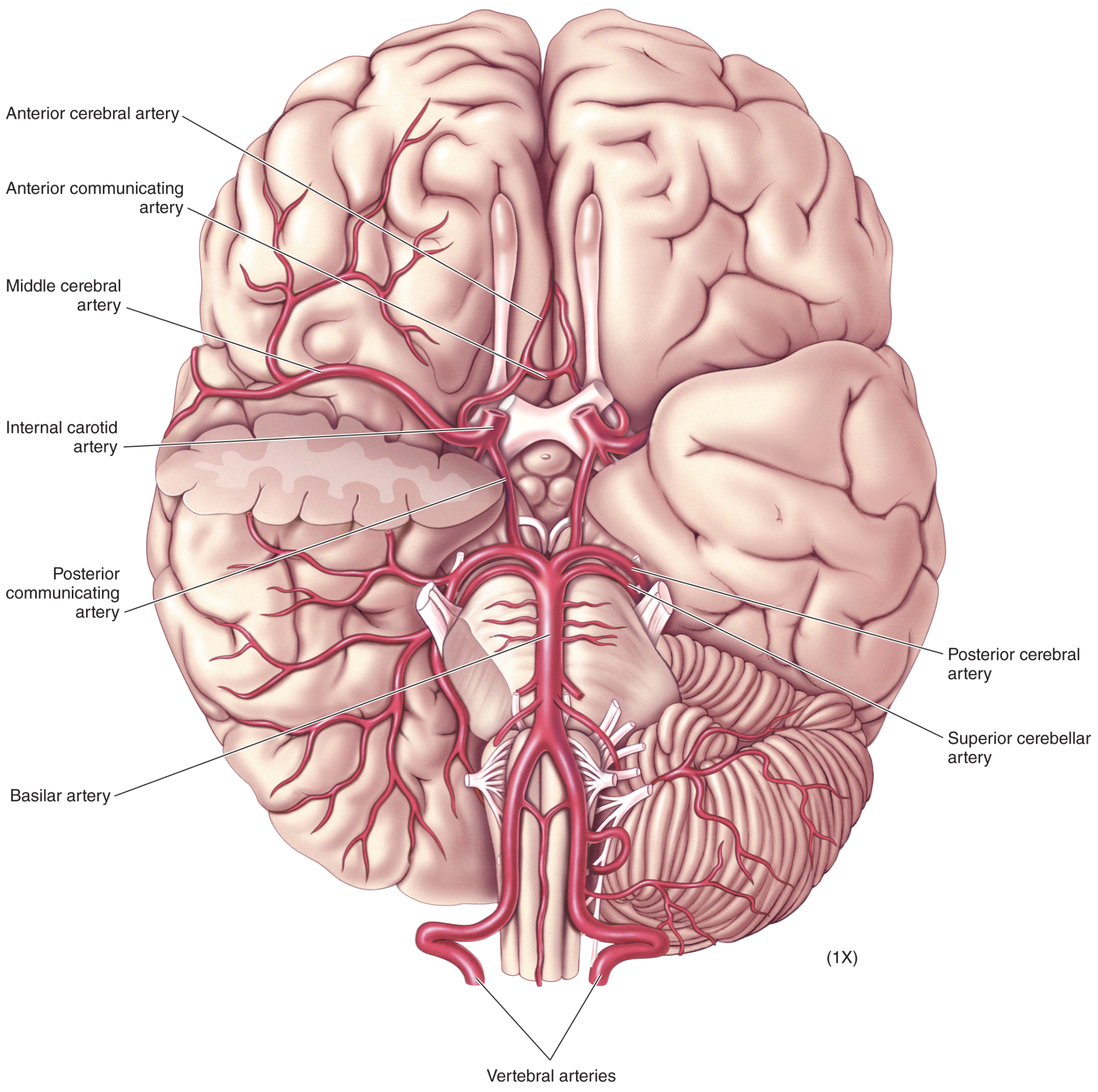
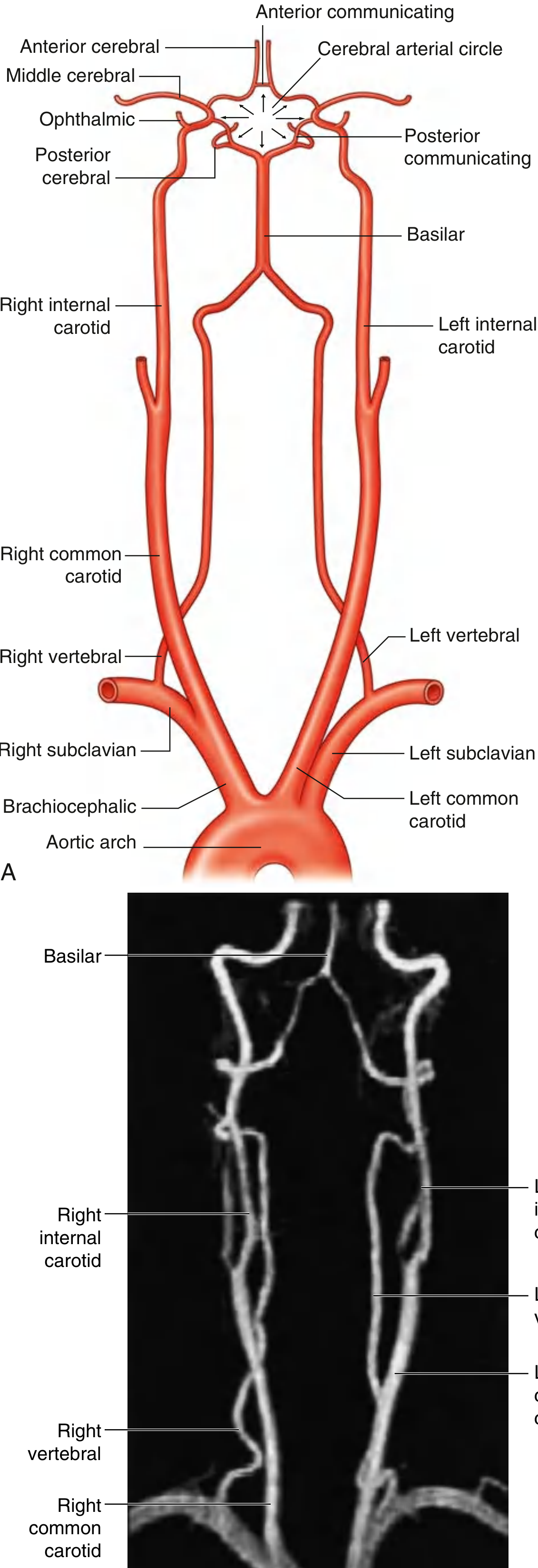
The brain is supplied by two pairs of arteries - the internal carotid arteries (anterior circulation) and the vertebral arteries (posterior/vertebrobasilar circulation) - which anastomose at the base of the brain to form the Circle of Willis.
Arterial Supply: Overview
1. Posterior (Vertebrobasilar) Circulation
Vertebral Arteries
- Each vertebral artery arises from the first part of the subclavian artery in the lower neck.
- Passes superiorly through the foramen transversarium of the upper six cervical vertebrae.
- Enters the cranial cavity through the foramen magnum.
- Inside the cranium, each vertebral artery gives off:
- Posterior inferior cerebellar artery (PICA) - supplies posterior inferior cerebellum and lateral medulla
- Posterior spinal artery - descends on the posterior cord surface (more commonly a branch of PICA)
- A branch that joins its partner to form the anterior spinal artery
- A small meningeal branch
Basilar Artery
- Formed by the union of the two vertebral arteries just inferior to the pons.
- Travels rostrally along the anterior surface of the pons.
- Branches of the basilar artery (caudal to rostral):
- Anterior inferior cerebellar arteries (AICA) - anterior inferior cerebellum, lower pons
- Pontine arteries (perforating branches) - pons
- Superior cerebellar arteries (SCA) - superior cerebellum and upper pons
- Terminates by bifurcating into the two posterior cerebral arteries (PCA)
2. Anterior (Carotid) Circulation
Internal Carotid Arteries (ICA)
- Arise as terminal branches of the common carotid arteries.
- Enter the skull via the carotid canal, traverse the cavernous sinus (forming the carotid siphon), and enter the cranial cavity.
- Inside the cranium, each ICA gives off:
- Ophthalmic artery - first major intracranial branch; supplies the orbit and eye
- Posterior communicating artery (PComm) - connects ICA to the PCA, completing the posterior part of the Circle of Willis
- Middle cerebral artery (MCA) - the largest branch
- Anterior cerebral artery (ACA) - terminal branch that runs medially
3. The Circle of Willis (Cerebral Arterial Circle)
The Circle of Willis is an anastomotic ring at the base of the brain formed by the interconnection of the carotid and vertebrobasilar systems.
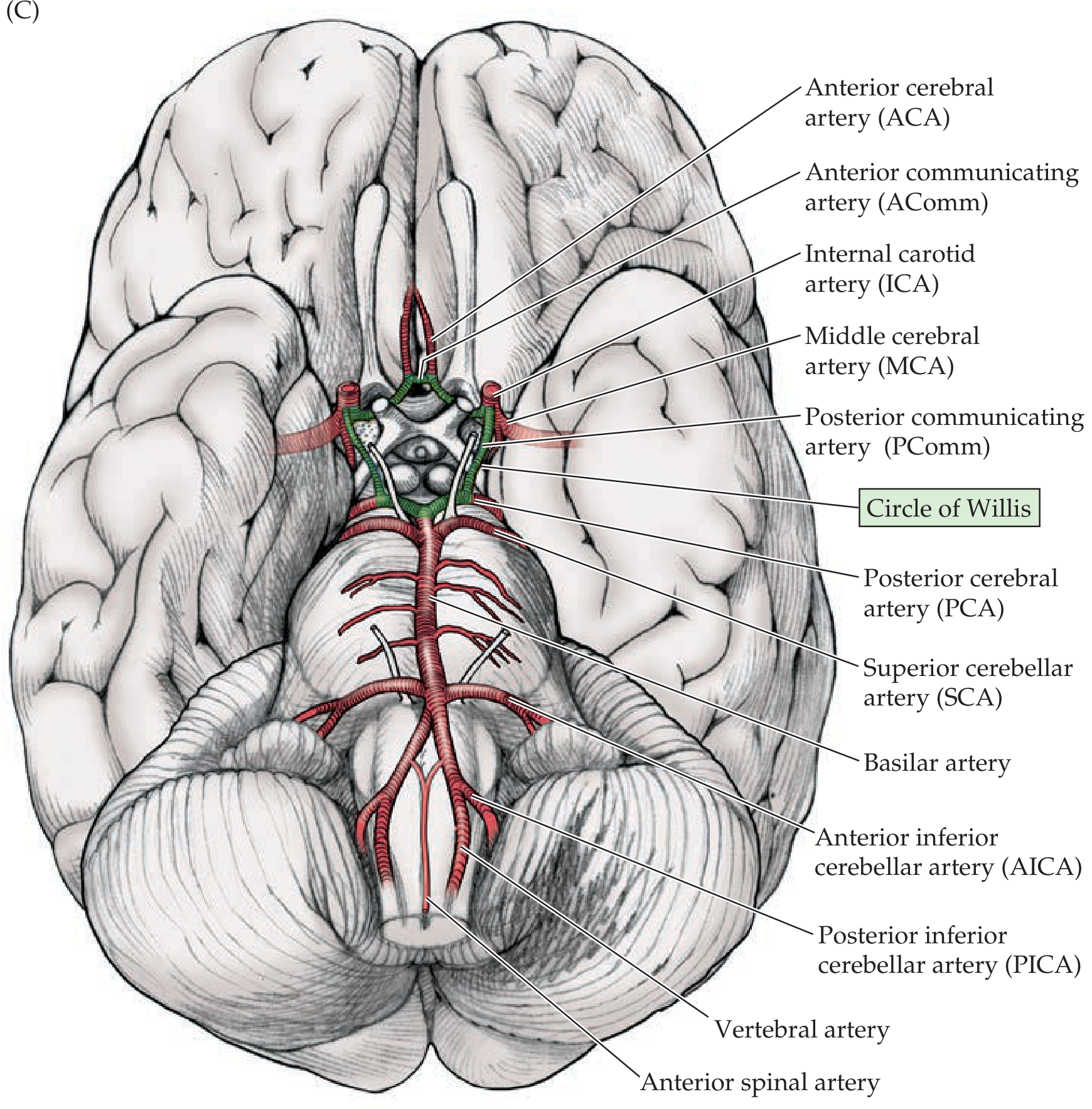
Components of the Circle of Willis:
| Component | Connects |
|---|---|
| Anterior communicating artery (AComm) | Left ACA ↔ Right ACA |
| Posterior communicating artery (PComm) - bilateral | ICA ↔ Posterior cerebral artery (PCA) |
| Anterior cerebral arteries (ACA) - bilateral | Part of the ring |
| Internal carotid arteries (ICA) - bilateral | Part of the ring |
| Posterior cerebral arteries (PCA) - bilateral | Part of the ring |
The circle allows collateral flow when one vessel is occluded, though in practice the vessels are often unequal in size and the collateral capacity is variable.
Common sites for aneurysm formation at the Circle of Willis: anterior communicating artery, posterior communicating artery, MCA bifurcation, and the basilar tip.
4. Cortical Territories
Anterior Cerebral Artery (ACA)
- Runs medially and anteriorly over the corpus callosum.
- Supplies the medial surface of the frontal and parietal lobes.
- Includes the leg/foot area of the primary motor and sensory cortex.
- ACA occlusion: contralateral leg weakness (with relative sparing of the arm and face).
Middle Cerebral Artery (MCA)
- The largest and most clinically significant branch.
- Supplies the entire lateral surface of the cerebral hemisphere.
- Also feeds deep structures via lenticulostriate (perforating) branches: basal ganglia, internal capsule.
- MCA occlusion: contralateral hemiparesis (arm > leg), hemisensory loss, aphasia (dominant hemisphere), hemineglect (non-dominant hemisphere).
Posterior Cerebral Artery (PCA)
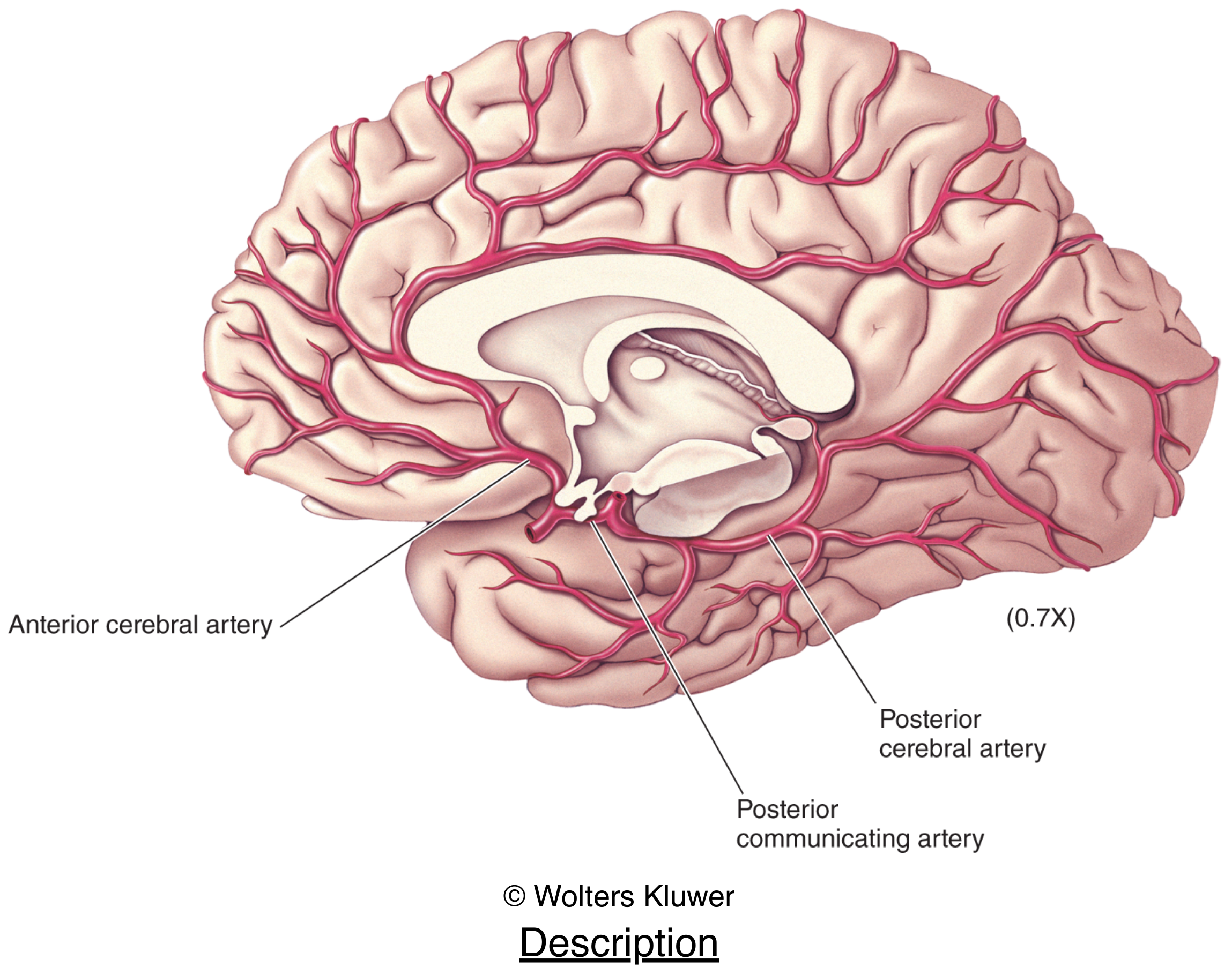
- Supplies the medial occipital lobe and inferior temporal lobe.
- PCA occlusion: contralateral homonymous hemianopia (with macular sparing if posterior pole has collateral supply).
- Also supplies the thalamus and midbrain.
5. Venous Drainage
Venous drainage of the brain is provided almost entirely by the internal jugular veins. Blood drains from cerebral veins into the dural venous sinuses, which ultimately drain into the internal jugular veins at the jugular foramina.
Key dural venous sinuses:
- Superior sagittal sinus - runs along the superior border of the falx cerebri; drains most of the cerebral cortex
- Inferior sagittal sinus - runs along the inferior free border of the falx cerebri
- Straight sinus - formed by the junction of the inferior sagittal sinus and great cerebral vein (of Galen)
- Confluence of sinuses (torcular Herophili) - where superior sagittal, straight, and occipital sinuses meet at the internal occipital protuberance
- Transverse sinuses - run laterally from the confluence
- Sigmoid sinuses - S-shaped continuation of the transverse sinuses, draining into the internal jugular veins
- Cavernous sinuses - on either side of the sella turcica; drain the ophthalmic veins and receive ICA within their walls
6. Clinical Correlates
| Vessel | Classic Stroke Syndrome |
|---|---|
| MCA | Contralateral hemiparesis (arm > leg), aphasia or hemineglect |
| ACA | Contralateral leg weakness, abulia, incontinence |
| PCA | Contralateral homonymous hemianopia, thalamic pain |
| PICA (lateral medullary/Wallenberg) | Ipsilateral face + contralateral body sensory loss, dysphagia, Horner's, vertigo |
| Basilar artery | "Locked-in" syndrome, coma, quadriparesis |
| Lacunar (lenticulostriate) | Pure motor or pure sensory stroke, ataxic hemiparesis |
A stroke (CVA) is defined as interruption of cerebral blood flow producing neurological deficit lasting >24 hours. A TIA resolves within 24 hours. Ischemic strokes (most common) are thrombotic or embolic - emboli from carotid atherosclerosis are the most frequent cause. Thrombolysis with tPA can be given within 3-4.5 hours of onset, and mechanical thrombectomy can extend this window further.
Sources:
- Gray's Anatomy for Students, pp. 1017-1023
- Neuroscience: Exploring the Brain, 5th Edition, pp. 705-708
- Neuroanatomy through Clinical Cases, 3rd Edition, pp. 70-71
This is a shared conversation. Sign in to Orris to start your own chat.