
Vitiligo
vitiligo depigmented patches clinical photograph
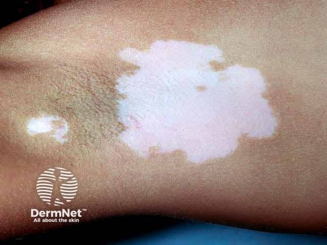
Clinical photography of axillary skin showing depigmented vitiligo patches. Modality: Clinical photograph, dermoscopy not used. The image depicts a large, well-demarcated, chalk-white macule/patch centered in the axillary fold with irregular but sharply defined borders. Adjacent skin exhibits relative hyperpigmentation surrounding the patch, consistent with repigmentation margins typical of vitiligo-affected areas. The surface appears smooth, non-scaly, and non-erythematous, with no crusting or ulceration. Hair within the depigmented areas may be reduced or absent, although follicular retention can occur. Lesions are unilateral and localized to the axilla in this view; a second smaller depigmented area is visible nearby. The clinical impression is consistent with localized vitiligo (leukoderma) presenting as hypopigmented to depigmented patches due to melanocyte destruction. The absence of scale distinguishes vitiligo from tinea versicolor or eczema; the uniform whiteness under standard lighting supports vitiligo rather than postinflammatory hypopigmentation. This image is relevant for educational descriptions of vitiligo morphology, differential diagnosis (hypopigmentation variants, pityriasis versicolor), and for demonstrating axillary involvement. Potential clinical utility includes patient counseling, Wood lamp correlation, and monitoring treatment response to topical corticosteroids, calcineurin inhibitors, or phototherapy over time. Image serves as a reference for clinicians evaluating depigmented lesions and educating patients about prognosis.
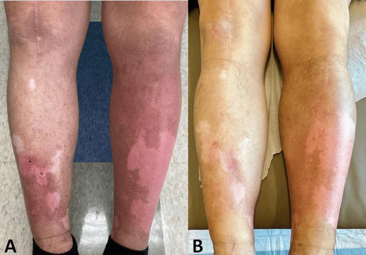
Clinical photograph comparison (A and B) of the lower extremities in a 60-year-old male, demonstrating pemetrexed-induced pseudocellulitis (PIP) and concurrent vitiligo. Image A shows bilateral, asymmetric, poorly demarcated erythematous patches and edema, most pronounced on the left shin. Superimposed are well-demarcated depigmented macules and patches consistent with vitiligo. Image B illustrates the clinical response following an eight-day prednisone taper, showing a significant reduction in diffuse erythema and localized swelling, while the underlying depigmented vitiligo patches remain stable. The photograph serves as a visual reference for differentiating drug-induced pseudocellulitis from bacterial cellulitis, characterized here by the lack of exquisite tenderness, bilateral involvement, and temporal association with pemetrexed infusion. The case highlights diagnostic features for oncologists and dermatologists managing chemotherapy-related cutaneous adverse drug reactions.
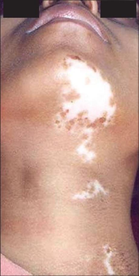
Clinical photograph of the anterior neck and submental region of a patient with medium-brown skin tone, demonstrating segmental vitiligo. The image shows several distinct, milky-white depigmented macules and patches. The largest patch is centrally located on the submental area and upper neck, characterized by irregular borders and follicular repigmentation (pigmented specks within the depigmented area). Smaller, satellite-like depigmented macules extend inferiorly along the midline of the neck in a linear or segmental distribution. There is a sharp contrast between the amelanotic (depigmented) areas and the surrounding normally pigmented skin. The photograph illustrates characteristic features used in dermatology to identify stable vitiligo and assess for signs of repigmentation following therapeutic interventions like PUVA or surgical grafting.
vitiligo AND treatment
vitiligo diagnosis treatment American Academy Dermatology
https://www.aad.org/public/diseases/a-z/vitiligo-treatment
Vitiligo
Typical features
- Smooth, non-scaly white patches, often on the face, hands, around the mouth or eyes, armpits, groin, elbows, knees, or genital area.
- Usually no symptoms, though patches can burn easily in sunlight or occasionally itch.
- Non-segmental vitiligo: the common form, often bilateral and relatively symmetrical.
- Segmental vitiligo: usually one-sided, following a localized pattern, and often stabilizes after initial spread.
Andrews' Diseases of the Skin, p. 1001.
Why it happens
Diagnosis
Treatment
- Topical corticosteroids, generally for limited periods due to risks such as skin thinning.
- Tacrolimus or pimecrolimus, especially useful on the face, neck, and skin folds.
- Topical ruxolitinib for eligible people with non-segmental vitiligo. In the US, it is FDA-approved for patients aged 12 years and older with limited involvement.
- Narrowband UVB phototherapy, often used for more extensive or active non-segmental disease.
- Excimer laser/light for selected localized patches.
- In stable, treatment-resistant localized vitiligo, dermatologists may consider surgical grafting techniques.
Everyday care
- Use broad-spectrum SPF 30 or higher on exposed skin and reapply as directed.
- Avoid sunburn and reduce friction or repeated trauma to skin where possible.
- Camouflage makeup or self-tanner can help appearance if desired.
- Seek support if it affects mood, confidence, work, or social life. The emotional impact can be substantial and deserves care.
Final treatment
vitiligo AND treatment
2025 vitiligo treatment guideline topical ruxolitinib narrowband UVB International Vitiligo Task Force
Final treatment plan for vitiligo
-
Limited vitiligo, especially face/neck
- A dermatologist may use tacrolimus ointment or pimecrolimus cream.
- A short, intermittent course of a topical corticosteroid may be used for thicker skin on the trunk or limbs, not continuously on thin facial or flexural skin.
- For eligible patients aged 12 years or older with non-segmental vitiligo involving limited body surface area, ruxolitinib 1.5% cream is a targeted option, typically applied twice daily under dermatology supervision. Dermatology 2-Volume Set, 5e, p. 1314.
-
Widespread or progressing non-segmental vitiligo
- Narrowband UVB phototherapy is the usual preferred treatment to both slow progression and encourage repigmentation.
- It is commonly combined with a topical treatment such as tacrolimus, a topical steroid, or ruxolitinib where appropriate.
- Benefit takes months; treatment response is often best on the face and trunk and poorer on hands, feet, and areas with white hairs.
-
Rapidly spreading disease
- A dermatologist may consider a short course of systemic corticosteroid treatment to arrest activity, alongside topical therapy and/or narrowband UVB. This is not a routine long-term treatment because of systemic risks.
-
Stable localized vitiligo that has not responded to medical treatment
- Excimer laser/light may suit small areas.
- Surgical grafting or melanocyte-keratinocyte transplantation can be considered only after the disease has been stable, usually for at least 12 months, and in specialist centers.
-
For everyone
- Broad-spectrum sunscreen SPF 30 or higher daily on exposed areas.
- Camouflage cosmetics or self-tanners if wanted.
- Screening for associated autoimmune thyroid disease should be considered, particularly if there are symptoms or family history.
- Address anxiety, depression, or social impact as part of treatment.

Let me know
vitiligo AND diagnosis
American Academy Dermatology vitiligo diagnosis Wood lamp acanthosis nigricans neck
What to do
- Arrange a visit with a dermatologist or primary-care clinician for an in-person exam.
- Ask whether you need screening for blood sugar such as fasting glucose and HbA1c, plus assessment for insulin resistance and related conditions.
- If you also have distinct white patches elsewhere, ask for a Wood lamp examination, which helps confirm or rule out vitiligo. The AAD explains Wood lamp use.
- Avoid applying steroid or “fairness” creams without a diagnosis.
- Keep the area dry, reduce friction from tight collars or chains, and avoid harsh scrubbing.