
Fungi superficial and deep micosis ...bsc nursing 3rd semester
superficial fungal infection tinea dermatophyte skin
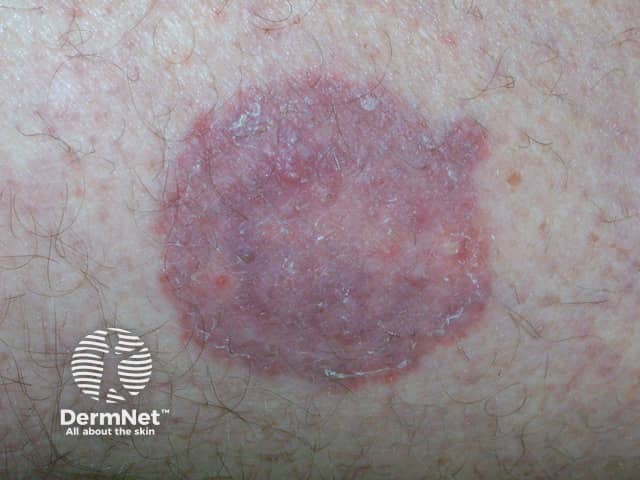
This image is a clinical dermatology photograph of tinea corporis (dermatophyte fungal infection) affecting cutaneous skin. Modality and technique: visible-light clinical photography, macro close-up, standard illumination, no special stains or contrast; acquisition from a superficial skin lesion for bedside documentation. Anatomical context: integumentary system; skin (epidermis/dermis) of the trunk or proximal extremities; localized circular to oval plaque. Visual features: annular erythematous plaque with a well‑defined, active border and central clearing; surface desquamation with fine scale; the border may appear slightly raised and pruritic in vivo; hue ranges from pink to violaceous in inflammatory areas; surrounding hair and adjacent skin show mild irritation without deep tissue involvement. Pathology/diagnosis signals: the classic ring-shaped lesion with central clearing is characteristic of tinea corporis; clinical impression supported by dermatophyte infection of keratinized tissue; laboratory confirmation may show hyphae on potassium hydroxide (KOH) preparation or positive fungal culture; histopathology shows hyperkeratosis and fungal elements in stratum corneum when biopsy is performed. Diagnostic significance and utility: high clinical probability for dermatophyte infection guides antifungal therapy; differential includes nummular dermatitis, psoriasis, granuloma annulare, pityriasis rosea; treatment implications include topical agents (terbinafine, naftifine) or systemic therapy for extensive disease; infection is contagious and may require contact precautions.
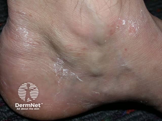
Clinical photography of the plantar foot demonstrating tinea pedis, a dermatophyte fungal infection. Modality: Dermatologic photograph using standard visible light, close-up view of the plantar surface and interdigital spaces. The image shows diffuse to patchy scaling and maceration with superficial fissuring along the plantar arch and between the toes, with mild surrounding erythema and no obvious edema. The epidermis is dry and hyperkeratotic with white flaky scales adherent to the skin. These features are characteristic of dermatophytosis (tinea pedis), often caused by Trichophyton species; the interdigital web spaces frequently harbor moisture and provide a portal for fungal invasion. In this view, the predominant pattern appears dry-type with fissuring rather than a vesiculobullous eruption. The diagnostic significance lies in confirming a superficial fungal infection and guiding therapy. Clinically, this image is relevant for dermatology, primary care and telemedicine education, and can be used to monitor response to antifungal therapy (e.g., terbinafine, azoles) and foot hygiene measures. Differential considerations include eczema, contact dermatitis, candidal intertrigo and psoriasis of the interdigital spaces. The image illustrates typical signs: scaling, maceration, fissuring, with a probable interdigital involvement on the plantar aspect. This photograph is valuable for educational demonstrations of dermatophyte infections and treatment outcomes.
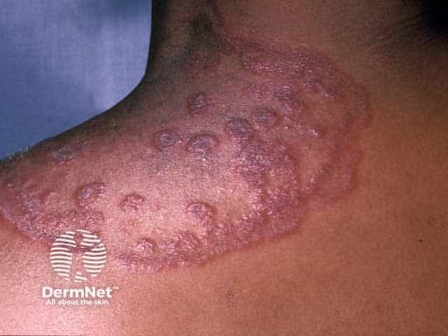
This image depicts a dermatology clinical photograph of tinea circinata (tinea corporis) involving the left shoulder/upper deltoid region in a patient with HIV infection. The modality is clinical photography; color-rendered skin tones demonstrate multiple annular, centripetally expanding plaques with raised, scaly, erythematous borders and central clearing. Lesions display discrete circular to oval morphology, with concentric rings in some plaques and variable postinflammatory hyperpigmentation at perilesional skin. The distribution is localized to the shoulder region, with superficial involvement of the epidermis and superficial dermis. The classic ring-shaped pattern, peripheral scale, and itch-scratch cycles are characteristic. In HIV-associated immunosuppression, dermatophytoses can be extensive, recurrent, or atypical in distribution; secondary bacterial colonization or satellite lesions may occur. The image highlights the differential diagnosis of nummular eczema, plaque psoriasis, pityriasis rosea, and granulomatous fungal infections; correlates with clinical history of pruritus and exposure. The diagnostic significance lies in recognizing dermatophyte infection requiring topical antifungals and/or systemic therapy in immunocompromised hosts. Potential clinical use cases include education on fungal skin infections in HIV, dermatology exam prep, and research on opportunistic dermatophytoses. The finding underscores the need for fungal culture or KOH microscopy to confirm dermatophyte species and guide therapy.
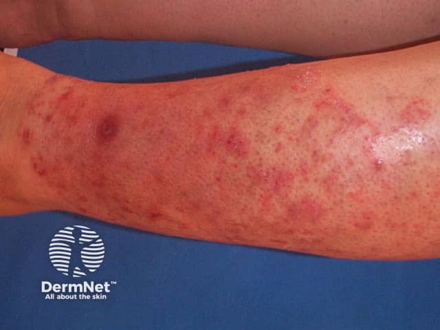
Clinical photography of a forearm displaying multiple annular, erythematous plaques with well-defined, advancing borders and central clearing, typical of tinea corporis (dermatophytosis). The lesions show fine superficial scaling and variable edema at the margins, with surrounding erythema and coalescence along the antebrachial region. This cutaneous fungal infection is usually acquired through contact with contaminated skin or fomites and commonly affects exposed extremities. The image emphasizes characteristic ring-shaped morphology (circinate lesions) with peripheral scale and a central clearing area, aiding visual recognition in dermatology and primary care settings. Diagnosis is supported by mycologic testing such as potassium hydroxide (KOH) prep revealing hyaline septate hyphae and by fungal culture identifying dermatophyte species (Trichophyton, Microsporum, Epidermophyton). Wood lamp examination may yield fluorescence for select organisms. Clinically, this finding is relevant for infectious disease, dermatology, and medical education, and it prompts antifungal therapy and hygiene measures. Differential considerations include nummular eczema, plaque psoriasis, pityriasis rosea, and less likely pityriasis versicolor. The image serves as an educational reference for recognition, differential diagnosis, and treatment planning in dermatology and general medicine. In educational contexts, this image supports case-based learning, clinical reasoning, and patient counseling about transmission, medication adherence, and skin care. Follow-up imaging may document treatment response.
candidiasis oral thrush Candida infection
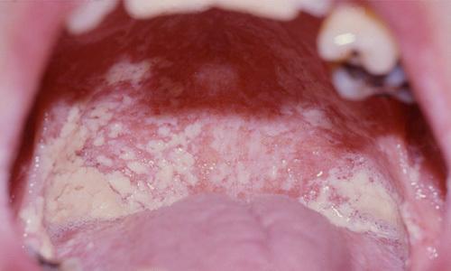
This clinical photograph shows a close-up intraoral view of a patient with extensive pseudomembranous oropharyngeal candidiasis (thrush). The oral cavity exhibits characteristic thick, white-to-creamy yellow plaques and curd-like patches distributed across the dorsal surface of the tongue, the hard and soft palates, and the buccal mucosa. The lesions consist of a mixture of small punctate spots and larger confluent patches with irregular, raised borders. Underneath and surrounding these white coatings, the mucosal tissue is markedly erythematous, indicating underlying inflammation. This presentation is highly characteristic of opportunistic fungal infection by Candida species, frequently seen in immunocompromised individuals, such as those with HIV/AIDS. The image demonstrates the hallmark clinical signs of oral thrush, which may be associated with symptoms like dysphagia and odynophagia if the infection extends into the esophagus.
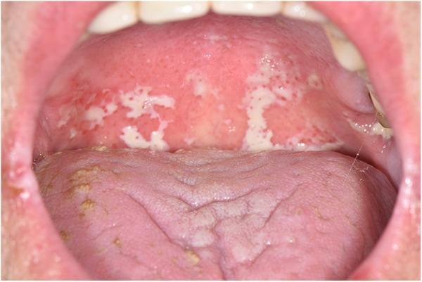
Clinical photograph of the oral cavity showing pseudomembranous candidiasis (oral thrush) in a cancer patient. The image primarily features the soft palate and the posterior oropharynx, which exhibit diffuse erythema (redness) and inflammation. Overlying this erythematous base are multiple, irregularly shaped, creamy-white pseudomembranous plaques characteristic of a fungal infection by Candida species. The plaques appear elevated and adherent but are typically scrapable in a clinical setting. The dorsum of the tongue is visible in the foreground, showing a pink, slightly fissured surface with scattered yellowish-tan patches and mild white coatings. This visual serves as a classic educational representation of opportunistic fungal infections in immunocompromised individuals, such as those undergoing oncology treatments. Key educational concepts include the identification of pseudomembranous lesions, differentiation from other forms of oral candidiasis, and the clinical presentation of oral mucositis superinfection.
histoplasmosis deep mycosis systemic fungal infection
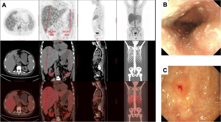
A multi-modal clinical imaging panel demonstrating systemic involvement of a fungal infection, likely Histoplasmosis. (A) Composite 18F-FDG PET/CT images in axial, coronal, sagittal, and maximum intensity projection (MIP) views. The images reveal significant hepatosplenomegaly (liver length 245.5 mm, spleen length 187.9 mm) with increased metabolic activity. There is also evidence of diffuse hyper-metabolism in the bone marrow and abdominal lymphadenopathy. (B) Upper gastrointestinal endoscopy image of the esophagus showing white, bean-curd-like plaques covering the mucosal surface, characteristic of fungal esophagitis (e.g., Candida). (C) Lower gastrointestinal endoscopy image of the colon depicting a protuberant, umbilicated lesion with central depression, erosion, and surrounding erythema, indicative of granulomatous mucosal inflammation from mycotic infection. The combination of findings illustrates systemic disseminated disease with multi-organ involvement including the reticuloendothelial system and gastrointestinal tract.
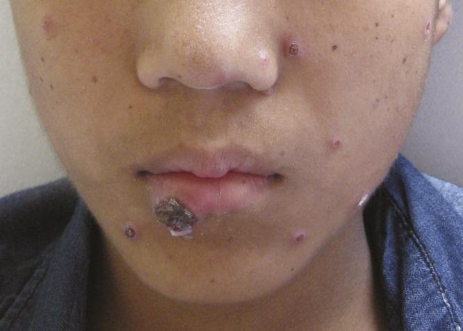
Clinical photograph of a young male patient's face demonstrating disseminated cutaneous lesions characteristic of fungal infection, specifically Histoplasmosis. The primary visible pathology consists of multiple small, discrete, erythematous papules distributed across the cheeks, nose, and chin. Several of these papules exhibit central umbilication or shallow ulceration with a necrotic or hemorrhagic center and a raised, red rim. A larger, more complex lesion is situated on the vermilion border of the lower lip, presenting as a prominent, irregular, brownish-black crusted plaque with associated whitish exudate, suggesting secondary ulceration or necrosis. The presentation illustrates the cutaneous manifestations of systemic fungal disease in the context of immunocompromise or severe systemic infection. This visual material is pedagogically useful for dermatology and infectious disease education regarding the morphology of miliary or disseminated fungal skin signs.
🍄 Fungi: Superficial and Deep Mycoses
BSc Nursing - 3rd Semester | Microbiology & Infectious Diseases
1. Introduction to Fungi (Mycology)
- Non-photosynthetic (no chlorophyll)
- Acquire nutrients from the external environment
- May be unicellular (yeasts) or multicellular (molds/hyphae)
Source: Sherris & Ryan's Medical Microbiology, 8th Ed.
2. Classification of Mycoses
| Type | Tissue Affected | Examples |
|---|---|---|
| Superficial | Stratum corneum, hair, nails | Tinea, Pityriasis versicolor |
| Subcutaneous | Dermis, subcutaneous tissue | Sporotrichosis, Chromoblastomycosis |
| Deep (Systemic) | Internal organs, disseminated | Histoplasmosis, Cryptococcosis, Blastomycosis |
| Opportunistic | Any tissue (immunocompromised) | Candidiasis, Aspergillosis |
"Cutaneous infections are divided into superficial (those that affect mostly the skin) and deep (those that are typically more invasive) mycoses." - Andrews' Diseases of the Skin
3. Superficial Mycoses
A. Dermatophytes (Tinea Infections)
- Microsporum - mainly affects hair and skin
- Trichophyton - affects hair, skin, and nails
- Epidermophyton - affects only skin and nails (never hair)
Types of Tinea (by body site):
| Name | Site | Key Features |
|---|---|---|
| Tinea capitis | Scalp | Common in children; patchy alopecia with scaling |
| Tinea corporis | Body/trunk | Classic ring-shaped (annular) lesion with central clearing |
| Tinea pedis | Feet ("Athlete's foot") | Scaling, maceration, fissuring between toes |
| Tinea cruris | Groin ("Jock itch") | Erythematous, scaly, pruritic patches in groin |
| Tinea unguium / Onychomycosis | Nails | Nail thickening, yellowing, brittleness |
| Tinea barbae | Beard area | Folliculitis-like; requires oral antifungals |
| Tinea faciei | Face | Frequently misdiagnosed; lacks classic annular rings |
| Tinea manuum | Hands | Scaling, hyperkeratosis |
Risk Factors for Dermatophyte Infections:
- Hot, humid climates
- Occlusive footwear
- Sweating and maceration
- Close contact with infected individuals
- Immunosuppression (e.g., topical steroids)
Diagnosis:
- KOH preparation: Scraping + 10% KOH - shows hyphae under microscope
- Wood's lamp: Fluorescence in Microsporum infections
- Fungal culture: On Sabouraud dextrose agar
- PAS stain (histology): Periodic acid-Schiff - 41-93% sensitive for onychomycosis
Treatment:
- Topical: Clotrimazole, miconazole, terbinafine, naftifine - for limited skin infections
- Oral: Griseofulvin, terbinafine, itraconazole - needed for tinea capitis, tinea barbae, onychomycosis, and extensive disease
- Note: Oral griseofulvin is NOT effective against non-dermatophyte molds
B. Pityriasis Versicolor (Tinea Versicolor)
- Caused by Malassezia species (formerly Pityrosporum)
- Superficial yeast infection of the skin
- Presents as hypo- or hyperpigmented macules on trunk and upper arms
- Common in tropical and humid climates
- Diagnosis: KOH prep shows "spaghetti and meatballs" pattern
- Treatment: Topical selenium sulfide, azole shampoos; oral fluconazole/itraconazole for widespread disease
C. Candidiasis (Cutaneous / Mucocutaneous)
- Oral thrush: White curd-like plaques on tongue/buccal mucosa (pseudomembranous candidiasis)
- Vulvovaginal candidiasis: Thick white discharge, pruritus, erythema
- Diaper dermatitis: Erythematous rash with satellite pustules in infants
- Intertrigo: Moist skin folds (groin, axilla, under breasts)
- Chronic mucocutaneous candidiasis: Rare immune deficiency; persistent infection of skin, nails, and mucous membranes
- Candidal paronychia: Inflammation of nail fold with brownish nail discoloration - common in diabetics and those with chronic wet exposure
- Antibiotic use (disrupts normal flora)
- Diabetes mellitus
- HIV/AIDS, immunosuppression
- Prolonged corticosteroid use
- Pregnancy
- Dentures, poor oral hygiene
- Oral: Nystatin suspension, oral fluconazole
- Topical: Clotrimazole, miconazole
- Systemic/severe: IV fluconazole or amphotericin B
4. Deep (Systemic) Mycoses
"Most deep cutaneous fungal infections are a manifestation of systemic infection from inhalation of aerosolized fungus." - Andrews' Diseases of the Skin
- Mold form in the environment (at 25°C/room temperature)
- Yeast form in the human body (at 37°C/body temperature)
A. Histoplasmosis
| Feature | Details |
|---|---|
| Causative organism | Histoplasma capsulatum |
| Mode of infection | Inhalation of spores from bird/bat droppings |
| Endemic area | Ohio and Mississippi River valleys (USA); tropical regions |
| Primary site | Lungs |
- Acute pulmonary: Flu-like illness; most cases self-limiting
- Chronic pulmonary: Resembles TB - cavities, fibrosis
- Disseminated: In immunocompromised (HIV); hepatosplenomegaly, lymphadenopathy, skin lesions
- Cutaneous: Erythematous papules, ulcers, nodules as part of disseminated disease
B. Coccidioidomycosis (Valley Fever)
| Feature | Details |
|---|---|
| Causative organism | Coccidioides immitis or C. posadasii |
| Mode of infection | Inhalation of arthroconidia from soil |
| Endemic area | Southwestern USA, Mexico (San Joaquin Valley) |
| Incubation period | 10 days to several weeks |
- 60% asymptomatic
- Acute pulmonary: Flu-like; hilar adenopathy on CXR
- Skin manifestations: Erythema nodosum (over shins) in ~30% of women, ~15% of men - tender nodules that turn purple-brown over weeks
- Disseminated: CNS involvement (meningitis) - most serious; more common in immunocompromised, pregnant women, Filipino, and Black patients
Source: Andrews' Diseases of the Skin, p. 359
C. Blastomycosis
| Feature | Details |
|---|---|
| Causative organism | Blastomyces dermatitidis |
| Mode of infection | Inhalation; also direct skin inoculation |
| Endemic area | Ohio-Mississippi Valley, Great Lakes region |
- Pulmonary: Flu-like, pneumonia, mimics TB
- Skin: Most common extrapulmonary site - verrucous (wart-like), crusted, ulcerated plaques; often on face and extremities
- Bone, CNS, genitourinary involvement possible
D. Sporotrichosis
| Feature | Details |
|---|---|
| Causative organism | Sporothrix schenckii |
| Mode of infection | Traumatic inoculation - thorn pricks, wood splinters ("Rose thorn disease") |
| Occupation risk | Gardeners, florists, farmers |
- Cutaneous lymphangitic (most common): Primary nodule at inoculation site → painless nodules track along lymphatics ("sporotrichoid spread")
- Fixed cutaneous: Lesion stays at inoculation site; no lymphatic spread
- Disseminated: Rare; lungs, bones, joints, CNS in immunocompromised
E. Cryptococcosis
| Feature | Details |
|---|---|
| Causative organism | Cryptococcus neoformans |
| Mode of infection | Inhalation of dried pigeon/bird droppings |
| Most at risk | HIV/AIDS patients (CD4 < 100) |
- Pulmonary: Mild, often self-limiting in immunocompetent
- Cryptococcal meningitis: Most feared complication - headache, fever, neck stiffness, altered mental status
- Skin lesions: Papules/nodules resembling molluscum contagiosum in disseminated disease
F. Aspergillosis
| Feature | Details |
|---|---|
| Causative organism | Aspergillus fumigatus (most common), A. flavus, A. niger |
| Mode of infection | Inhalation of ubiquitous airborne conidia |
| Most at risk | Neutropenic patients, transplant recipients, corticosteroid users |
- Allergic bronchopulmonary aspergillosis (ABPA): In asthma/cystic fibrosis patients
- Aspergilloma ("fungus ball"): Grows in pre-existing lung cavities (TB cavities)
- Invasive pulmonary aspergillosis: Life-threatening in immunocompromised; angioinvasion, hemorrhage, necrosis
- Cutaneous: In burn victims - can colonize eschar; may disseminate
5. Opportunistic Fungi
| Fungus | Key Disease |
|---|---|
| Candida spp. | Mucocutaneous and systemic candidiasis |
| Aspergillus | Invasive aspergillosis |
| Mucor, Rhizopus (Zygomycetes) | Mucormycosis - rhinocerebral, pulmonary |
| Pneumocystis jirovecii | PCP pneumonia (HIV, CD4 <200) |
6. Antifungal Drugs - Overview
| Class | Drug | Mechanism of Action | Used For |
|---|---|---|---|
| Polyenes | Amphotericin B, Nystatin | Binds ergosterol → membrane pores | Severe systemic; Nystatin for mucosal |
| Azoles | Fluconazole, Itraconazole, Voriconazole, Ketoconazole | Inhibit ergosterol synthesis (CYP51) | Wide spectrum; most fungal infections |
| Allylamines | Terbinafine | Inhibit squalene epoxidase | Dermatophytes, onychomycosis |
| Echinocandins | Caspofungin, Micafungin | Inhibit glucan synthesis (cell wall) | Invasive Candida and Aspergillus |
| Antimetabolite | Flucytosine (5-FC) | Inhibits DNA/RNA synthesis | Combination with Ampho B for Cryptococcus |
| Griseofulvin | Griseofulvin | Disrupts microtubule polymerization | Dermatophytes (older drug) |
7. Key Nursing Implications
- Isolation: Standard precautions; ensure good hand hygiene to prevent spread of dermatophytes
- Patient education: Skin hygiene - keep skin dry, change socks daily, avoid sharing personal items (combs, towels)
- Medication adherence: Antifungal courses can be long (weeks to months for onychomycosis); counsel patients on completing therapy
- Monitor side effects:
- Amphotericin B: Nephrotoxicity, electrolyte imbalances (K+, Mg2+), infusion-related fever/chills (premedicate with paracetamol/antihistamine)
- Fluconazole/Azoles: Hepatotoxicity, drug interactions (CYP450)
- Itraconazole: Negative inotropic effect - avoid in heart failure
- Immunocompromised patients (HIV, transplant, cancer): Watch closely for opportunistic fungal infections; low threshold for investigation
- Diabetes control: Reduces recurrence of candidal and dermatophyte infections
- Specimen collection: Properly collect skin scrapings, nail clippings, or swabs for fungal culture before starting antifungals
8. Quick Summary Table
| Feature | Superficial Mycoses | Deep Mycoses |
|---|---|---|
| Tissue invaded | Stratum corneum, hair, nails | Internal organs, viscera |
| Common pathogens | Trichophyton, Microsporum, Epidermophyton, Candida, Malassezia | Histoplasma, Coccidioides, Blastomyces, Cryptococcus, Aspergillus |
| Mode of entry | Contact, direct skin | Inhalation (mostly); inoculation |
| Host immune | Normal host affected | Often immunocompromised (or endemic exposure) |
| Systemic spread | Rare | Frequent (especially in immunocompromised) |
| Treatment | Topical or short oral antifungals | Prolonged systemic antifungals |
| Life-threatening | Rarely | Yes, especially with dissemination |
Sources: Andrews' Diseases of the Skin - Clinical Dermatology; Sherris & Ryan's Medical Microbiology, 8th Edition; Fitzpatrick's Dermatology
Now create pdf of it including images for final examination
mkdir -p /tmp/workspace/fungi-notes/images && ls /tmp/workspace/fungi-notes/
cd /tmp/workspace/fungi-notes/images && \ wget -q -O tinea_corporis.jpg "https://cdn.orris.care/cdss_images/DermNetNZ_1760031352474_e9484726-3d13-4c35-ab37-ce0170fac122.jpg" && \ wget -q -O tinea_pedis.jpg "https://cdn.orris.care/cdss_images/DermNetNZ_1760031388440_a7240925-8188-4fe2-bc0c-19fa2cd962ff.jpg" && \ wget -q -O oral_thrush.jpg "https://cdn.orris.care/cdss_images/pmc_clinical_VQA_174cdd5a05e9c760c847cb6310e852f49bf410da3df9f7ec100fb386e96770e3.jpg" && \ wget -q -O histoplasmosis.jpg "https://cdn.orris.care/cdss_images/pmc_clinical_VQA_7c25641eb3bf41d77c3532aa9b493b4e4e7ffdc84d05a9cabfd45e3e58fdbdcf.jpg" && \ wget -q -O tinea_corporis2.jpg "https://cdn.orris.care/cdss_images/DermNetNZ_1760031958554_884bb3ac-5808-4393-8771-ac9ee1ec26f6.jpg" && \ ls -lh
cd /tmp/workspace/fungi-notes/images && \ python3 -c " import urllib.request images = { 'tinea_corporis.jpg': 'https://cdn.orris.care/cdss_images/DermNetNZ_1760031352474_e9484726-3d13-4c35-ab37-ce0170fac122.jpg', 'tinea_pedis.jpg': 'https://cdn.orris.care/cdss_images/DermNetNZ_1760031388440_a7240925-8188-4fe2-bc0c-19fa2cd962ff.jpg', 'oral_thrush.jpg': 'https://cdn.orris.care/cdss_images/pmc_clinical_VQA_174cdd5a05e9c760c847cb6310e852f49bf410da3df9f7ec100fb386e96770e3.jpg', 'histoplasmosis.jpg': 'https://cdn.orris.care/cdss_images/pmc_clinical_VQA_7c25641eb3bf41d77c3532aa9b493b4e4e7ffdc84d05a9cabfd45e3e58fdbdcf.jpg', 'tinea_hiv.jpg': 'https://cdn.orris.care/cdss_images/DermNetNZ_1760031958554_884bb3ac-5808-4393-8771-ac9ee1ec26f6.jpg', } for fname, url in images.items(): try: urllib.request.urlretrieve(url, fname) print(f'Downloaded: {fname}') except Exception as e: print(f'Failed {fname}: {e}') " ls -lh
~/fungi-notes/generate_pdf.py
from reportlab.lib.pagesizes import A4
from reportlab.lib import colors
from reportlab.lib.styles import getSampleStyleSheet, ParagraphStyle
from reportlab.lib.units import cm, mm
from reportlab.platypus import (
SimpleDocTemplate, Paragraph, Spacer, Table, TableStyle,
Image, HRFlowable, KeepTogether, PageBreak
)
from reportlab.lib.enums import TA_CENTER, TA_LEFT, TA_JUSTIFY
from reportlab.platypus import Flowable
from reportlab.pdfbase import pdfmetrics
from reportlab.pdfbase.ttfonts import TTFont
import os
OUTPUT = "/tmp/workspace/fungi-notes/Fungi_Superficial_Deep_Mycoses_BSc_Nursing.pdf"
IMG_DIR = "/tmp/workspace/fungi-notes/images"
# ── Colour palette ──────────────────────────────────────────────────────────
DARK_BLUE = colors.HexColor("#1A3A5C")
MED_BLUE = colors.HexColor("#2E6DA4")
LIGHT_BLUE = colors.HexColor("#D6E8F7")
ACCENT_TEAL = colors.HexColor("#17A589")
LIGHT_TEAL = colors.HexColor("#D1F2EB")
GOLD = colors.HexColor("#F0B429")
LIGHT_GOLD = colors.HexColor("#FEF9E7")
PALE_GREY = colors.HexColor("#F4F6F7")
WHITE = colors.white
RED_DARK = colors.HexColor("#C0392B")
LIGHT_RED = colors.HexColor("#FADBD8")
doc = SimpleDocTemplate(
OUTPUT,
pagesize=A4,
rightMargin=1.8*cm, leftMargin=1.8*cm,
topMargin=2*cm, bottomMargin=2*cm,
title="Fungi: Superficial and Deep Mycoses",
author="BSc Nursing 3rd Semester"
)
W, H = A4
CONTENT_W = W - 3.6*cm
styles = getSampleStyleSheet()
# Custom styles
def S(name, **kw):
return ParagraphStyle(name, **kw)
cover_title = S("CoverTitle", fontSize=28, textColor=WHITE,
leading=34, alignment=TA_CENTER, fontName="Helvetica-Bold",
spaceAfter=6)
cover_sub = S("CoverSub", fontSize=14, textColor=LIGHT_BLUE,
leading=20, alignment=TA_CENTER, fontName="Helvetica",
spaceAfter=4)
cover_info = S("CoverInfo", fontSize=11, textColor=GOLD,
leading=16, alignment=TA_CENTER, fontName="Helvetica-Oblique")
h1 = S("H1", fontSize=16, textColor=WHITE, leading=20,
fontName="Helvetica-Bold", spaceBefore=14, spaceAfter=4,
leftIndent=0, borderPad=6)
h2 = S("H2", fontSize=13, textColor=DARK_BLUE, leading=17,
fontName="Helvetica-Bold", spaceBefore=10, spaceAfter=4)
h3 = S("H3", fontSize=11, textColor=MED_BLUE, leading=15,
fontName="Helvetica-Bold", spaceBefore=7, spaceAfter=3)
body = S("BodyText2", fontSize=9.5, textColor=colors.HexColor("#2C2C2C"),
leading=14, alignment=TA_JUSTIFY, fontName="Helvetica",
spaceBefore=3, spaceAfter=3)
bullet = S("Bullet2", fontSize=9.5, textColor=colors.HexColor("#2C2C2C"),
leading=14, fontName="Helvetica",
leftIndent=14, firstLineIndent=-10, spaceBefore=1)
note_style = S("Note", fontSize=9, textColor=colors.HexColor("#555555"),
leading=13, fontName="Helvetica-Oblique",
leftIndent=8, spaceBefore=2)
caption_style = S("Caption", fontSize=8.5, textColor=colors.HexColor("#555"),
leading=12, fontName="Helvetica-Oblique", alignment=TA_CENTER,
spaceBefore=2, spaceAfter=6)
class ColorBox(Flowable):
"""A coloured banner used as section heading background."""
def __init__(self, text, bg=DARK_BLUE, fg=WHITE, fontsize=14, height=26):
super().__init__()
self.text = text
self.bg = bg
self.fg = fg
self.fontsize = fontsize
self.height = height
self.width = CONTENT_W
def draw(self):
self.canv.setFillColor(self.bg)
self.canv.roundRect(0, 0, self.width, self.height, 4, fill=1, stroke=0)
self.canv.setFillColor(self.fg)
self.canv.setFont("Helvetica-Bold", self.fontsize)
self.canv.drawString(10, 7, self.text)
def wrap(self, *args):
return self.width, self.height + 4
def section_box(text, bg=DARK_BLUE, fg=WHITE, fontsize=14, height=28):
return ColorBox(text, bg=bg, fg=fg, fontsize=fontsize, height=height)
def sub_section(text):
return ColorBox(text, bg=MED_BLUE, fg=WHITE, fontsize=12, height=24)
def sub_sub(text):
return ColorBox(text, bg=ACCENT_TEAL, fg=WHITE, fontsize=11, height=22)
def hr(color=MED_BLUE):
return HRFlowable(width="100%", thickness=1.2, color=color,
spaceAfter=4, spaceBefore=4)
def sp(h=6):
return Spacer(1, h)
def p(text, style=None):
return Paragraph(text, style or body)
def b(text):
return Paragraph(f"• {text}", bullet)
def tbl(data, col_widths, style_cmds, row_heights=None):
t = Table(data, colWidths=col_widths, rowHeights=row_heights)
base = [
("FONTNAME", (0,0), (-1,0), "Helvetica-Bold"),
("FONTSIZE", (0,0), (-1,-1), 8.5),
("ROWBACKGROUNDS", (0,0), (-1,-1), [PALE_GREY, WHITE]),
("BACKGROUND", (0,0), (-1, 0), DARK_BLUE),
("TEXTCOLOR", (0,0), (-1, 0), WHITE),
("ALIGN", (0,0), (-1,-1), "LEFT"),
("VALIGN", (0,0), (-1,-1), "MIDDLE"),
("GRID", (0,0), (-1,-1), 0.4, colors.HexColor("#B0BEC5")),
("TOPPADDING", (0,0), (-1,-1), 5),
("BOTTOMPADDING",(0,0),(-1,-1), 5),
("LEFTPADDING", (0,0), (-1,-1), 6),
] + style_cmds
t.setStyle(TableStyle(base))
return t
def img(fname, caption, width=None, height=None):
path = os.path.join(IMG_DIR, fname)
if not os.path.exists(path):
return [p(f"[Image: {caption}]")]
w = width or 7*cm
h = height or 5*cm
im = Image(path, width=w, height=h)
return [im, Paragraph(caption, caption_style)]
def hdr_p(text, style=h1):
return Paragraph(text, style)
# ═══════════════════════════════════════════════════════════════════
# BUILD STORY
# ═══════════════════════════════════════════════════════════════════
story = []
# ── COVER PAGE ────────────────────────────────────────────────────
cover_data = [[
Paragraph("FUNGI", cover_title),
Paragraph("Superficial & Deep Mycoses", cover_title),
sp(8),
Paragraph("BSc Nursing — 3rd Semester", cover_sub),
Paragraph("Microbiology & Infectious Diseases", cover_sub),
sp(16),
Paragraph("Final Examination Notes", cover_info),
Paragraph("Based on: Andrews' Diseases of the Skin | Sherris & Ryan's Medical Microbiology, 8e", cover_info),
]]
cover_tbl = Table(cover_data, colWidths=[CONTENT_W])
cover_tbl.setStyle(TableStyle([
("BACKGROUND", (0,0), (-1,-1), DARK_BLUE),
("ALIGN", (0,0), (-1,-1), "CENTER"),
("VALIGN", (0,0), (-1,-1), "MIDDLE"),
("TOPPADDING", (0,0), (-1,-1), 40),
("BOTTOMPADDING",(0,0),(-1,-1), 40),
("ROUNDEDCORNERS", [8]),
]))
story.append(cover_tbl)
story.append(PageBreak())
# ── 1. INTRODUCTION ───────────────────────────────────────────────
story.append(section_box("1. Introduction to Fungi (Mycology)"))
story.append(sp(6))
story.append(p("Fungi are <b>eukaryotic organisms</b> — they possess a nucleus, mitochondria, and endoplasmic reticulum, making them more complex than bacteria. They are non-photosynthetic and acquire nutrients from the external environment. Fungi may be unicellular (yeasts) or multicellular (molds/hyphae). The diseases caused by fungi are called <b>mycoses</b>."))
story.append(sp(4))
story.append(p("Of more than 90,000 known fungal species, <b>fewer than 200</b> cause disease in humans. Fungal infections tend to be <b>subacute or chronic</b>, often relapsing over time — unlike the acute presentation of most bacterial or viral infections. They are becoming increasingly common, especially among immunocompromised patients."))
story.append(sp(8))
# Key facts box
kf_data = [
[Paragraph("<b>KEY FACTS</b>", S("kf_h", fontSize=9, textColor=WHITE, fontName="Helvetica-Bold", leading=12))],
[Paragraph("• Fungi are eukaryotes — they respond differently to antibiotics than bacteria", bullet)],
[Paragraph("• Cell wall contains <b>ergosterol</b> (not cholesterol) — target of most antifungals", bullet)],
[Paragraph("• Dimorphic fungi = mold at 25°C (environment), yeast at 37°C (body)", bullet)],
[Paragraph("• Mycoses are classified: superficial, subcutaneous, systemic, opportunistic", bullet)],
]
kf_tbl = Table(kf_data, colWidths=[CONTENT_W])
kf_tbl.setStyle(TableStyle([
("BACKGROUND", (0,0), (0,0), DARK_BLUE),
("BACKGROUND", (0,1), (0,-1), LIGHT_BLUE),
("TOPPADDING", (0,0), (-1,-1), 5),
("BOTTOMPADDING",(0,0),(-1,-1), 5),
("LEFTPADDING", (0,0), (-1,-1), 8),
("BOX", (0,0), (-1,-1), 1, MED_BLUE),
]))
story.append(kf_tbl)
story.append(sp(10))
# ── 2. CLASSIFICATION ─────────────────────────────────────────────
story.append(section_box("2. Classification of Mycoses"))
story.append(sp(6))
class_data = [
[Paragraph("<b>Type</b>", S("th", fontSize=9, textColor=WHITE, fontName="Helvetica-Bold")),
Paragraph("<b>Tissue Affected</b>", S("th", fontSize=9, textColor=WHITE, fontName="Helvetica-Bold")),
Paragraph("<b>Examples</b>", S("th", fontSize=9, textColor=WHITE, fontName="Helvetica-Bold")),
Paragraph("<b>Host Status</b>", S("th", fontSize=9, textColor=WHITE, fontName="Helvetica-Bold"))],
["Superficial", "Stratum corneum, hair, nails", "Tinea, Pityriasis versicolor, Candida", "Normal host"],
["Subcutaneous", "Dermis, subcutaneous tissue", "Sporotrichosis, Chromoblastomycosis", "Inoculation via trauma"],
["Deep / Systemic", "Internal organs, disseminated", "Histoplasmosis, Cryptococcosis, Blastomycosis", "Endemic exposure / immunocomp."],
["Opportunistic", "Any tissue", "Candidiasis, Aspergillosis, Mucormycosis", "Immunocompromised"],
]
story.append(tbl(class_data,
[3.5*cm, 4.5*cm, 6.5*cm, 4.3*cm],
[("BACKGROUND", (0,2), (-1,2), LIGHT_TEAL),
("BACKGROUND", (0,3), (-1,3), LIGHT_GOLD)]))
story.append(sp(10))
# ── 3. SUPERFICIAL MYCOSES ────────────────────────────────────────
story.append(section_box("3. Superficial Mycoses"))
story.append(sp(6))
story.append(p("Superficial mycoses are <b>limited to the stratum corneum, hair, and nails</b>. They do NOT invade living tissue and rarely cause systemic disease in healthy hosts. They are the most common fungal infections worldwide."))
story.append(sp(8))
# 3A. Dermatophytes
story.append(sub_section("3A. Dermatophytes (Tinea Infections)"))
story.append(sp(6))
story.append(p("<b>Three genera</b> are responsible for all dermatophyte infections:"))
story.append(b("<b>Microsporum</b> — mainly affects hair and skin (e.g., M. canis from cats/dogs)"))
story.append(b("<b>Trichophyton</b> — affects hair, skin, AND nails (most common genus; T. rubrum is #1 in USA)"))
story.append(b("<b>Epidermophyton</b> — affects skin and nails ONLY (never hair)"))
story.append(sp(8))
# Tinea types table
story.append(hdr_p("Types of Tinea (by body site)", h3))
story.append(sp(4))
tinea_data = [
[Paragraph("<b>Name</b>", S("th2", fontSize=9, textColor=WHITE, fontName="Helvetica-Bold")),
Paragraph("<b>Site</b>", S("th2", fontSize=9, textColor=WHITE, fontName="Helvetica-Bold")),
Paragraph("<b>Key Features</b>", S("th2", fontSize=9, textColor=WHITE, fontName="Helvetica-Bold")),
Paragraph("<b>Treatment</b>", S("th2", fontSize=9, textColor=WHITE, fontName="Helvetica-Bold"))],
["Tinea capitis", "Scalp", "Patchy alopecia + scaling; common in children", "Oral griseofulvin / terbinafine"],
["Tinea corporis", "Body/trunk", "Annular lesion — ring with central clearing + scale", "Topical azole / terbinafine"],
["Tinea pedis", "Feet (Athlete's foot)", "Scaling, maceration, fissuring between toes", "Topical terbinafine"],
["Tinea cruris", "Groin (Jock itch)", "Erythematous, scaly, pruritic patches in groin", "Topical azole"],
["Tinea unguium / Onychomycosis", "Nails", "Nail thickening, yellowing, brittleness", "Oral terbinafine / itraconazole"],
["Tinea barbae", "Beard area", "Folliculitis-like; usually T. rubrum or T. mentagrophytes", "Oral antifungals"],
["Tinea faciei", "Face", "Lacks classic rings; photosensitive; mimics LE", "Topical or oral antifungals"],
["Tinea manuum", "Hands", "Scaling, hyperkeratosis of palm", "Topical azole / terbinafine"],
]
story.append(tbl(tinea_data,
[3.8*cm, 3.2*cm, 6.2*cm, 5.6*cm],
[("BACKGROUND", (0,1), (-1,1), LIGHT_BLUE),
("BACKGROUND", (0,3), (-1,3), LIGHT_BLUE),
("BACKGROUND", (0,5), (-1,5), LIGHT_BLUE),
("BACKGROUND", (0,7), (-1,7), LIGHT_BLUE)]))
story.append(sp(10))
# Clinical images — tinea
story.append(hdr_p("Clinical Images — Tinea Infections", h3))
story.append(sp(4))
img_row = [
[Image(os.path.join(IMG_DIR,"tinea_corporis.jpg"), width=8.5*cm, height=6.5*cm),
Image(os.path.join(IMG_DIR,"tinea_pedis.jpg"), width=8.5*cm, height=6.5*cm)],
[Paragraph("Fig. 1 — Tinea corporis: classic annular erythematous plaque with central clearing and peripheral scale. KOH prep confirms septate hyphae.", caption_style),
Paragraph("Fig. 2 — Tinea pedis (Athlete's foot): scaling, maceration and fissuring of the plantar foot and interdigital spaces.", caption_style)],
]
img_tbl = Table(img_row, colWidths=[CONTENT_W/2, CONTENT_W/2])
img_tbl.setStyle(TableStyle([
("ALIGN", (0,0), (-1,-1), "CENTER"),
("VALIGN", (0,0), (-1,-1), "TOP"),
("TOPPADDING", (0,0), (-1,-1), 4),
("BOTTOMPADDING", (0,0), (-1,-1), 4),
]))
story.append(img_tbl)
story.append(sp(6))
img_row2 = [
[Image(os.path.join(IMG_DIR,"tinea_hiv.jpg"), width=8.5*cm, height=6.5*cm),
Table([
[Paragraph("<b>Diagnosis of Tinea</b>", S("dh", fontSize=10, textColor=WHITE, fontName="Helvetica-Bold", leading=14))],
[Paragraph("<b>KOH Preparation</b> — scraping + 10% KOH; shows septate hyphae under microscopy (most common/cheap)", body)],
[Paragraph("<b>Wood's Lamp</b> — UV fluorescence for Microsporum spp. (green fluorescence)", body)],
[Paragraph("<b>Fungal Culture</b> — Sabouraud dextrose agar; 1-4 weeks; gold standard for species ID", body)],
[Paragraph("<b>PAS Stain (histology)</b> — Periodic acid-Schiff; 41-93% sensitive for nail infections", body)],
[Paragraph("<b>PCR</b> — Rapid, highly sensitive; increasingly used", body)],
], colWidths=[CONTENT_W/2 - 0.5*cm],
style=TableStyle([
("BACKGROUND", (0,0), (-1,0), MED_BLUE),
("BACKGROUND", (0,1), (-1,-1), LIGHT_BLUE),
("TOPPADDING", (0,0), (-1,-1), 5),
("BOTTOMPADDING", (0,0), (-1,-1), 5),
("LEFTPADDING", (0,0), (-1,-1), 8),
("BOX", (0,0), (-1,-1), 1, MED_BLUE),
("LINEBELOW", (0,0), (-1,-1), 0.3, colors.HexColor("#B0BEC5")),
]))
],
[Paragraph("Fig. 3 — Tinea corporis in HIV patient: extensive, atypical, coalescing annular plaques on shoulder due to immunosuppression.", caption_style),
""],
]
img_tbl2 = Table(img_row2, colWidths=[CONTENT_W/2, CONTENT_W/2])
img_tbl2.setStyle(TableStyle([
("ALIGN", (0,0), (-1,-1), "CENTER"),
("VALIGN", (0,0), (-1,-1), "TOP"),
]))
story.append(img_tbl2)
story.append(sp(10))
# Risk Factors
story.append(sub_sub("Risk Factors for Dermatophyte Infections"))
story.append(sp(4))
rf_data = [
[Paragraph("<b>Factor</b>", S("th3", fontSize=9, textColor=WHITE, fontName="Helvetica-Bold")),
Paragraph("<b>Mechanism</b>", S("th3", fontSize=9, textColor=WHITE, fontName="Helvetica-Bold"))],
["Hot, humid climate", "Promotes fungal growth and skin maceration"],
["Occlusive footwear / athletics", "Maceration of feet — increases tinea pedis risk"],
["Diabetes mellitus", "Impaired immunity + glucose-rich environment"],
["Immunosuppression (steroids, HIV)", "Larger lesions, widespread or invasive infection"],
["Close contact (schools, dormitories)", "Person-to-person spread, especially tinea capitis"],
["Broken skin barrier (ichthyosis)", "Reduced mechanical defence against fungi"],
["Blood type A", "Slightly more prone to chronic dermatophyte disease"],
]
story.append(tbl(rf_data, [5.5*cm, 13.3*cm], []))
story.append(sp(10))
# 3B. Pityriasis Versicolor
story.append(sub_section("3B. Pityriasis Versicolor (Tinea Versicolor)"))
story.append(sp(6))
story.append(p("<b>Causative organism:</b> Malassezia species (formerly Pityrosporum) — a lipophilic yeast normally commensal on human skin."))
story.append(b("Hypo- or hyperpigmented (or salmon-pink) macules on trunk and upper arms"))
story.append(b("Typically young adults; worse in tropical, humid climates"))
story.append(b("Asymptomatic or mildly pruritic; fine branny scale when scratched"))
story.append(b("<b>KOH preparation:</b> Classic 'spaghetti and meatballs' pattern — hyphae + spores"))
story.append(b("<b>Treatment:</b> Topical selenium sulfide 2.5%, azole shampoo (ketoconazole); oral fluconazole/itraconazole for widespread disease"))
story.append(sp(10))
# 3C. Candidiasis
story.append(sub_section("3C. Candidiasis (Cutaneous / Mucocutaneous)"))
story.append(sp(6))
story.append(p("Candida is normally part of the oral, GI, and vaginal flora. Disease occurs when <b>host defences are impaired</b>. C. albicans is the most common species; non-albicans (C. glabrata, C. tropicalis, C. krusei) are increasing."))
story.append(sp(6))
cand_forms = [
[Paragraph("<b>Clinical Form</b>", S("th4", fontSize=9, textColor=WHITE, fontName="Helvetica-Bold")),
Paragraph("<b>Presentation</b>", S("th4", fontSize=9, textColor=WHITE, fontName="Helvetica-Bold")),
Paragraph("<b>Key Notes</b>", S("th4", fontSize=9, textColor=WHITE, fontName="Helvetica-Bold"))],
["Oral thrush (pseudomembranous)", "White curd-like plaques on tongue/buccal mucosa; wipe off leaving erythematous base", "Most common in infants, elderly, immunocompromised"],
["Vulvovaginal candidiasis", "Thick white cottage cheese-like discharge, pruritus, erythema", "Common in pregnancy, DM, antibiotic use"],
["Diaper dermatitis", "Erythematous rash with satellite pustules in infants", "Moist occlusive environment"],
["Candidal intertrigo", "Moist red rash in skin folds (groin, axilla, under breasts)", "Obese patients, diabetics"],
["Candidal paronychia", "Inflamed nail fold; brownish discoloured nails", "Common in diabetics and those with chronic wet hands"],
["Chronic mucocutaneous candidiasis", "Persistent skin, nail, and mucous membrane infection", "Rare; underlying immune deficiency"],
["Oesophageal candidiasis", "Dysphagia, odynophagia; white plaques on oesophagoscopy", "AIDS-defining illness; CD4 <100"],
]
story.append(tbl(cand_forms, [4.5*cm, 6.5*cm, 7.8*cm],
[("BACKGROUND", (0,1), (-1,1), LIGHT_TEAL),
("BACKGROUND", (0,3), (-1,3), LIGHT_TEAL),
("BACKGROUND", (0,5), (-1,5), LIGHT_TEAL),
("BACKGROUND", (0,7), (-1,7), LIGHT_TEAL)]))
story.append(sp(8))
# Candida image
cand_img_row = [
[Image(os.path.join(IMG_DIR,"oral_thrush.jpg"), width=8.5*cm, height=6*cm),
Table([
[Paragraph("<b>Candida Predisposing Factors</b>", S("pdh", fontSize=10, textColor=WHITE, fontName="Helvetica-Bold", leading=14))],
[Paragraph("1. Antibiotic use — disrupts protective normal flora", body)],
[Paragraph("2. Diabetes mellitus — glucose-rich secretions", body)],
[Paragraph("3. HIV/AIDS, immunosuppression", body)],
[Paragraph("4. Prolonged corticosteroid / chemotherapy", body)],
[Paragraph("5. Pregnancy — hormonal changes", body)],
[Paragraph("6. Dentures, poor oral hygiene", body)],
[Paragraph("7. Neonates — immature immune system", body)],
[Paragraph("8. Malnutrition, extreme ages", body)],
], colWidths=[CONTENT_W/2 - 0.5*cm],
style=TableStyle([
("BACKGROUND", (0,0), (-1,0), ACCENT_TEAL),
("BACKGROUND", (0,1), (-1,-1), LIGHT_TEAL),
("TOPPADDING", (0,0), (-1,-1), 5),
("BOTTOMPADDING", (0,0), (-1,-1), 5),
("LEFTPADDING", (0,0), (-1,-1), 8),
("BOX", (0,0), (-1,-1), 1, ACCENT_TEAL),
("LINEBELOW", (0,0), (-1,-1), 0.3, colors.HexColor("#B0BEC5")),
]))
],
[Paragraph("Fig. 4 — Oral thrush (pseudomembranous candidiasis): thick white curd-like plaques on tongue and buccal mucosa in an immunocompromised patient (HIV/AIDS).", caption_style), ""],
]
cand_tbl = Table(cand_img_row, colWidths=[CONTENT_W/2, CONTENT_W/2])
cand_tbl.setStyle(TableStyle([
("ALIGN", (0,0), (-1,-1), "CENTER"),
("VALIGN", (0,0), (-1,-1), "TOP"),
]))
story.append(cand_tbl)
story.append(sp(6))
story.append(p("<b>Treatment of Candidiasis:</b>"))
story.append(b("Oral/mucosal (mild): Nystatin suspension, clotrimazole lozenges"))
story.append(b("Systemic (moderate-severe): Oral or IV fluconazole"))
story.append(b("Resistant / non-albicans species: Echinocandins (caspofungin) or amphotericin B"))
story.append(b("Topical (skin/nails): Clotrimazole, miconazole cream"))
story.append(PageBreak())
# ── 4. DEEP MYCOSES ───────────────────────────────────────────────
story.append(section_box("4. Deep (Systemic) Mycoses", bg=RED_DARK))
story.append(sp(6))
story.append(p('"Most deep cutaneous fungal infections are a manifestation of systemic infection from inhalation of aerosolized fungus." <i>— Andrews\' Diseases of the Skin</i>'))
story.append(sp(4))
story.append(p("The major deep mycoses are caused by <b>dimorphic fungi</b>:"))
dimorphic = [
[Paragraph("<b>Temperature</b>", S("dmt", fontSize=9, textColor=WHITE, fontName="Helvetica-Bold")),
Paragraph("<b>Form</b>", S("dmt", fontSize=9, textColor=WHITE, fontName="Helvetica-Bold")),
Paragraph("<b>Location</b>", S("dmt", fontSize=9, textColor=WHITE, fontName="Helvetica-Bold"))],
["25°C (room / environment)", "Mold (hyphae + conidia)", "Soil, environment"],
["37°C (human body)", "Yeast (budding)", "Lungs, bloodstream, organs"],
]
story.append(tbl(dimorphic, [5*cm, 6*cm, 7.8*cm], [("BACKGROUND", (0,0), (-1,0), RED_DARK)]))
story.append(sp(4))
story.append(p("<b>Important principle:</b> Always order a chest X-ray when investigating a patient with deep mycosis (except classic inoculation types such as sporotrichosis, mycetoma, chromoblastomycosis)."))
story.append(sp(10))
# 4A. Histoplasmosis
story.append(sub_section("4A. Histoplasmosis"))
story.append(sp(4))
histo_info = [
[Paragraph("<b>Feature</b>", S("hi", fontSize=9, textColor=WHITE, fontName="Helvetica-Bold")),
Paragraph("<b>Details</b>", S("hi", fontSize=9, textColor=WHITE, fontName="Helvetica-Bold"))],
["Causative organism", "Histoplasma capsulatum (dimorphic fungus)"],
["Mode of infection", "Inhalation of spores from bird or bat droppings"],
["Endemic area", "Ohio and Mississippi River valleys (USA); tropical regions worldwide"],
["Primary site", "Lungs — then dissemination possible"],
["High-risk group", "HIV patients (CD4 <150), cave explorers, construction workers"],
]
story.append(tbl(histo_info, [5*cm, 13.8*cm], [("BACKGROUND", (0,0), (-1,0), MED_BLUE)]))
story.append(sp(6))
story.append(p("<b>Clinical Forms:</b>"))
story.append(b("<b>Acute pulmonary:</b> Flu-like illness; most cases self-limiting in immunocompetent; fever, cough, myalgia"))
story.append(b("<b>Chronic pulmonary:</b> Resembles TB — upper lobe cavities, fibrosis; chronic cough, haemoptysis"))
story.append(b("<b>Disseminated histoplasmosis:</b> In HIV/AIDS — hepatosplenomegaly, lymphadenopathy, bone marrow involvement, skin lesions"))
story.append(b("<b>Cutaneous manifestations:</b> Erythematous papules, ulcers, nodules with central necrosis — always sign of systemic disease"))
story.append(sp(8))
histo_img_row = [
[Image(os.path.join(IMG_DIR,"histoplasmosis.jpg"), width=8.5*cm, height=6.5*cm),
Table([
[Paragraph("<b>Histoplasmosis Diagnosis</b>", S("hdh", fontSize=10, textColor=WHITE, fontName="Helvetica-Bold", leading=14))],
[Paragraph("• Urine/serum antigen (most sensitive for disseminated)", body)],
[Paragraph("• Histopathology — intracellular yeast INSIDE macrophages (Wright/Giemsa stain)", body)],
[Paragraph("• Blood culture — takes weeks; less practical acutely", body)],
[Paragraph("• Serology (complement fixation) — useful in chronic/endemic disease", body)],
[Paragraph("• Bronchoalveolar lavage (BAL) for pulmonary form", body)],
[sp(6)],
[Paragraph("<b>Treatment</b>", S("hdh2", fontSize=10, textColor=DARK_BLUE, fontName="Helvetica-Bold", leading=14))],
[Paragraph("• Mild-moderate: <b>Itraconazole</b> for 12 weeks", body)],
[Paragraph("• Severe/disseminated: <b>Amphotericin B</b> (induction) then itraconazole", body)],
[Paragraph("• HIV patients: Lifelong itraconazole suppressive therapy", body)],
], colWidths=[CONTENT_W/2 - 0.5*cm],
style=TableStyle([
("BACKGROUND", (0,0), (-1,0), MED_BLUE),
("BACKGROUND", (0,1), (-1,5), LIGHT_BLUE),
("BACKGROUND", (0,6), (-1,6), WHITE),
("BACKGROUND", (0,7), (-1,7), LIGHT_GOLD),
("BACKGROUND", (0,8), (-1,-1), LIGHT_GOLD),
("TOPPADDING", (0,0), (-1,-1), 5),
("BOTTOMPADDING", (0,0), (-1,-1), 5),
("LEFTPADDING", (0,0), (-1,-1), 8),
("BOX", (0,0), (-1,-1), 1, MED_BLUE),
("LINEBELOW", (0,0), (-1,-1), 0.3, colors.HexColor("#B0BEC5")),
]))
],
[Paragraph("Fig. 5 — Disseminated cutaneous histoplasmosis: multiple erythematous papules with central ulceration/necrosis on the face — a cutaneous sign of systemic fungal disease.", caption_style), ""],
]
histo_tbl = Table(histo_img_row, colWidths=[CONTENT_W/2, CONTENT_W/2])
histo_tbl.setStyle(TableStyle([
("ALIGN", (0,0), (-1,-1), "CENTER"),
("VALIGN", (0,0), (-1,-1), "TOP"),
]))
story.append(histo_tbl)
story.append(sp(10))
# 4B. Coccidioidomycosis
story.append(sub_section("4B. Coccidioidomycosis (Valley Fever)"))
story.append(sp(4))
story.append(p("<b>Causative organism:</b> Coccidioides immitis or C. posadasii | <b>Infection:</b> Inhalation of arthroconidia from soil | <b>Endemic:</b> Southwestern USA (San Joaquin Valley), Mexico | <b>Incubation:</b> 10 days to several weeks"))
story.append(sp(4))
story.append(b("<b>60% are asymptomatic</b>"))
story.append(b("<b>Acute pulmonary:</b> Flu-like illness; hilar adenopathy and infiltrates on chest X-ray"))
story.append(b("<b>Erythema nodosum:</b> Tender red-purple nodules over shins — occurs in ~30% of women, ~15% of men 2-3 weeks after infection (sign of good immune response)"))
story.append(b("<b>Disseminated (severe):</b> CNS meningitis (most dangerous), bone/joint involvement; more common in immunocompromised, pregnant women, Filipino and Black patients"))
story.append(b("<b>Diagnosis:</b> Serology (complement fixation), KOH of sputum showing spherules, culture"))
story.append(b("<b>Treatment:</b> Fluconazole or itraconazole (mild-moderate); Amphotericin B (severe/meningeal)"))
story.append(sp(10))
# 4C. Blastomycosis
story.append(sub_section("4C. Blastomycosis"))
story.append(sp(4))
story.append(p("<b>Causative organism:</b> Blastomyces dermatitidis | <b>Infection:</b> Inhalation; also direct skin inoculation | <b>Endemic:</b> Ohio-Mississippi Valley, Great Lakes region"))
story.append(sp(4))
story.append(b("Pulmonary: Flu-like illness or pneumonia; mimics TB or lung cancer on X-ray"))
story.append(b("<b>Skin (most common extrapulmonary site):</b> Verrucous (wart-like), crusted, ulcerated plaques — typically on face and extremities"))
story.append(b("Bone, CNS, genitourinary involvement also possible"))
story.append(b("<b>Diagnosis:</b> Broad-based budding yeast on KOH/histology; culture; serology (unreliable)"))
story.append(b("<b>Treatment:</b> Itraconazole (mild-moderate); Amphotericin B (severe/CNS)"))
story.append(sp(10))
# 4D. Sporotrichosis
story.append(sub_section("4D. Sporotrichosis ('Rose Thorn Disease')"))
story.append(sp(4))
story.append(p("<b>Causative organism:</b> Sporothrix schenckii | <b>Mode:</b> <b>Traumatic inoculation</b> — thorn pricks, wood splinters | <b>Occupation risk:</b> Gardeners, florists, farmers, miners"))
story.append(sp(4))
story.append(b("<b>Cutaneous lymphangitic (most common):</b> Primary painless ulcerated nodule at inoculation site, then nodules track along lymphatics proximally ('sporotrichoid spread') — classic ascending lymphangitis"))
story.append(b("<b>Fixed cutaneous:</b> Lesion stays at inoculation site; no lymphatic spread"))
story.append(b("<b>Disseminated (rare):</b> Lungs, bones, joints, CNS — only in severely immunocompromised"))
story.append(b("<b>Diagnosis:</b> Fungal culture of biopsy or exudate — gold standard; microscopy may miss organism"))
story.append(b("<b>Treatment:</b> Oral itraconazole (first line); supersaturated potassium iodide (SSKI) for cutaneous forms; Amphotericin B for disseminated"))
story.append(sp(10))
# 4E. Cryptococcosis
story.append(sub_section("4E. Cryptococcosis"))
story.append(sp(4))
story.append(p("<b>Causative organism:</b> Cryptococcus neoformans (encapsulated yeast) | <b>Mode:</b> Inhalation of dried pigeon/bird droppings | <b>Most at risk:</b> HIV/AIDS patients with CD4 <100"))
story.append(sp(4))
story.append(b("Pulmonary: Mild, often self-limiting in immunocompetent"))
story.append(b("<b>Cryptococcal meningitis (most feared):</b> Headache, fever, neck stiffness, photophobia, altered mental status — may be indolent; can cause raised intracranial pressure"))
story.append(b("Skin lesions: Papules/nodules resembling molluscum contagiosum in disseminated disease"))
story.append(b("<b>Diagnosis:</b> India ink preparation of CSF (encapsulated yeast with wide capsule), Latex agglutination for capsular antigen (very sensitive)"))
story.append(b("<b>Treatment:</b> Amphotericin B + flucytosine (induction, 2 weeks) → Fluconazole (consolidation + maintenance)"))
story.append(sp(10))
# 4F. Aspergillosis
story.append(sub_section("4F. Aspergillosis"))
story.append(sp(4))
story.append(p("<b>Causative organism:</b> Aspergillus fumigatus (most common), A. flavus, A. niger | <b>Mode:</b> Inhalation of ubiquitous airborne conidia | Fungal hyphae show <b>acute-angle (45°) branching</b>"))
story.append(sp(4))
asp_forms = [
[Paragraph("<b>Form</b>", S("af", fontSize=9, textColor=WHITE, fontName="Helvetica-Bold")),
Paragraph("<b>Patient</b>", S("af", fontSize=9, textColor=WHITE, fontName="Helvetica-Bold")),
Paragraph("<b>Features</b>", S("af", fontSize=9, textColor=WHITE, fontName="Helvetica-Bold"))],
["ABPA (Allergic bronchopulmonary)", "Asthma / cystic fibrosis patients", "Eosinophilia, elevated IgE, fleeting pulmonary infiltrates"],
["Aspergilloma (fungus ball)", "Pre-existing lung cavities (post-TB)", "Mass in cavity; haemoptysis; 'air crescent sign' on CT"],
["Invasive pulmonary aspergillosis", "Neutropenic / transplant / steroid users", "Angioinvasion, necrosis, haemorrhage; high mortality"],
["Cutaneous aspergillosis", "Burn victims / immunocompromised", "Colonises burn eschar; can disseminate to systemic disease"],
]
story.append(tbl(asp_forms, [5*cm, 5*cm, 8.8*cm],
[("BACKGROUND", (0,1), (-1,1), LIGHT_RED),
("BACKGROUND", (0,3), (-1,3), LIGHT_RED)]))
story.append(sp(4))
story.append(b("<b>Diagnosis:</b> CT chest (halo sign in invasive disease), serum galactomannan antigen, bronchoscopy + BAL culture, tissue biopsy"))
story.append(b("<b>Treatment:</b> Voriconazole (drug of choice for invasive); Amphotericin B (alternative); Itraconazole (ABPA/aspergilloma)"))
story.append(PageBreak())
# ── 5. OPPORTUNISTIC FUNGI ────────────────────────────────────────
story.append(section_box("5. Opportunistic Fungi (Immunocompromised Hosts)", bg=colors.HexColor("#6C3483")))
story.append(sp(6))
story.append(p("These fungi cause disease almost exclusively in <b>immunocompromised hosts</b> — HIV/AIDS, organ transplant, prolonged corticosteroid use, haematological malignancy, neutropenia."))
story.append(sp(6))
oppo_data = [
[Paragraph("<b>Fungus</b>", S("op", fontSize=9, textColor=WHITE, fontName="Helvetica-Bold")),
Paragraph("<b>Key Disease</b>", S("op", fontSize=9, textColor=WHITE, fontName="Helvetica-Bold")),
Paragraph("<b>Key Diagnostic Clue</b>", S("op", fontSize=9, textColor=WHITE, fontName="Helvetica-Bold")),
Paragraph("<b>Treatment</b>", S("op", fontSize=9, textColor=WHITE, fontName="Helvetica-Bold"))],
["Candida spp.", "Mucocutaneous & systemic candidiasis", "Pseudohyphae on KOH; Germ tube test for C. albicans", "Fluconazole / echinocandin"],
["Aspergillus", "Invasive aspergillosis", "Halo sign on CT; galactomannan antigen", "Voriconazole"],
["Mucor / Rhizopus (Zygomycetes)", "Rhinocerebral / pulmonary mucormycosis", "Ribbon-like non-septate hyphae; angioinvasion, tissue necrosis", "Amphotericin B + surgery"],
["Pneumocystis jirovecii", "PCP pneumonia", "CD4 <200; bilateral interstitial infiltrates on CXR; LDH elevated", "Co-trimoxazole (TMP-SMX)"],
["Cryptococcus neoformans", "Meningitis / pulmonary", "India ink; latex agglutination antigen", "Ampho B + flucytosine → fluconazole"],
]
story.append(tbl(oppo_data, [3.5*cm, 4.5*cm, 6.5*cm, 4.3*cm],
[("BACKGROUND", (0,0), (-1,0), colors.HexColor("#6C3483")),
("BACKGROUND", (0,2), (-1,2), colors.HexColor("#EDE7F6")),
("BACKGROUND", (0,4), (-1,4), colors.HexColor("#EDE7F6"))]))
story.append(sp(10))
# ── 6. ANTIFUNGAL DRUGS ───────────────────────────────────────────
story.append(section_box("6. Antifungal Drugs — Overview", bg=ACCENT_TEAL))
story.append(sp(6))
drug_data = [
[Paragraph("<b>Class</b>", S("dr", fontSize=9, textColor=WHITE, fontName="Helvetica-Bold")),
Paragraph("<b>Drug(s)</b>", S("dr", fontSize=9, textColor=WHITE, fontName="Helvetica-Bold")),
Paragraph("<b>Mechanism of Action</b>", S("dr", fontSize=9, textColor=WHITE, fontName="Helvetica-Bold")),
Paragraph("<b>Key Uses</b>", S("dr", fontSize=9, textColor=WHITE, fontName="Helvetica-Bold")),
Paragraph("<b>Side Effects</b>", S("dr", fontSize=9, textColor=WHITE, fontName="Helvetica-Bold"))],
["Polyenes", "Amphotericin B\nNystatin", "Binds ergosterol → membrane pores → cell lysis",
"Severe systemic mycoses\nNystatin: topical/mucosal only",
"Nephrotoxicity, hypokalaemia, infusion reactions (fever/chills)"],
["Azoles", "Fluconazole\nItraconazole\nVoriconazole\nKetoconazole",
"Inhibit CYP51 enzyme → block ergosterol synthesis",
"Wide spectrum; most fungal infections",
"Hepatotoxicity; drug interactions (CYP450); itraconazole: avoid in heart failure"],
["Allylamines", "Terbinafine", "Inhibit squalene epoxidase → ergosterol deficiency",
"Dermatophytes; onychomycosis",
"GI upset; hepatotoxicity (rare); taste disturbance"],
["Echinocandins", "Caspofungin\nMicafungin\nAnidulafungin",
"Inhibit 1,3-beta-D-glucan synthase → cell wall disruption",
"Invasive Candida; refractory Aspergillus; neutropenic patients",
"Well tolerated; mild hepatotoxicity; histamine release"],
["Antimetabolite", "Flucytosine (5-FC)", "Converted to 5-FU inside cell → inhibits DNA/RNA synthesis",
"COMBINATION with Ampho B for Cryptococcal meningitis",
"Bone marrow suppression; GI toxicity; resistance if used alone"],
["Griseofulvin", "Griseofulvin", "Disrupts microtubule polymerization → inhibits mitosis",
"Dermatophytes (hair/nail); older drug; NOT for non-dermatophytes",
"Photosensitivity; headache; GI upset; teratogenic"],
]
story.append(tbl(drug_data, [2.8*cm, 3.2*cm, 4.5*cm, 4.5*cm, 3.8*cm],
[("BACKGROUND", (0,0), (-1,0), ACCENT_TEAL),
("BACKGROUND", (0,2), (-1,2), LIGHT_TEAL),
("BACKGROUND", (0,4), (-1,4), LIGHT_TEAL),
("BACKGROUND", (0,6), (-1,6), LIGHT_TEAL),
("FONTSIZE", (0,0), (-1,-1), 8),
("LEADING", (0,0), (-1,-1), 11),
("TOPPADDING", (0,0), (-1,-1), 4),
("BOTTOMPADDING", (0,0), (-1,-1), 4),]))
story.append(sp(10))
# ── 7. NURSING IMPLICATIONS ───────────────────────────────────────
story.append(section_box("7. Key Nursing Implications", bg=colors.HexColor("#1E8449")))
story.append(sp(6))
nursing_data = [
[Paragraph("<b>Area</b>", S("nu", fontSize=9, textColor=WHITE, fontName="Helvetica-Bold")),
Paragraph("<b>Nursing Action / Rationale</b>", S("nu", fontSize=9, textColor=WHITE, fontName="Helvetica-Bold"))],
["Infection Control", "Standard precautions; hand hygiene; avoid sharing personal items (towels, combs) to prevent dermatophyte spread"],
["Patient Education", "Keep skin dry; change socks daily; wear breathable footwear; maintain good oral hygiene; control blood glucose"],
["Medication Adherence", "Antifungal courses are long (weeks–months for onychomycosis); counsel patients never to stop early; explain that nail regrowth takes time"],
["Monitor Ampho B", "Nephrotoxicity — monitor urine output, BUN, creatinine, K+, Mg2+; pre-medicate with paracetamol + antihistamine for infusion reactions; give IV fluids before each dose"],
["Monitor Azoles (fluconazole, itraconazole)", "Hepatotoxicity — monitor LFTs; warn about drug-drug interactions (statins, warfarin, phenytoin via CYP450); itraconazole has negative inotropic effect — avoid in heart failure"],
["Immunocompromised Patients", "Low threshold for investigating fungal infections; watch for unusual presentations; promptly report fever in neutropenic patients"],
["Specimen Collection", "Collect skin scrapings, nail clippings, swabs or BAL BEFORE starting antifungals to preserve culture sensitivity"],
["Diabetes Management", "Tight glycaemic control reduces risk of recurrent candidal and dermatophyte infections — educate patient on this link"],
["Nutritional Support", "Ensure adequate nutrition — malnutrition impairs cell-mediated immunity, increasing susceptibility to fungal infections"],
]
story.append(tbl(nursing_data, [5*cm, 13.8*cm],
[("BACKGROUND", (0,2), (-1,2), colors.HexColor("#D5F5E3")),
("BACKGROUND", (0,4), (-1,4), colors.HexColor("#D5F5E3")),
("BACKGROUND", (0,6), (-1,6), colors.HexColor("#D5F5E3")),
("BACKGROUND", (0,8), (-1,8), colors.HexColor("#D5F5E3"))]))
story.append(sp(10))
# ── 8. COMPARISON TABLE ───────────────────────────────────────────
story.append(section_box("8. Quick Comparison: Superficial vs Deep Mycoses", bg=DARK_BLUE))
story.append(sp(6))
comp_data = [
[Paragraph("<b>Feature</b>", S("cm", fontSize=9, textColor=WHITE, fontName="Helvetica-Bold")),
Paragraph("<b>Superficial Mycoses</b>", S("cm", fontSize=9, textColor=WHITE, fontName="Helvetica-Bold")),
Paragraph("<b>Deep (Systemic) Mycoses</b>", S("cm", fontSize=9, textColor=WHITE, fontName="Helvetica-Bold"))],
["Tissue invaded", "Stratum corneum, hair, nails only", "Internal organs (lungs, CNS, liver, bone marrow)"],
["Common pathogens", "Trichophyton, Microsporum, Epidermophyton, Candida, Malassezia", "Histoplasma, Coccidioides, Blastomyces, Cryptococcus, Aspergillus"],
["Mode of entry", "Direct contact with infected person/animal/soil", "Mainly inhalation of conidia; some by inoculation"],
["Host immune status", "Normal host commonly affected", "Immunocompromised OR endemic exposure"],
["Systemic spread", "Rare (only in immunocompromised)", "Frequent; defines the disease in many cases"],
["Life-threatening", "Rarely (exception: immunocompromised)", "Yes — especially meningitis, disseminated disease"],
["Treatment duration", "Days to weeks (topical); weeks (oral)", "Months; lifelong suppression in HIV patients"],
["Key investigations", "KOH prep, Wood's lamp, culture", "CXR, CT, serology/antigen, biopsy, blood culture"],
]
story.append(tbl(comp_data, [4.5*cm, 6.5*cm, 7.8*cm],
[("BACKGROUND", (0,2), (-1,2), LIGHT_BLUE),
("BACKGROUND", (0,4), (-1,4), LIGHT_BLUE),
("BACKGROUND", (0,6), (-1,6), LIGHT_BLUE),
("BACKGROUND", (0,8), (-1,8), LIGHT_BLUE)]))
story.append(sp(10))
# ── 9. MNEMONICS ──────────────────────────────────────────────────
story.append(section_box("9. Exam Mnemonics & Memory Aids", bg=GOLD))
story.append(sp(6))
mnem_data = [
[Paragraph("<b>Topic</b>", S("mn", fontSize=9, textColor=WHITE, fontName="Helvetica-Bold")),
Paragraph("<b>Mnemonic / Memory Aid</b>", S("mn", fontSize=9, textColor=WHITE, fontName="Helvetica-Bold"))],
["Dermatophyte genera (3)", "MET — Microsporum, Epidermophyton, Trichophyton"],
["Epidermophyton rule", "'E' for Epidermophyton = Everything EXCEPT hair (nails + skin only)"],
["Dimorphic fungi", "\"Mold at Cold (25°C), Yeast at Heat (37°C)\" — think: Mold in the Cold, Yeast in the Beast"],
["Tinea types", "Captain (capitis), Cory (corporis), Pedro (pedis), Cruz (cruris), Uncle (unguium), Barb (barbae), Frank (faciei), Manny (manuum)"],
["Pityriasis versicolor KOH", "'Spaghetti and Meatballs' — spaghetti = hyphae, meatballs = spores"],
["Cryptococcus diagnosis", "India INK = CrYptococcus (both 'y' sounding)"],
["Sporotrichosis occupations", "RAFTER — Rose growers, Agricultural workers, Florists, Timber workers, Excavators, Recreational hikers"],
["Ampho B side effects", "FUNDS — Fever/infusion reactions, Under-potassium (hypokalaemia), Nephrotoxicity, Desoxycholate formulation toxic, Shake well (liposomal form safer)"],
["Antifungal targets", "Ergosterol = TARGET of polyenes AND azoles (different mechanisms); Glucan = target of echinocandins; Microtubule = target of griseofulvin"],
]
story.append(tbl(mnem_data, [5*cm, 13.8*cm],
[("BACKGROUND", (0,0), (-1,0), colors.HexColor("#B7770D")),
("BACKGROUND", (0,2), (-1,2), LIGHT_GOLD),
("BACKGROUND", (0,4), (-1,4), LIGHT_GOLD),
("BACKGROUND", (0,6), (-1,6), LIGHT_GOLD),
("BACKGROUND", (0,8), (-1,8), LIGHT_GOLD)]))
story.append(sp(10))
# ── FOOTER NOTE ───────────────────────────────────────────────────
footer_data = [[
Paragraph(
"Sources: Andrews' Diseases of the Skin — Clinical Dermatology | "
"Sherris & Ryan's Medical Microbiology, 8th Edition | "
"Fitzpatrick's Dermatology | Robbins & Cotran Pathologic Basis of Disease. "
"Prepared for BSc Nursing 3rd Semester — Final Examination 2026.",
note_style)
]]
footer_tbl = Table(footer_data, colWidths=[CONTENT_W])
footer_tbl.setStyle(TableStyle([
("BACKGROUND", (0,0), (-1,-1), LIGHT_BLUE),
("BOX", (0,0), (-1,-1), 0.5, MED_BLUE),
("TOPPADDING", (0,0), (-1,-1), 6),
("BOTTOMPADDING", (0,0), (-1,-1), 6),
("LEFTPADDING", (0,0), (-1,-1), 8),
]))
story.append(footer_tbl)
# ── BUILD ──────────────────────────────────────────────────────────
doc.build(story)
print(f"PDF created: {OUTPUT}")
cd /tmp/workspace/fungi-notes && python3 generate_pdf.py 2>&1
ls -lh /tmp/workspace/fungi-notes/Fungi_Superficial_Deep_Mycoses_BSc_Nursing.pdf
Fungi Superficial Deep Mycoses BSc Nursing
PDF Document · PDF
| Section | Content |
|---|---|
| Cover Page | Titled, colour-coded, with subject and sources |
| 1. Introduction | Fungal biology, key facts box (ergosterol, dimorphism) |
| 2. Classification | 4-type table — superficial, subcutaneous, deep, opportunistic |
| 3A. Dermatophytes | All 8 tinea types table + 3 clinical photos (tinea corporis, pedis, HIV patient) |
| 3B. Pityriasis Versicolor | Malassezia, spaghetti-meatballs KOH |
| 3C. Candidiasis | 7 clinical forms table + oral thrush photo + predisposing factors box |
| 4A-4F. Deep Mycoses | Histoplasmosis (with skin lesion photo), Coccidioidomycosis, Blastomycosis, Sporotrichosis, Cryptococcosis, Aspergillosis |
| 5. Opportunistic Fungi | PCP, Mucormycosis, Cryptococcus comparison table |
| 6. Antifungal Drugs | Full table — mechanism, uses, side effects (Polyenes, Azoles, Allylamines, Echinocandins, Griseofulvin, Flucytosine) |
| 7. Nursing Implications | 9-point nursing care table |
| 8. Comparison Table | Superficial vs Deep at a glance |
| 9. Mnemonics | 9 exam memory aids including MET, RAFTER, FUNDS, dimorphic fungi rule |