
Toxocariasis. Fascioliasis. Etiology, epidemiology, pathogenesis, sign and symptoms, diagnosis, differential diagnosis, treatment, prevention
Reading File
Finding Sources
Finding Sources
Reading File
Reading File
Reading File
Reading File
Good content retrieved. Let me now read the rest of the Fascioliasis content from Goldman-Cecil and Tietz, plus get the Red Book content on Toxocariasis prevention, and search for images.
Reading File
Reading File
Reading File
Searching PubMed
toxocariasis treatment albendazole
Searching PubMed
fascioliasis triclabendazole treatment
Reading File
Reading File
I now have comprehensive information from authoritative sources. Let me also fetch the Fasciola image and search for a Toxocara image.
Loading Image
Searching Images
Toxocara canis visceral larva migrans eosinophilic granuloma liver
TOXOCARIASIS
Etiology
Toxocariasis is caused by Toxocara canis (dog roundworm) and, less commonly, Toxocara cati (cat roundworm) — nematode parasites whose natural definitive hosts are dogs and cats, especially puppies and kittens. Most human cases result from T. canis. Humans are accidental (dead-end) hosts; the larvae cannot develop into adult worms in the human body.
— Goldman-Cecil Medicine, p. 3470
Epidemiology
- Infection is ubiquitous worldwide. In the US, approximately 5% of the population ≥6 years has serologic evidence of infection.
- Visceral larva migrans (VLM) typically affects children 2–7 years old; ocular larva migrans (OLM) typically manifests in older children and adolescents (5–10 years).
- Higher risk in dog owners, people living in poverty, and in hot/humid regions where eggs survive longer in soil.
- OLM is more common in southern US states.
- Transmission occurs through ingestion of embryonated eggs from contaminated soil (sandboxes, playgrounds where dogs/cats defecate). Direct animal contact is not required — eggs are not infectious immediately when shed; they require 2–4 weeks in soil to become infective but may persist long-term.
— Red Book 2021, p. 1179; Goldman-Cecil Medicine, p. 3470
Pathogenesis
After ingestion by a human, embryonated eggs hatch in the intestine, releasing larvae that penetrate the intestinal wall and enter the vasculature. Unlike in the animal host, larvae in humans cannot complete their life cycle — they migrate through the body (lungs, liver, CNS, eyes) but do not mature into adult worms. When larvae eventually die, they trigger significant immediate-type and delayed-type hypersensitivity reactions, forming eosinophilic granulomas. VLM and OLM appear to be mutually exclusive syndromes.
Key point: The inflammatory response to larval migration — not direct larval toxicity — is responsible for most tissue damage.
— Goldman-Cecil Medicine, p. 3470
Signs and Symptoms
Most infections are asymptomatic. Clinical disease depends on the affected organ and degree of host inflammatory response.
1. Covert Toxocariasis
- Simple persistent eosinophilia with minimal or no symptoms
- May persist for years during ongoing migratory phase
2. Visceral Larva Migrans (VLM)
- Low-grade fever, cough, wheezing (pulmonary infiltrates)
- Hepatosplenomegaly, right upper quadrant pain
- Malaise, urticaria, lymphadenopathy
- Less common: myocarditis, nephritis, rash
- Lab: leukocytosis, marked eosinophilia, hypergammaglobulinemia, elevated isohemagglutinins to A/B blood group antigens
- Symptoms appear gradually, resolve over 4–8 weeks
3. Neurotoxocariasis
- Eosinophilic meningoencephalitis, space-occupying lesions, myelitis, cerebral vasculitis
- Seizures, encephalopathy, neuropsychiatric symptoms
4. Ocular Larva Migrans (OLM)
- Typically unilateral vision loss in older children
- Uveitis, endophthalmitis, retinal granulomas, strabismus
- Usually without systemic signs of infection
— Red Book 2021, p. 1179; Goldman-Cecil Medicine, p. 3470
Diagnosis
| Test | Notes |
|---|---|
| CBC | Leukocytosis with marked eosinophilia; anemia |
| Serology (ELISA) | ELISA for anti-Toxocara antibodies (serum or vitreous fluid); available via CDC. Does not distinguish past from active infection; less sensitive for OLM |
| Immunoblot | Used to confirm ELISA; improves specificity |
| Recombinant larval antigen ELISA | Improved sensitivity/specificity; not yet widely available |
| Imaging (US/CT/MRI liver) | Diffuse nodular lesions <2 cm; multiple hypodense nodules; helpful in VLM |
| CT/Fluorescein angiography | For OLM — differentiate from retinoblastoma |
| Aqueous/vitreous anti-Toxocara antibodies | Elevated relative to serum in OLM |
| Liver biopsy | Rarely indicated; microscopically diagnostic but low sensitivity |
| PCR | Not currently available |
— Red Book 2021, p. 1180; Goldman-Cecil Medicine, p. 3470
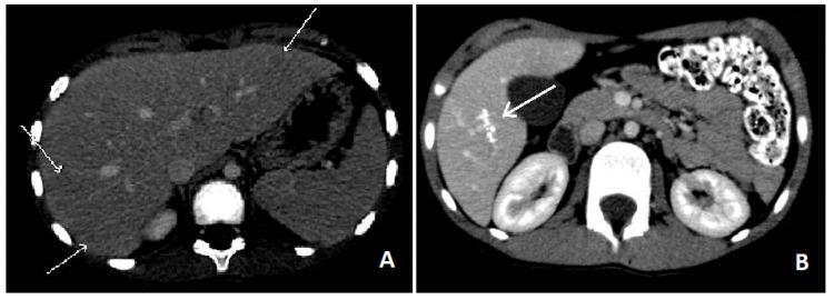
CT abdomen: Acute VLM showing multiple small hypodense nodules scattered in liver parenchyma (left), with resolution to residual calcific granulomas on follow-up (right)
Differential Diagnosis
- Asthma / allergic bronchitis — persistent eosinophilic pulmonary disease
- Löffler syndrome (other helminth migration)
- Hepatic metastases — CT lesions can mimic metastatic disease
- Retinoblastoma — OLM with intraocular mass
- Tuberculosis — focal intraretinal lesions
- Other helminth infections: ascariasis, hookworm, strongyloidiasis, baylisascariasis (Baylisascaris procyonis — raccoon roundworm, causes more severe neurologic disease)
- Hypereosinophilic syndrome
- Eosinophilic leukemia
Treatment
| Condition | Drug | Regimen |
|---|---|---|
| Visceral toxocariasis | Albendazole (preferred) | 400 mg twice daily × 5 days |
| Visceral toxocariasis (alternative) | Mebendazole | Per dosing guidelines |
| Severe (CNS, myocarditis) | Albendazole + corticosteroids | Prednisone 60 mg/day × 5 days concurrently |
| Ocular toxocariasis | Albendazole + corticosteroids (oral/topical) | Anti-inflammatory control; surgical therapy for complications |
- Albendazole is FDA-approved (though not specifically for this indication); safe in children ≥1 year.
- No person-to-person transmission — standard precautions only.
— Red Book 2021, p. 1180; Goldman-Cecil Medicine, p. 3470
Prevention
- Proper disposal of dog and cat feces
- Regular veterinary deworming of dogs, cats, especially puppies and kittens
- Cover sandboxes when not in use to prevent animal defecation
- Wash hands after contact with soil; wash vegetables thoroughly
- No post-exposure prophylaxis is recommended
— Red Book 2021, p. 1181
FASCIOLIASIS
Etiology
Fascioliasis is a zoonosis caused by the liver fluke:
- Fasciola hepatica (adult: 30 × 13 mm) — cosmopolitan distribution
- Fasciola gigantica (adult: up to 75 × 20 mm) — Africa and Asia only
Natural definitive hosts: cattle, sheep, and goats. Humans are incidental hosts.
— Goldman-Cecil Medicine, p. 3470
Epidemiology
- WHO estimates 2.4–17 million people infected in >70 countries
- Both species found in Africa and Asia; only F. hepatica in the Americas, Europe, and Oceania
- Highest prevalence rates (>60%) reported in Peru and Bolivia
- In non-endemic countries (e.g., USA), cases are primarily imported in immigrants and returning travelers; rare autochthonous cases exist
- Women have higher incidence and more severe infections than men
- Estimated ~90,000 DALYs lost annually
— Goldman-Cecil Medicine, p. 3470; Tietz Textbook of Laboratory Medicine, p. 3598
Life Cycle & Pathogenesis
Complete life cycle:
- Eggs passed in feces → deposited in water
- Miracidia hatch (9–14 days) → invade freshwater snail (Lymnaea spp.) — first intermediate host
- In snail: develop through sporocysts → rediae → cercariae (4–7 weeks)
- Free-swimming cercariae leave snail → encyst as metacercariae on aquatic plants (watercress, water lettuce, alfalfa, mint, parsley, khat)
- Human infection: ingestion of metacercariae-contaminated plants or water
- Metacercariae excyst in the duodenum → penetrate bowel wall → migrate through peritoneal cavity → penetrate liver capsule (4 weeks)
- Larvae migrate through liver parenchyma → reach hepatic biliary ducts (3–5 months) → mature into adult flukes
- Adults lay eggs → passed via bile into intestine → excreted in feces
Pathologic response: Larval migration causes a track of eosinophilic inflammation and necrosis. Larvae may die and leave cavities filled with necrotic debris → replaced by scar/calcification. In the bile ducts, adults cause chronic inflammation, hyperplasia, fibrosis, and biliary obstruction.
— Goldman-Cecil Medicine, p. 3470; Tietz Textbook, p. 3599
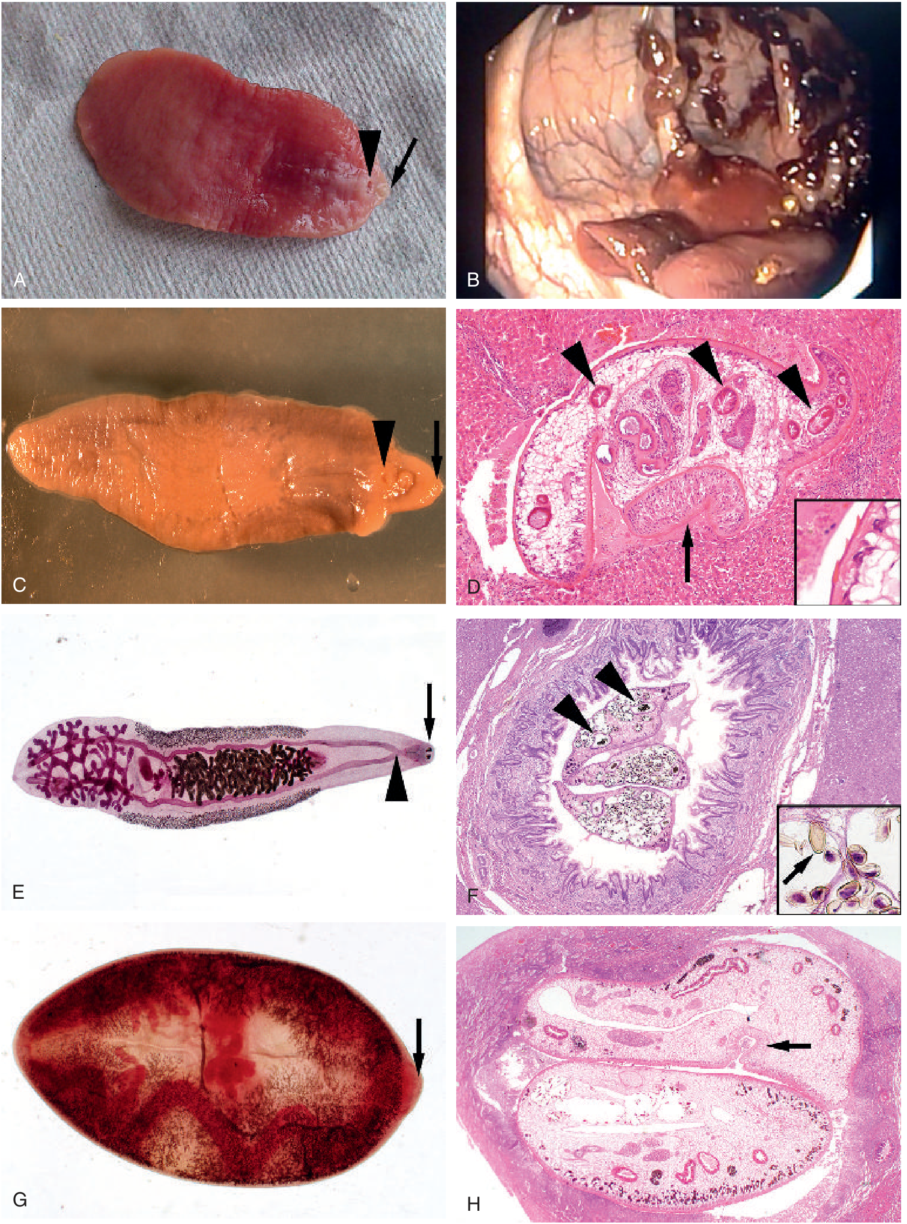
Intestinal and hepatic trematodes. (C) Adult F. hepatica with cone-shaped anterior end, oral sucker (arrow), ventral sucker (arrowhead); up to 3 cm long. (D) F. hepatica migrating through liver parenchyma showing intestinal branches (arrowheads) in cross-section, ventral sucker (arrow) (H&E 100×), with cuticular spines inset (1000×). — Tietz Textbook of Laboratory Medicine
Signs and Symptoms
Disease is biphasic:
Acute (Invasive/Migratory) Phase
Begins when larvae penetrate the bowel wall and migrate through the liver (weeks to months after infection):
- Prolonged fever, malaise
- Hepatomegaly, right upper quadrant / epigastric pain
- Marked peripheral eosinophilia (hallmark)
- Anorexia, weight loss, nausea, vomiting
- Cough, urticaria, lymphadenopathy, arthralgias
- Acute diarrhea (2–5 days, before liver invasion)
- Rare: subcapsular liver hematoma (intense hemorrhage)
- Ectopic fascioliasis: larvae reach subcutaneous tissue, pancreas, eye, brain, stomach wall
- Note: Hyperbilirubinemia is notably absent in the acute phase
Chronic (Biliary) Phase
After adult flukes establish in bile ducts:
- Colicky right upper quadrant and epigastric pain (biliary obstruction)
- Eosinophilia may be absent in ~50% of chronic cases
- Elevated serum alkaline phosphatase
- Bacterial superinfection → cholangitis
- Complications: hemobilia, liver abscess, liver fibrosis, cholelithiasis
- Adults survive in bile ducts for up to 13 years
— Goldman-Cecil Medicine, p. 3470
Diagnosis
Acute Phase
| Test | Notes |
|---|---|
| Serology (Fas2 ELISA / cathepsin L1 ELISA) | Sensitivity 92%, specificity 84%; antibodies detectable within 2–4 weeks of infection. First-line test in acute phase |
| Immunoblot | Confirms positive/equivocal ELISA; cross-reactivity with schistosomiasis may occur |
| CT / MRI / Ultrasound | Track-like hypodense lesions, hepatomegaly, subcapsular hematoma, serpiginous tunneling lesions 2–10 mm; lesions shift in position over time (distinguishing feature from metastases); enhancement of Glisson capsule |
| Liver biopsy | May show flukes migrating through parenchyma; used when serology/CT unavailable |
| Stool exam | Negative during acute phase (adults not yet in ducts) |
Chronic Phase
| Test | Notes |
|---|---|
| Stool microscopy | Eggs detected by Lumbreras rapid sedimentation or Kato-Katz technique; ≥3 stool examinations preferred |
| Egg morphology | Oval, yellow-brown, thin-shelled, 130–150 μm × 63–90 μm; small often indistinct operculum; among the largest eggs in human parasitology |
| ERCP | Can visualize adult flukes in bile duct; allows retrieval and removal |
| Serology | May remain positive >1 year after treatment |
Important caveats:
- Spurious egg passage can occur after eating infected sheep/cattle liver — correlate with clinical presentation
- F. hepatica and F. buski eggs are essentially indistinguishable microscopically
- In the USA, serology available only at the CDC
— Goldman-Cecil Medicine, p. 3470; Tietz Textbook, p. 3599–3600
Differential Diagnosis
Acute phase:
- Acute cholecystitis — similar RUQ pain and fever, but eosinophilia distinguishes fascioliasis
- Hepatic metastases — CT lesions appear similar; in fascioliasis lesions change in position, attenuation, and shape over time
- Hepatic abscess (amoebic or bacterial)
- Schistosomiasis (Katayama fever)
- Other causes of eosinophilic hepatitis: drug reactions, other helminthiases
Chronic phase:
- Cholelithiasis / cholangitis from other causes
- Cholangiocarcinoma (unlike Clonorchis / Opisthorchis, fascioliasis does not cause cholangiocarcinoma)
- Primary sclerosing cholangitis
- Other liver flukes: Clonorchis sinensis, Opisthorchis spp.
Treatment
| Regimen | Details |
|---|---|
| Triclabendazole (drug of choice) | 10 mg/kg orally on 2 consecutive days; cure rate >90% |
| Monitoring cure | Stool examinations negative for ≥3 months |
| Praziquantel | Not effective against Fasciola — a critical distinction from other flukes |
| Treatment failure | Multiple rounds of triclabendazole; some infections are resistant |
| Serology post-treatment | May take >1 year to resolve — not used to confirm cure |
| Biliary obstruction | Dead parasites can cause obstruction requiring surgical or ERCP intervention |
FDA approval: Triclabendazole approved for fascioliasis in patients ≥6 years old.
— Goldman-Cecil Medicine, p. 3470; Tietz Textbook, p. 3599
Prevention
- Avoid eating raw aquatic plants (watercress, water lettuce) from endemic or potentially contaminated areas
- Treat water used for washing vegetables in endemic regions
- Control of intermediate host snails (Lymnaea spp.) in livestock-rearing areas
- Veterinary control of fascioliasis in cattle and sheep (main reservoir hosts)
- Health education in endemic communities (Bolivia, Peru, Africa, Asia)
- Washing vegetables does not reliably destroy metacercariae — avoidance is the safest approach
Side-by-Side Comparison
| Feature | Toxocariasis | Fascioliasis |
|---|---|---|
| Causative organism | Toxocara canis, T. cati (nematode) | Fasciola hepatica, F. gigantica (trematode) |
| Animal reservoir | Dogs, cats | Cattle, sheep, goats |
| Transmission | Ingestion of embryonated eggs in soil | Ingestion of metacercariae on aquatic plants |
| Intermediate host | None (eggs directly infective) | Freshwater snail (Lymnaea spp.) |
| Human role | Accidental dead-end host | Accidental dead-end host |
| Primary organs | Liver, lungs, CNS, eyes | Liver, bile ducts |
| Eosinophilia | Prominent | Prominent (acute); absent in ~50% chronic |
| Imaging | Hepatic nodules <2 cm | Track-like/serpiginous hypodense lesions |
| Key serology | ELISA (CDC) | Fas2 / cathepsin L1 ELISA (CDC) |
| Stool exam | Not useful (no eggs in humans) | Positive in chronic phase; negative acutely |
| Treatment | Albendazole 400 mg BID × 5 days | Triclabendazole 10 mg/kg × 2 days |
| Praziquantel | Not used | Ineffective |
| Corticosteroids | For severe/ocular/CNS disease | Not standard |
Sources: Red Book 2021: Report of the Committee on Infectious Diseases, p. 1179–1181 | Goldman-Cecil Medicine International Edition, p. 3470 | Tietz Textbook of Laboratory Medicine, 7th Edition, p. 3598–3600Both conditions are covered in full detail above. Here is a brief orientation to the two sections:
Toxocariasis is a nematode infection (roundworm larvae of dogs/cats) transmitted by ingesting soil-contaminated embryonated eggs. It produces four distinct syndromes — covert, visceral larva migrans (VLM), ocular larva migrans (OLM), and neurotoxocariasis — all driven by eosinophilic granuloma formation around migrating larvae that cannot mature in humans. Diagnosis rests on serology (ELISA) and imaging; stool exam is useless since adults never develop. Treatment is albendazole ± corticosteroids for severe/ocular disease.
Fascioliasis is a trematode (liver fluke) infection with a two-phase course: an acute invasive/migratory phase (fever, hepatomegaly, intense eosinophilia, shifting CT lesions) and a chronic biliary phase (colicky pain, obstruction, cholangitis, eosinophilia often absent). A key clinical pearl: praziquantel does not work — the only effective drug is triclabendazole. Diagnosis in the acute phase relies on serology (Fas2 ELISA); in the chronic phase, stool microscopy (Kato-Katz) identifies the large operculate eggs.
This is a shared conversation. Sign in to Orris to start your own chat.