What is ecg
Reading File
Finding Sources
Searching the Web
what is an ECG electrocardiogram overview
Reading File
Finding Sources
Searching Images
ECG electrocardiogram normal waveform PQRST
Reading File
Loading Image
Loading Image
Reading File
Here is a comprehensive overview of the ECG (Electrocardiogram):
ECG (Electrocardiogram)
An electrocardiogram (ECG, or EKG) is a graphical record of the electrical activity of the heart over time. Because body fluids are good conductors (the body acts as a volume conductor), fluctuations in electrical potential generated by the heart's action potentials can be detected at the body surface using electrodes and recorded as a waveform.
How It Works
The heart generates electrical impulses that spread in a predictable sequence:
- SA node fires → spreads to both atria → atrial contraction → produces the P wave
- Impulse reaches the AV node → deliberate slow conduction (creates a pause) → represented by the PR interval
- Impulse travels down the His bundle → left/right bundle branches → Purkinje fibers → depolarizes ventricular muscle → produces the QRS complex
- Ventricles then repolarize → produces the T wave
The ECG Waveform (PQRSTU)
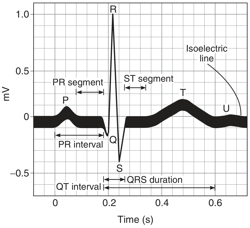
| Wave / Interval | Meaning |
|---|---|
| P wave | Atrial depolarization (atria contracting) |
| PR interval | AV conduction time (normal: 0.12–0.20 s) |
| QRS complex | Ventricular depolarization (ventricles contracting); normal duration ≤ 0.10 s |
| ST segment | Plateau of ventricular action potential; isoelectric in normal ECG |
| T wave | Ventricular repolarization |
| QT interval | Total ventricular electrical activity (normal: ~0.40–0.43 s) |
| U wave | Inconstant; may reflect slow repolarization in some ventricular myocytes |
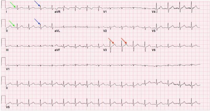
The 12-Lead ECG
A standard clinical ECG records from 12 different viewpoints (leads) of the heart:
Bipolar Limb Leads (standard leads)
- Lead I — Left arm (+) vs Right arm (−)
- Lead II — Left leg (+) vs Right arm (−)
- Lead III — Left leg (+) vs Left arm (−)
Augmented Unipolar Limb Leads
- aVR — Right arm
- aVL — Left arm
- aVF — Left foot (inferior view)
Precordial (Chest) Leads
- V1–V6 — Placed across the chest wall; view the heart from anterior and lateral perspectives
What the ECG Is Used For
- Detecting arrhythmias (abnormal heart rhythms)
- Diagnosing myocardial infarction (heart attack) — ST elevation/depression, Q waves
- Identifying conduction blocks — prolonged PR, widened QRS
- Assessing electrolyte abnormalities — e.g., hypokalemia causes flat T waves and U waves
- Monitoring drug effects — e.g., prolonged QT from certain medications
- Evaluating ventricular hypertrophy, pericarditis, pulmonary embolism, and more
Key Principles
- Upward deflection = depolarization moving toward the active electrode
- Downward deflection = depolarization moving away from the active electrode
- Einthoven's triangle: electrodes on both arms and the left leg form an equilateral triangle with the heart at center — the basis of the standard limb leads
Sources: Ganong's Review of Medical Physiology, 26th ed.; Miller's Anesthesia, 10th ed.
This is a shared conversation. Sign in to Orris to start your own chat.