explain ctg with pics abnormalities

Reading File
Finding Sources
Finding Sources
Finding Sources
Finding Sources
Reading File
Finding Sources
Finding Sources
Reading File
Reading File
Reading File
Finding Sources
Searching the Web

CTG cardiotocography abnormalities types explained with images 2024

Reading Web Page

https://zerotofinals.com/obgyn/labouranddelivery/ctg

Loading Image
Loading Image
Searching the Web

CTG early late variable deceleration diagram image sinusoidal pattern

Loading Image
Loading Image
Loading Image
Loading Image
Searching the Web

CTG fetal heart rate monitoring decelerations types diagram site:wikimedia.org OR site:commons.wikimedia.org

Finding Sources
Reading File
Reading File
Loading Image
Loading Image
Searching the Web

CTG normal abnormal fetal heart rate tracing image reassuring non-reassuring NICE classification

Loading Image
Loading Image

explain ctg in easy way with pictures abnormalities

I already have all the information and images needed from the previous research session. Let me put together a clear, easy-to-understand guide with pictures now.

🫀 CTG (Cardiotocography) - Easy Guide with Pictures

What is a CTG?

CTG is a machine that records two things at the same time on a paper strip:
  • Top line = Fetal Heart Rate (FHR) - measured in beats per minute (bpm)
  • Bottom line = Uterine Contractions - pressure of the womb tightening
Two sensors are placed on the mother's belly:
  • One over the fetal heart (Doppler ultrasound)
  • One at the fundus (to detect contractions)

📊 How to Read a CTG - The 5 Features (DR C BraVaDo)

LetterFeatureWhat to look for
DRDefine RiskIs it high or low risk?
CContractionsHow many per 10 min? (normal ≤5)
BraBaseline RateNormal = 110-160 bpm
VaVariabilityNormal = 5-25 bpm fluctuation
DDecelerationsAny dips? What type?
oOxytocin/AccelerationsAny heart rate spikes? (good sign)

1. BASELINE RATE

The average FHR over 10 minutes, ignoring accelerations and decelerations.
FindingRateMeaning
Normal110-160 bpmHealthy fetus
Tachycardia>160 bpmFever, infection, hypoxia, drugs
Bradycardia<110 bpmFetal distress, cord compression, maternal drugs

2. VARIABILITY

This is how much the heart rate "wiggles" up and down around the baseline. A healthy, awake baby's heart rate should bounce around constantly - it should NOT be a flat line.
VariabilityRangeMeaning
Normal5-25 bpmHealthy CNS + cardiovascular system
Reduced<5 bpm for >40-90 minHypoxia, sleep cycle, drugs (opioids, MgSO4)
AbsentFlat lineSerious - possible severe hypoxia/acidosis
Increased/Saltatory>25 bpm for >25 minAcute hypoxia, cord compression
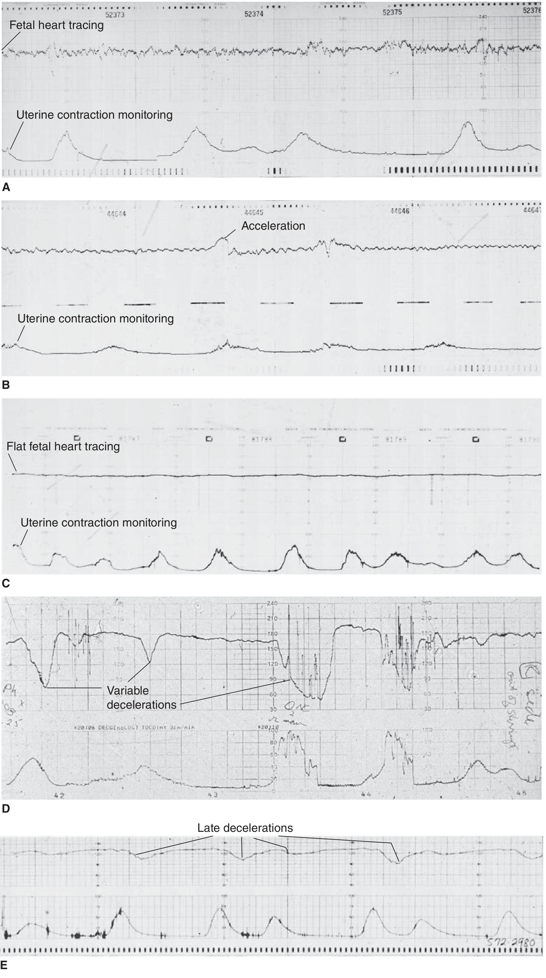
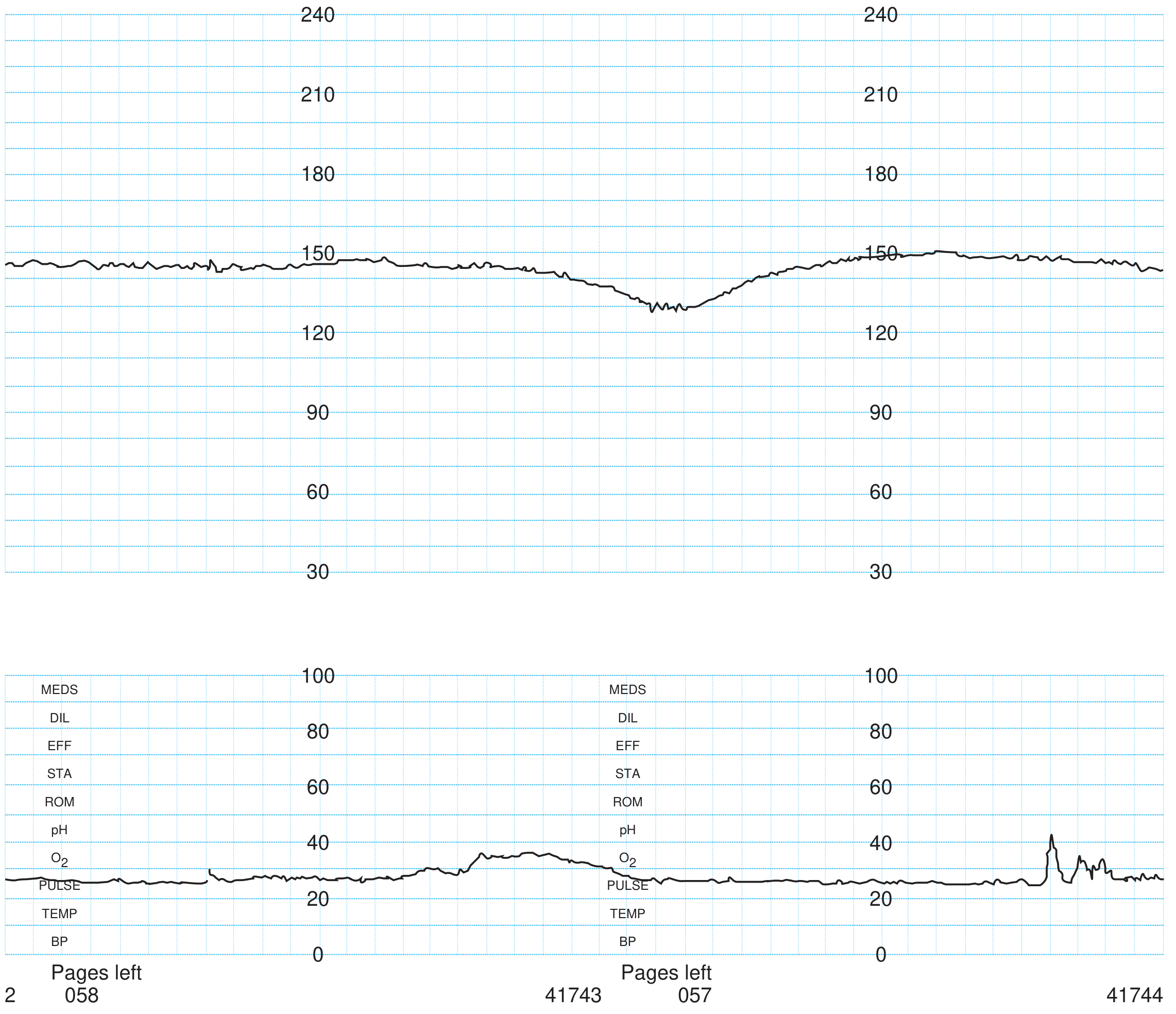
Here is a real CTG tracing showing several patterns - good variability (A, B), flat/absent variability (C), variable decelerations (D), and late decelerations (E):
CTG tracing showing variability, accelerations, variable and late decelerations
A = Good variability (normal, reassuring). B = Good variability with accelerations (very reassuring). C = Flat/poor variability - may indicate fetal hypoxia. D = Variable decelerations (cord compression). E = Late decelerations (uteroplacental insufficiency).

3. ACCELERATIONS

Temporary rises in FHR of ≥15 bpm lasting ≥15 seconds.
  • Presence = REASSURING - means fetus is not acidotic
  • Caused by fetal movement stimulating the sympathetic nervous system
  • Form the basis of the Non-Stress Test (NST)
  • Reactive NST = at least 2 accelerations in 20-30 minutes

4. DECELERATIONS - The Most Important Part ⚠️

Decelerations = drops in FHR of >15 bpm lasting >15 seconds.
There are 4 types - each with a different cause and significance:

🟢 Type 1: EARLY Decelerations (Benign)

FeatureDetail
ShapeGradual, smooth, uniform dip
TimingMirror the contraction - start and end WITH it
NadirCoincides with the peak of contraction
CauseFetal head compression → vagal stimulation
SignificanceNormal/benign - not pathological
Think of it as: the baby's head gets squeezed during a contraction, slows the heart briefly, then recovers. No fetal distress.

🔴 Type 2: LATE Decelerations (Most Ominous)

FeatureDetail
ShapeGradual, smooth dip
TimingStart AFTER the contraction begins, recover AFTER it ends
NadirOccurs >30 seconds after peak of contraction
CauseUteroplacental insufficiency → fetal hypoxia
SignificancePathological - indicates fetal compromise
Think of it as: the placenta isn't delivering enough oxygen. When the womb contracts, blood flow drops even further, and the heart slows - but only AFTER the contraction.
Causes: maternal hypotension, pre-eclampsia, abruption, postmaturity, IUGR, oxytocin hyperstimulation

🟡 Type 3: VARIABLE Decelerations (Most Common)

FeatureDetail
ShapeAbrupt, sharp, V-shaped or W-shaped
TimingVariable - no consistent relationship to contractions
Onset to nadir<30 seconds (abrupt)
Duration15 seconds to <2 minutes
CauseUmbilical cord compression
SignificanceIsolated = not concerning; Persistent = worrying
Reassuring "shoulders" = small accelerations just before and after the dip (fetus compensating well)
Atypical/concerning features:
  • Drop to <60 bpm
  • Duration >60 seconds
  • Slow return to baseline
  • Loss of variability during the deceleration
  • No shoulders

🔴 Type 4: PROLONGED Decelerations

FeatureDetail
Duration2-10 minutes with drop >15 bpm
CauseCord prolapse, maternal hypotension, uterine rupture, abruption
SignificanceAlways abnormal - requires immediate action
Drop >10 minutes = new baseline (bradycardia)

5. SINUSOIDAL PATTERN 🚨

CTG prediction app showing hypoxia-compensated vs hypoxia-decompensated patterns
A smooth, sine wave-like pattern, ≥10 minutes with:
  • Regular oscillations at 2-5 cycles/min
  • Amplitude 5-15 bpm
  • No accelerations
  • No short-term variability
Causes: Severe fetal anaemia (Rh isoimmunization, fetal-maternal haemorrhage), severe hypoxia
This is a MEDICAL EMERGENCY - requires immediate delivery

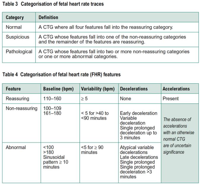
📋 CTG CLASSIFICATION (NICE Guidelines)

NICE CTG classification table showing normal, suspicious and pathological categories
CategoryDefinitionAction
NormalALL features reassuringContinue monitoring
Suspicious1 non-reassuring feature (rest reassuring)Correct reversible causes, closer monitoring
Pathological2+ non-reassuring OR 1+ abnormal featuresUrgent action - consider delivery

Feature Classification Quick Reference:

FeatureReassuringNon-ReassuringAbnormal
Baseline110-160 bpm100-109 or 161-180<100 or >180, sinusoidal ≥10 min
Variability5-25 bpm<5 for 40-90 min or >25 for 15-25 min<5 for ≥90 min
DecelerationsNoneEarly; Variable <90 minLate; Atypical variable; Prolonged >3 min
AccelerationsPresentAbsent (uncertain significance)-

🛠️ Management When CTG is Abnormal

Correct reversible causes first (ALSO mnemonic):
LetterAction
AAssess maternal position (left lateral)
LLower oxytocin / stop if running
SStop pushing / maternal effort
OOxygen (though evidence is mixed)
Then escalate:
  • Suspicious → continue monitoring, repeat VE
  • Pathological → consider fetal blood sampling (scalp pH <7.20 = acidosis) or expedite delivery
  • Sinusoidal / prolonged bradycardia → emergency caesarean section

🧠 Quick Memory Summary

Normal CTG = "CAVE"
C - Contractions ≤5 per 10 min
A - Accelerations present
V - Variability 5-25 bpm  
E - no dEcelerations (or only early ones)

Baseline: 110-160 bpm
Sources: Tintinalli's Emergency Medicine (McGraw-Hill), Mulholland & Greenfield's Surgery, Creasy & Resnik's Maternal-Fetal Medicine, NICE Guidelines 2017 (updated), ZeroToFinals CTG

pics of ctg

Searching the Web

CTG fetal heart rate tracing images normal late deceleration variable sinusoidal bradycardia

Finding Sources
Reading File
Loading Image
Loading Image
Loading Image
Loading Image
Loading Image
Finding Sources
Reading File
Reading File
Reading File
Reading File
Loading Image
Loading Image
Loading Image
Reading File
Loading Image
Loading Image
Here is a complete visual guide to CTG tracings - all images sourced directly from medical textbooks (Creasy & Resnik's Maternal-Fetal Medicine and Textbook of Family Medicine).

📸 CTG Pictures - All Key Patterns


✅ 1. NORMAL CTG

Normal CTG fetal heart rate pattern - baseline ~130 bpm with normal variability and no decelerations
What you're seeing:
  • Top strip: Fetal heart rate running nicely around 130 bpm
  • The line has a healthy "wobble" (that's good variability, 5-25 bpm)
  • Bottom strip: Smooth uterine contractions, 2-3 min apart
  • No dips in the heart rate = reassuring, healthy baby

❌ 2. LATE DECELERATIONS (Uteroplacental Insufficiency)

Late deceleration CTG - fetal heart rate dips after peak of contraction, slow return to baseline
What you're seeing:
  • Baseline around 150 bpm, nearly flat (minimal variability - already worrying)
  • The heart rate dips AFTER the contraction peaks and returns slowly
  • The gap between contraction peak and heart rate nadir is the key sign
  • Cause: Placenta cannot deliver enough oxygen during contractions
  • Significance: PATHOLOGICAL - may indicate fetal acidosis

🔴 3. FETAL TACHYCARDIA (>160 bpm)

CTG showing fetal tachycardia - baseline heart rate around 170-180 bpm with contractions
What you're seeing:
  • Baseline running consistently around 170-180 bpm (well above the normal 110-160)
  • Some variability is preserved here
  • Causes: Maternal fever, chorioamnionitis, fetal hypoxia, β-mimetic drugs, maternal anxiety
  • If tachycardia + decelerations together = high concern for hypoxia

⚠️ 4. PROLONGED BRADYCARDIA

CTG showing prolonged fetal bradycardia - heart rate drops suddenly then recovers after treatment
What you're seeing:
  • Heart rate starts normal (~120-130 bpm), then suddenly drops to below 60 bpm
  • The drop lasts several minutes (prolonged bradycardia)
  • Note on the tracing: "Pit off, O2 6L/min, side" - showing treatment was given
  • Heart rate eventually recovers at the end of the strip
  • Cause here: Oxytocin hyperstimulation (too many contractions, not enough recovery time)
  • Key point: Variability was maintained throughout, meaning the brain still had enough oxygen

⚠️ 5. ABSENT VARIABILITY (Flat Line CTG)

CTG showing absent fetal heart rate variability - completely flat line around 120 bpm
What you're seeing:
  • Heart rate is a completely flat line around 115-120 bpm - no wiggling at all
  • The circled "7.28" is the scalp blood pH - normal in this case
  • This patient was on magnesium sulfate and narcotics (drugs can flatten the trace)
  • Key teaching point: Absent variability is NOT always hypoxia - drugs, fetal sleep, and CNS depression can all cause it
  • When variability is absent WITHOUT a known cause = urgent concern

🔶 6. VARIABLE DECELERATIONS (Cord Compression)

From the multi-panel CTG (shown in the previous session):
Multi-panel CTG showing variable decelerations, poor variability, late decelerations, and accelerations
What you're seeing (5 panels A-E):
PanelPatternMeaning
AGood variability, no decelerationsNormal - reassuring
BGood variability + brief accelerationsVery reassuring - healthy baby
CFlat fetal heart tracing (poor variability)Non-reassuring - possible hypoxia
DSharp V-shaped variable decelerationsCord compression - no fixed relation to contractions
ELate decelerationsUteroplacental insufficiency

🚨 7. SINUSOIDAL PATTERN (Medical Emergency)

Sinusoidal CTG pattern - smooth sine wave undulation without any variability or accelerations
The classic sinusoidal pattern looks like a perfectly smooth, regular wave - like this:
  • Smooth, undulating sine-wave at 2-5 cycles/minute
  • Amplitude 5-15 bpm
  • No accelerations at all
  • No short-term variability (completely regular, no beat-to-beat changes)
  • Must persist for ≥10-20 minutes
Causes: Severe fetal anaemia (Rh isoimmunisation, fetal-maternal haemorrhage), severe hypoxia Action: Immediate delivery

📋 QUICK VISUAL COMPARISON TABLE

PatternWhat it looks likeCauseAction
NormalWiggly line 110-160 bpm, no dipsHealthy fetusContinue monitoring
TachycardiaLine stuck above 160 for >10 minFever, infection, drugsFind and treat cause
BradycardiaLine stuck below 110 for >10 minHypoxia, cord prolapseUrgent review
Early decelDip mirrors contraction exactlyHead compressionBenign - no action
Late decelDip AFTER contraction, slow returnPlacental insufficiencyPathological - act now
Variable decelAbrupt V-shape, no fixed timingCord compressionWatch closely
Absent variabilityCompletely flat lineDrugs / hypoxia / sleepIdentify cause urgently
SinusoidalSmooth perfect wave, no variationSevere anaemia/hypoxiaEmergency delivery

Sources: Creasy & Resnik's Maternal-Fetal Medicine, Textbook of Family Medicine 9e, ACOG guidelines

show levels of cervical lymph nodes with pic

Finding Sources
Searching the Web

cervical lymph node levels I II III IV V VI anatomy diagram image

Loading Image
Loading Image
Finding Sources
Finding Sources
Loading Image
Searching the Web

cervical lymph node levels primary tumour drainage chart oral cavity oropharynx thyroid

Reading File
Loading Image
Loading Image
Excellent - I have all the best images. Here is the complete visual guide:

🗺️ Cervical Lymph Node Levels - Complete Visual Guide


📌 Overview Diagram - All Levels at a Glance

Cervical lymph node levels diagram showing levels Ia, Ib, II, III, IV, Va, Vb, VI and VII on a lateral neck illustration
Classic Radiopaedia diagram (F. Gaillard, CC-NC-SA) showing all lymph node levels on the lateral neck. Key landmarks: hyoid bone separates levels I/II from III, and cricoid cartilage separates III from IV/V.

🎨 Detailed Colour-Coded Map (with all sublevels)

Detailed colour-coded cervical lymph node map showing all sublevels IA, IB, IIA, IIB, III, IVA, IVB, VA, VB, VC, VI, VIIA, VIIB, VIII, IX, XA, XB with legend
This extended map from Radiology Assistant shows the full modern classification including retropharyngeal (VII), parotid (VIII), buccofacial (IX), and occipital (X) nodes.

🏷️ What Each Level Contains & Drains

Reference table showing cervical lymph node levels IA through VI with anatomic correlation, location boundaries, and primary sites drained

🔍 Level-by-Level Breakdown


🟠 Level I - Submental & Submandibular (Anterior Triangle)

SublevelNameLocationDrains
IaSubmentalBetween the two anterior digastric bellies, above hyoidFloor of mouth, tip of tongue, lower lip, chin
IbSubmandibularAround submandibular gland, under mandibleOral cavity (lips, cheek, nose, anterior tongue, gingiva)
Clinical clue: Enlarged Level I node = suspect oral cavity cancer (floor of mouth, tongue, lower lip)

🟢 Level II - Upper Jugular (Upper 1/3 of internal jugular vein)

SublevelNameBoundaryKey structure
IIaAnterior to spinal accessory nerveSkull base to hyoidMost common site of nodal metastasis from H&N cancers
IIbPosterior to spinal accessory nerveSkull base to hyoidLess commonly involved; NPC metastasis
Borders: Skull base (above) → Hyoid bone (below) → SCM (lateral) → Sternohyoid (medial)
Clinical clue: Most head & neck cancers drain here first. Enlarged Level II = oropharynx, larynx, nasopharynx, oral cavity, major salivary glands

🟡 Level III - Middle Jugular (Middle 1/3 of internal jugular vein)

Borders: Hyoid bone (above) → Cricoid cartilage (below) → SCM (lateral) → Sternohyoid (medial)
Contains the jugulo-omohyoid node (landmark node for tongue cancer)
Clinical clue: Enlarged Level III = oropharynx, larynx, hypopharynx, oral cavity

🔵 Level IV - Lower Jugular (Lower 1/3 of internal jugular vein)

Borders: Cricoid (above) → Clavicle (below) → SCM (lateral) → Sternohyoid (medial)
Important: The thoracic duct enters on the LEFT side at Level IV
Clinical clue: Enlarged Level IV = larynx, hypopharynx, thyroid, cervical oesophagus Virchow's node = left Level IV/supraclavicular = abdominal/thoracic malignancy (gastric, lung, breast)

🔵 Level V - Posterior Triangle

SublevelBoundaryDrains
VaAbove cricoid (superior half)Nasopharynx, oropharynx, posterior scalp
VbBelow cricoid (inferior half)Posterior scalp, skin of posterior neck
Borders: Posterior border of SCM (anterior) → Anterior border of trapezius (posterior) → Clavicle (inferior)
Contains the spinal accessory nerve running through this level
Clinical clue: Enlarged Level V = nasopharyngeal carcinoma (NPC), thyroid, posterior scalp skin cancers

🟣 Level VI - Anterior (Central) Compartment

Borders: Hyoid (above) → Suprasternal notch (below) → Carotid arteries bilaterally (lateral)
Contains:
  • Prelaryngeal (Delphian) node
  • Pretracheal nodes
  • Paratracheal nodes
  • Perithyroidal nodes
Clinical clue: Enlarged Level VI = thyroid cancer (most specific!), larynx (subglottic/glottic), trachea, hypopharynx, cervical oesophagus

⬛ Level VII - Superior Mediastinal (sometimes added)

Borders: Suprasternal notch (above) → Aortic arch (below)
Clinical clue: Thyroid cancer (especially advanced), trachea, oesophagus

🩺 Drainage Patterns Diagram

Neck lymph node drainage diagram showing which primary tumour sites drain to which cervical lymph node levels - including thyroid, larynx, oral cavity, oropharynx, and nasopharynx

📐 Anatomical Zones with Level Names

Neck zones diagram showing lateral and central compartments with level names: Level I submandibular/submental, Level II superior spinal accessory/superior jugular/jugulo-digastric, Level III midjugular, Level IV jugulo-omohyoid/inferior jugular, Level V inferior spinal accessory/transverse cervical, Level VI pretracheal/paratracheal, Level VII infraclavicular/anterior mediastinal

🧠 Quick Memory Tricks

Level I   = "I" am under the chin / jaw  → Oral cavity
Level II  = "II" is high up               → ALL head & neck cancers drain here
Level III = "III" is in the middle        → Oropharynx, larynx
Level IV  = "IV" is low (near clavicle)   → Thyroid, oesophagus, subglottic
Level V   = "V" is at the back            → Nasopharynx, posterior scalp
Level VI  = "VI" is in front (central)    → Thyroid (most specific!)

⚠️ Clinical Importance - Which Level = Which Cancer?

Enlarged LevelMost Likely Primary
IOral cavity, floor of mouth, lip
IIOropharynx, tonsil, base of tongue, NPC
IIILarynx, oropharynx, hypopharynx
IVThyroid, hypopharynx, cervical oesophagus
VNasopharynx, posterior scalp
VIThyroid (papillary), larynx (subglottic)
Left IV supraclavicular (Virchow's)Stomach, lung, breast (infra-diaphragmatic)
Sources: Color Atlas of Human Anatomy Vol. 2 (Thieme), Radiopaedia Cervical Nodes, Radiology Assistant Node Map, AJCC/ENT guidelines
This is a shared conversation. Sign in to Orris to start your own chat.