
Gastric cancer
I'll start by reading the medical library skill, then search for comprehensive gastric cancer content in parallel.
Reading File
Now I'll run parallel searches across the library for gastric cancer content.
Finding Sources
Finding Sources
Searching PubMed
"gastric cancer"[MeSH Terms] AND ("diagnosis" OR "treatment")
Excellent results. Now I'll read the most comprehensive sources in parallel - Current Surgical Therapy, Bailey & Love, Sabiston, and Yamada's.
Now I'll read more detailed content from Bailey & Love and also from Yamada's gastroenterology text, focusing on pathology, staging, and management.
Reading File
Reading File
Finding Sources
Finding Sources
Excellent content gathered. Let me now read the detailed pathology, staging, and management sections from the most comprehensive sources.
Reading File
Reading File
Reading File
I have excellent, comprehensive content. Let me fetch the key images and read a bit more on treatment.
Loading Image
Loading Image
Reading File
I now have comprehensive content from multiple authoritative surgical and gastroenterology textbooks. Let me compile the full answer.
Gastric Cancer
Epidemiology
Gastric cancer is the sixth most common malignancy globally, responsible for over 1 million new cases and more than 750,000-780,000 deaths annually. It is a leading cause of cancer-related deaths worldwide, second only to lung cancer in mortality. Geographic variation is striking:
- Japan: ~70 per 100,000/year
- Eastern Europe: ~40 per 100,000/year
- UK: ~15 per 100,000/year
- USA: ~10 per 100,000/year (approximately 26,500 new cases/year)
Men are more affected than women. The disease is more common and carries higher mortality in Blacks, Asian Americans, and Hispanics compared with Whites. The overall 5-year survival approaches 90% in Japan due to screening programs, but only 10-30% in Western countries where late-stage presentation is common.
An important epidemiological shift is occurring in the West: incidence of distal gastric cancer is falling ~1% per year, while proximal (cardia/GOJ) cancers are increasing, linked to obesity and GERD rather than H. pylori. - Bailey and Love's Short Practice of Surgery 28th Ed.; Sabiston Textbook of Surgery
Aetiology and Risk Factors
Environmental Factors
- H. pylori infection - by far the most important risk factor globally, associated with ~75% of all gastric cancers. Classified as a WHO Group 1 (definite) carcinogen. Confers ~6-fold increased risk. Predominantly linked to distal (non-cardia) gastric cancer.
- Epstein-Barr virus (EBV) - linked to EBV-positive molecular subtype
- Diet: excessive salt intake, N-nitroso compounds, smoked/processed foods; deficiency of antioxidants
- Tobacco smoking and alcohol
- Obesity (especially for proximal/cardia cancers)
- Gastroesophageal reflux disease
Precancerous Conditions
- Chronic atrophic gastritis and intestinal metaplasia
- Pernicious anaemia and gastric atrophy
- Gastric adenomatous polyps
- Previous peptic ulcer surgery (Billroth II, Polya gastrectomy) - ~4x increased risk due to bile reflux and intestinal metaplasia
Genetic and Familial Factors
- Hereditary Diffuse Gastric Cancer (HDGC) - autosomal dominant, 1-3% of all gastric cancers. Caused by loss-of-function mutation in CDH1 gene (chromosome 16q), encoding E-cadherin. Lifetime risk of gastric cancer: 37-42% in men, 25-33% in women. Also associated with lobular breast cancer (up to 60% of females) and cleft lip/palate.
- Other syndromes: Lynch syndrome, Li-Fraumeni syndrome, hereditary breast-ovarian cancer syndrome, polyp-associated syndromes
- Familial gastric cancer defined as two first/second-degree relatives with gastric cancer before age 50, or three relatives at any age
- Current Surgical Therapy 14e; Sabiston Textbook of Surgery
Pathology and Classification
Histological Subtypes
Adenocarcinoma comprises 90-95% of all gastric malignancies. Others: primary gastric lymphoma (2-8%), gastrointestinal stromal tumour (GIST, <1%), neuroendocrine tumour (<1%).
Lauren Classification (1965) - Most Widely Used
| Feature | Intestinal Type | Diffuse Type |
|---|---|---|
| Architecture | Glandular, cohesive | Poorly cohesive, infiltrative |
| Cells | Well-differentiated | Signet ring cells |
| Association | H. pylori, environmental factors, metaplasia | CDH1 mutations, younger patients |
| Metastasis | Haematogenous, hepatic | Submucosal lymphatic, transmural |
| Prognosis | Better | Worse |
| Epidemiology | Increasing with age | More common in women, younger |
A small proportion are of mixed morphology.
Borrmann Classification (Gross Appearance, 1926)
- Type I - Polypoid
- Type II - Fungating (ulcerated, raised borders)
- Type III - Ulcerating (infiltrating margins)
- Type IV - Diffusely infiltrating (linitis plastica)
Types III and IV are commonly incurable.
TCGA Molecular Subtypes (The Cancer Genome Atlas)
Four molecular subtypes:
- EBV-positive (~9%) - PIK3CA mutations, PD-L1 overexpression
- Microsatellite unstable (MSI) (~22%) - hypermutation, best prognosis
- Chromosomal instability (CIN) (~50%) - TP53 mutations, HER2 amplification; most common
- Genomically stable (GS) (~20%) - CDH1/RHOA mutations; diffuse type
- Bailey and Love's 28th Ed.; Current Surgical Therapy 14e
Early vs. Advanced Gastric Cancer
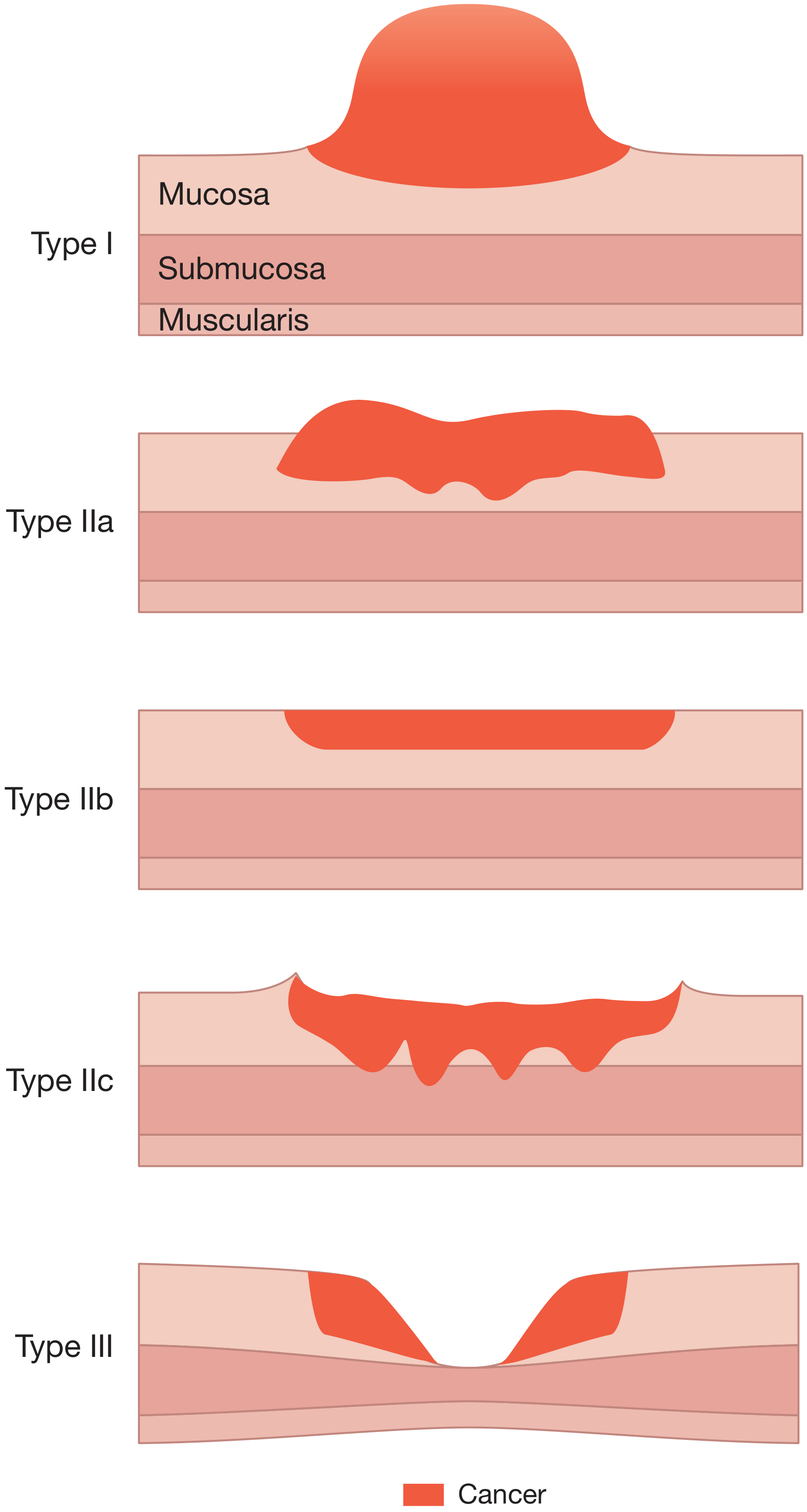
Early gastric cancer (EGC) is defined as cancer limited to the mucosa and submucosa, regardless of lymph node status (T1, any N). 5-year survival ~90%. In Japan, ~1/3 of diagnosed gastric cancers are EGC due to screening. In the West, EGC is rarely diagnosed.
Advanced gastric cancer involves the muscularis propria or beyond (T2+).
Clinical Features
Early gastric cancer: No specific features; indistinguishable from benign dyspepsia. High index of suspicion required.
Advanced gastric cancer:
- Early satiety, epigastric fullness, bloating
- Weight loss (can be profound)
- Dysphagia (proximal tumours)
- Vomiting, gastric outlet obstruction (pyloric tumours)
- Iron-deficiency anaemia (tumour bleeding; ~40% of patients have anaemia at diagnosis)
- Non-metastatic effects: Trousseau's sign (thrombophlebitis), DVT
Signs of metastatic disease (eponymous):
- Virchow's node - left supraclavicular lymphadenopathy
- Sister Mary Joseph node - peri-umbilical nodule (peritoneal spread)
- Krukenberg tumour - ovarian drop metastases
- Blumer's shelf - peritoneal metastases in pouch of Douglas (rectal exam)
- Bailey and Love's 28th Ed.; Current Surgical Therapy 14e
Spread of Disease
- Lymphatic - most common route; regional then para-aortic nodes
- Blood - haematogenous, typically to liver, lungs, bone
- Transcoelomic - peritoneal cavity (peritoneal carcinomatosis in 14-43% synchronously)
- Direct - adjacent structures (pancreas, liver, colon)
Peritoneal carcinomatosis accounts for 35% of synchronous metastases and is associated with a median survival approaching 0% if untreated.
Diagnosis and Staging
Diagnostic Modalities
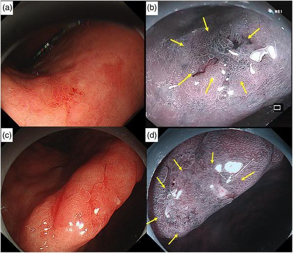
- Upper GI endoscopy - primary diagnostic tool; allows biopsy. Narrow Band Imaging (NBI) and magnified endoscopy improve detection of subtle mucosal changes.
- Endoscopic ultrasound (EUS) - gold standard for local T and N staging
- CT chest/abdomen/pelvis with contrast - systemic staging
- PET/CT - if no M1 disease evident on CT
- Diagnostic laparoscopy with peritoneal washings - all patients with suspected locoregional/advanced disease; positive cytology = M1
Molecular Testing
- HER2/neu - if metastatic disease suspected (targeted therapy eligibility)
- Microsatellite status (MSI/MSS) and PD-L1 expression - for unresectable disease (immunotherapy eligibility)
- CDH1 mutation testing - in young patients, genetic counselling recommended
TNM Staging (AJCC/UICC)
Based on depth of tumour invasion (T), number of involved lymph nodes (N), and presence of distant metastasis (M). Approximately 50% of patients present with advanced disease; of those with locoregional disease, 70-80% have lymph node involvement.
- Current Surgical Therapy 14e; Sabiston Textbook of Surgery
Treatment
Resectable Disease (Curative Intent)
Surgery is the only curative treatment. Radical gastrectomy (total or subtotal) with adequate lymphadenectomy is the procedure of choice. D2 lymphadenectomy (removal of second-tier nodes around principal arterial trunks) is standard in Japan and increasingly adopted in the West.
Perioperative chemotherapy significantly improves survival. Standard regimens:
- FLOT (5-FU, leucovorin, oxaliplatin, docetaxel) - current preferred perioperative regimen in Europe
- FOLFOX or XELOX (capecitabine + oxaliplatin)
- ECF (epirubicin, cisplatin, 5-FU) - older regimen
- Given 3-6 months pre-operatively, then restaging, then continued post-operatively
- Radiation may be added (+/- in perioperative setting)
All patients should be presented to a multidisciplinary tumour board (MDT).
Locally Advanced / Metastatic Disease
- Systemic chemotherapy is the first-line standard of care
- HER2-positive tumours: add trastuzumab (ToGA trial)
- MSI-high / PD-L1 positive: immunotherapy (pembrolizumab, nivolumab)
- A 2025 meta-analysis (PMID 40032744) found that immunotherapy or targeted therapy vs. best supportive care improved progression-free survival and quality of life in advanced gastric cancer
Peritoneal Disease - CRS + HIPEC
- Cytoreductive surgery (CRS) with hyperthermic intraperitoneal chemotherapy (HIPEC) is considered for patients with:
- Limited peritoneal disease (Peritoneal Cancer Index [PCI] <6-10)
- Good response to systemic therapy
- Good performance status, age <70
- No liver/extraperitoneal metastases
- NIPS/BISIC (neoadjuvant intraperitoneal + systemic chemotherapy) can be used to downstage disease before CRS + HIPEC
Endoscopic Treatment
- Endoscopic mucosal resection (EMR) and endoscopic submucosal dissection (ESD) - for select EGC limited to mucosa, well-differentiated, without lymphovascular invasion or lymph node involvement
Screening
- Japan and Korea: National screening with upper endoscopy and barium radiography - explains higher proportion of EGC diagnosed and better survival figures
- Western countries: No universal screening. Selective endoscopy for patients with: gastric polyps, pernicious anaemia, genetic disorders (CDH1 mutation carriers), family history
Prognosis
| Stage | Approximate 5-Year Survival |
|---|---|
| Early (T1) | ~90% |
| Locoregional, resected | 30-50% (West) |
| Advanced/metastatic | <15% |
| Peritoneal carcinomatosis (untreated) | ~0% |
Japan achieves >80% 5-year survival in national databases compared with <50% in the US, partially explained by the higher proportion of EGC detected through screening, differences in molecular subtypes, and surgical expertise.
Key Points Summary
- H. pylori is responsible for ~75% of gastric cancers and is a WHO Group 1 carcinogen
- Lauren classification (intestinal vs. diffuse) and TCGA molecular subtypes guide prognosis and therapy
- The only curative treatment is surgical resection; perioperative FLOT chemotherapy improves survival
- HER2 testing and MSI/PD-L1 testing are mandatory in metastatic disease for targeted therapy and immunotherapy eligibility
- Early diagnosis is key; early gastric cancer has ~90% 5-year survival
- CDH1 mutation carriers (HDGC) should be offered prophylactic total gastrectomy
Sources: Current Surgical Therapy 14e; Bailey and Love's Short Practice of Surgery 28th Ed.; Sabiston Textbook of Surgery; Mulholland & Greenfield's Surgery 7e; Harrison's Principles of Internal Medicine 22E
Recent evidence note: A 2025 systematic review and meta-analysis (PMID 40032744) confirmed that immunotherapy/targeted therapy improves PFS over best supportive care in advanced gastric cancer. A 2024 meta-analysis (PMID 38085005) supports adjuvant/postoperative chemotherapy in locally advanced disease.
This is a shared conversation. Sign in to Orris to start your own chat.