

690f6a59-419e-4c27-a15c-bafad5de871d.jpg
Ans all the questions serially according medical parasitology with reference book the ans should be easy and all answers in English And describe in bangla
Reading File
Now let me search the medical library for Cryptosporidium content comprehensively.
Finding Sources
Finding Sources
Reading File
Reading File
Reading File
Reading File
Reading File
Reading File
Finding Sources
Reading File
Finding Sources
Reading File
Finding Sources
Reading File
Finding Sources
Reading File
Finding Sources
Loading Image
Finding Sources
I now have comprehensive information from the textbooks. Let me compile the complete answers to all 9 questions in English with Bangla descriptions.
Card No. 05 - Opportunistic Intestinal Protozoa: Cryptosporidium & Balantidium coli
Reference: Jawetz, Melnick & Adelberg's Medical Microbiology 28th Ed. | Medical Microbiology 9e | Tietz Textbook of Laboratory Medicine 7th Ed. | Henry's Clinical Diagnosis and Management by Laboratory Methods
Q1. What is the clinical significance of Cryptosporidium parvum in recent years? Why is it so named?
ENGLISH:
Cryptosporidium parvum has gained major clinical significance in recent decades for the following reasons:
- AIDS epidemic: With the rise of HIV/AIDS, C. parvum emerged as a life-threatening opportunistic pathogen. In immunocompromised patients (especially those with CD4+ count <200), it causes severe, profuse, chronic watery diarrhea (5-10 bowel movements/day) that can last months to years and may contribute to death.
- Large waterborne outbreaks: The 1993 Milwaukee outbreak infected over 400,000 people through contaminated municipal water. The organism is resistant to chlorination, making standard water purification ineffective.
- Zoonotic importance: Cattle and other ruminants are major reservoirs for C. parvum, making it a classic zoonotic pathogen.
- Immunocompetent hosts: Even in normal individuals, it causes self-limited watery diarrhea lasting 1-2 weeks with spontaneous resolution.
- Reportable disease in the USA.
Why is it so named?
The name Cryptosporidium comes from Greek:
- "Kryptos" = hidden
- "Sporos" = seed/spore
The name refers to the fact that the oocysts are hidden within the cell membrane (intracellular but extracytoplasmic - they sit just under the brush border of the intestinal epithelium, not deep in the cytoplasm). The species name parvum means "small" - referring to the tiny oocyst size of only 4-6 μm.
বাংলা বর্ণনা:
Cryptosporidium parvum সাম্প্রতিক বছরগুলোতে অত্যন্ত গুরুত্বপূর্ণ পরজীবী হয়ে উঠেছে কারণ:
- AIDS রোগীদের মধ্যে: যাদের রোগ প্রতিরোধ ক্ষমতা কম (বিশেষত CD4+ <200), তাদের মধ্যে এটি তীব্র দীর্ঘস্থায়ী পানির মতো ডায়রিয়া তৈরি করে, যা মাসের পর মাস চলতে পারে এবং মৃত্যুর কারণ হতে পারে।
- ১৯৯৩ সালে Milwaukee-এ বিশাল প্রাদুর্ভাব: ৪ লাখেরও বেশি মানুষ দূষিত পানি পান করে আক্রান্ত হয়েছিল। এই জীবাণু ক্লোরিন দিয়ে মারা যায় না।
- পশু থেকে মানুষে সংক্রমণ (zoonosis): গরু ও অন্যান্য গবাদিপশু হলো প্রধান আধার।
নামকরণ: Kryptos (গ্রিক) = লুকানো, Sporos = বীজ/স্পোর। কারণ এটি অন্ত্রের কোষের আবরণের নিচে লুকিয়ে থাকে। Parvum মানে "ছোট" - কারণ এর oocyst মাত্র ৪-৬ μm।
Q2. Describe the peculiarities of morphology and life cycle of Cryptosporidium parvum
ENGLISH:
Peculiarities of Morphology:
- Oocyst size: Very small - only 4-6 μm in diameter (smallest coccidian oocyst)
- Intracellular but extracytoplasmic location: Unique among parasites - it lives inside the intestinal epithelial cell but outside the cytoplasm, just beneath the cell membrane at the brush border (apical portion of enterocytes). This is a key distinguishing feature.
- Two types of oocysts:
- Thick-walled oocysts (80%): Passed in stool, survive in environment, are infectious
- Thin-walled oocysts (20%): Remain inside the host and cause autoinfection (re-infect host cells)
- Sporulated oocysts: Each oocyst contains 4 sporozoites (no sporocysts - unlike other coccidia)
- Staining: Stain pink/red with modified acid-fast stain (Ziehl-Neelsen); also stained with auramine-rhodamine fluorescence
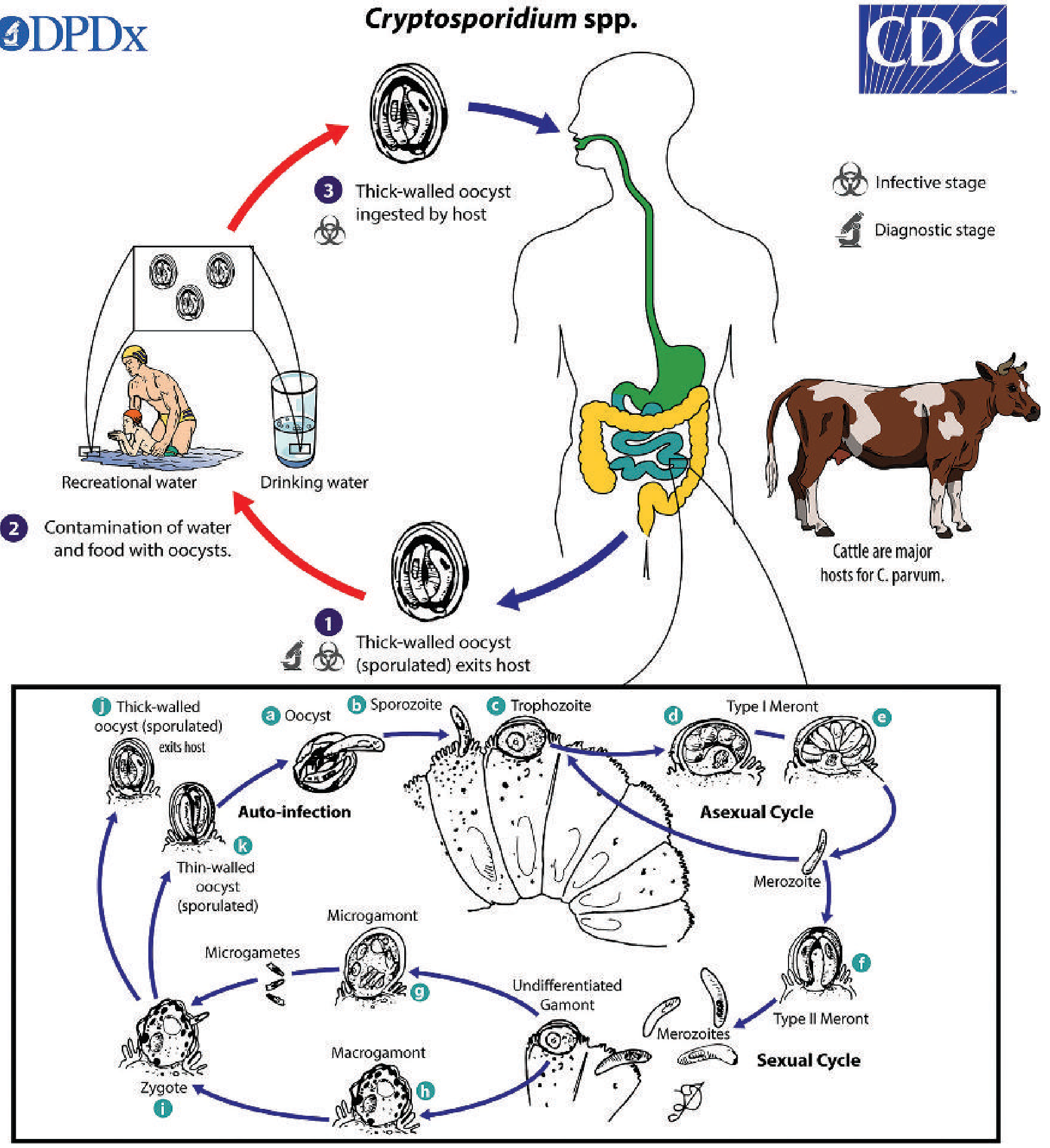
Peculiarities of Life Cycle (CDC diagram above):
The life cycle is monoxenous (single host) and involves both asexual and sexual cycles:
Steps:
- Ingestion: Thick-walled sporulated oocysts ingested via contaminated food/water (infective dose = as few as 30 oocysts)
- Excystation: In small intestine, 4 sporozoites excyst from the oocyst
- Attachment: Sporozoites attach to brush border of intestinal epithelial cells (intracellular but extracytoplasmic)
- Trophozoite formation: Sporozoites develop into trophozoites
- Asexual cycle (Merogony):
- Type I meronts: produce 8 merozoites → infect new cells
- Type II meronts: produce 4 merozoites → enter sexual cycle
- Sexual cycle (Gametogony):
- Type II merozoites → Undifferentiated gamonts → Microgamonts (male) and Macrogamonts (female)
- Fusion of microgametes + macrogamete → Zygote
- Sporogony: Zygote → Thick-walled oocysts (shed in stool) OR Thin-walled oocysts (autoinfection within same host)
Key peculiarity: The thin-walled oocyst causes autoinfection within the same host - this explains the severe, fulminating infections in immunocompromised patients who cannot clear the parasite.
বাংলা বর্ণনা:
গঠনগত বিশেষত্ব:
- Oocyst মাত্র ৪-৬ μm - সবচেয়ে ছোট coccidian oocyst
- অনন্য অবস্থান: অন্ত্রের কোষের ভেতরে কিন্তু cytoplasm-এর বাইরে (intracellular extracytoplasmic) - brush border-এ থাকে
- দুই ধরনের oocyst:
- মোটা দেওয়ালের (৮০%): মলে বের হয়, পরিবেশে টিকে থাকে
- পাতলা দেওয়ালের (২০%): হোস্টের ভেতরে থেকে autoinfection তৈরি করে
- প্রতিটি oocyst-এ ৪টি sporozoite থাকে (কোনো sporocyst নেই)
জীবনচক্রের বিশেষত্ব:
এটি একটিমাত্র হোস্টে (monoxenous) সম্পূর্ণ জীবনচক্র সম্পন্ন করে - যৌন ও অযৌন উভয় পদ্ধতিতে। পাতলা-দেওয়াল oocyst দিয়ে একই হোস্টের মধ্যে বারবার সংক্রমণ হয় (autoinfection), যার কারণে রোগ প্রতিরোধ ক্ষমতাহীন রোগীতে রোগ অত্যন্ত মারাত্মক হয়।
Q3. Write about the pathogenesis of Cryptosporidiosis
ENGLISH:
-
Entry & attachment: After ingestion, sporozoites excyst and attach to the brush border (apical membrane) of small intestinal epithelial cells - particularly the jejunum and ileum. They are intracellular but extracytoplasmic.
-
Villous injury: The parasite causes villous blunting, crypt hyperplasia, and mucosal inflammation. This leads to:
- Malabsorption
- Decreased surface area for absorption
-
Secretory diarrhea: The primary mechanism is secretory diarrhea (not invasive) - the organisms trigger secretion of chloride and water into the intestinal lumen.
-
In immunocompetent hosts: Mild, self-limited watery diarrhea lasting 1-2 weeks. The immune system (especially T-cell mediated immunity) clears the infection.
-
In immunocompromised hosts (AIDS, transplant, etc.):
- Massive, profuse watery diarrhea (up to 15-20 L/day in severe AIDS)
- The thin-walled oocysts cause autoinfection, perpetuating the cycle
- Parasite can spread beyond the intestine to: gallbladder (biliary cryptosporidiosis - causing cholangitis), pancreas, respiratory tract (lungs)
- Can cause cholangiopathy - thickening of bile duct walls
-
Mechanism of immune evasion: The extracytoplasmic location makes it resistant to many intracellular killing mechanisms.
বাংলা বর্ণনা:
১. Sporozoite ক্ষুদ্রান্ত্রের (জেজুনাম ও ইলিয়াম) epithelial কোষের brush border-এ আটকে যায়।
২. এটি villus blunting ও প্রদাহ তৈরি করে → malabsorption ও secretory diarrhea।
৩. রোগ প্রতিরোধ ক্ষমতাসম্পন্ন ব্যক্তিতে: ১-২ সপ্তাহের হালকা পানিযুক্ত ডায়রিয়া, নিজে নিজে ভালো হয়।
৪. AIDS/ইমিউনোকম্প্রোমাইজড রোগীতে: ব্যাপক পানির মতো ডায়রিয়া, autoinfection-এর কারণে থামে না; পিত্তথলি, অগ্ন্যাশয় ও ফুসফুসে ছড়াতে পারে।
Q4. What are opportunistic intestinal pathogens? Why are they so called?
ENGLISH:
Definition: Opportunistic pathogens are organisms that are normally harmless (or cause only mild, self-limited illness) in healthy immunocompetent individuals but cause severe, life-threatening disease in persons with impaired immunity.
Why "opportunistic"? - They "take the opportunity" of a weakened immune system to cause disease. When host defenses are compromised, these organisms multiply uncontrolled.
Opportunistic intestinal protozoa include:
| Organism | Disease |
|---|---|
| Cryptosporidium parvum/hominis | Cryptosporidiosis |
| Cystoisospora (Isospora) belli | Isosporiasis |
| Cyclospora cayetanensis | Cyclosporiasis |
| Microsporidia spp. | Microsporidiosis |
Common features making them "opportunistic":
- In immunocompetent hosts: mild or subclinical infection, self-limited, resolves in 1-3 weeks
- In immunocompromised hosts (HIV/AIDS, organ transplants, primary immunodeficiency, cancer chemotherapy):
- Chronic, prolonged, severe watery diarrhea (lasting months to years)
- Malabsorption and wasting
- Spread beyond intestine (extraintestinal disease)
- Can be life-threatening
- All are intracellular pathogens that replicate inside enterocytes
- All undergo autoinfection via thin-walled oocysts (especially Cryptosporidium)
- For Cryptosporidium - sporulation occurs inside the host (endogenous sporulation), enabling autoinfection. In Cyclospora and Isospora, sporulation occurs outside the host.
Why they are specifically "opportunistic intestinal pathogens": Because the intestinal immune surveillance (IgA, T-cells, cytokines) is the primary defense against them. When this is compromised (especially CD4+ T cells in AIDS), these parasites escape control.
বাংলা বর্ণনা:
সংজ্ঞা: Opportunistic pathogens হলো এমন জীবাণু যারা সুস্থ মানুষে সামান্য বা কোনো ক্ষতি করে না, কিন্তু যখন শরীরের রোগ প্রতিরোধ ক্ষমতা কমে যায় (যেমন AIDS, organ transplant, ক্যান্সারের চিকিৎসা), তখন এরা মারাত্মক রোগ তৈরি করে।
"Opportunistic" নামের কারণ: এরা দুর্বল ইমিউন সিস্টেমের "সুযোগ" নিয়ে রোগ তৈরি করে।
প্রধান opportunistic intestinal protozoa: Cryptosporidium, Isospora/Cystoisospora, Cyclospora, Microsporidia।
বৈশিষ্ট্য: এরা সবাই intracellular পরজীবী। সুস্থ মানুষে সল্পমেয়াদী ডায়রিয়া, কিন্তু AIDS রোগীতে মাসের পর মাস ব্যাপক ডায়রিয়া হয় যা মৃত্যুর কারণ হতে পারে।
Q5. What is the clinical significance of Cryptosporidium? How can cryptosporidiasis be diagnosed in the laboratory?
ENGLISH:
Clinical Significance:
- Immunocompetent individuals: Self-limited watery diarrhea, 5-10 bowel movements/day, lasting 9-23 days. Symptoms: malaise, fever, anorexia, abdominal cramps. Resolves spontaneously.
- Immunocompromised individuals:
- Profound, potentially life-threatening profuse watery diarrhea
- Can persist for months to years
- Biliary cryptosporidiosis (cholangitis, cholangiopathy)
- Respiratory cryptosporidiosis (pneumonia)
- Associated with high mortality in AIDS patients
- Public health: Major cause of waterborne outbreaks (Milwaukee 1993: 400,000+)
- Children: Common cause of acute diarrhea in day-care settings
Laboratory Diagnosis:
1. Microscopy (Gold standard):
- Modified acid-fast stain (Ziehl-Neelsen or Kinyoun): Oocysts stain pink/red on blue background; 4-6 μm diameter. This is the most commonly used method.
- Modified safranin stain: More uniform staining
- Auramine-rhodamine fluorescence stain: Higher sensitivity but lower specificity
- Concentration methods help: Formalin-ethyl acetate sedimentation or Sheather's sugar flotation
2. Antigen Detection (Immunological):
- Direct Fluorescent Antibody (DFA) test: Highly sensitive, uses monoclonal antibodies; can simultaneously detect Cryptosporidium and Giardia
- ELISA / EIA (Enzyme Immunoassay): Detect fecal antigen; commercially available kits; greater sensitivity than microscopy
- Rapid immunochromatographic tests: Lateral flow assays for quick diagnosis
3. Molecular Methods (NAATs):
- PCR / Multiplex GI PCR panels: Most sensitive method; FDA-approved assays available; can differentiate species; becoming widely used
- Can detect infection even at very low parasite loads
4. Histology:
- Intestinal biopsy: H&E stain shows organisms at the apical brush border of enterocytes
- Used mainly for research or when stool tests negative
Note: Culture and serology are NOT clinically useful for routine diagnosis.
বাংলা বর্ণনা:
ক্লিনিক্যাল গুরুত্ব:
- সুস্থ মানুষে: ৯-২৩ দিনের self-limited পানিযুক্ত ডায়রিয়া
- AIDS/ইমিউনোকম্প্রোমাইজড রোগীতে: দীর্ঘস্থায়ী মারাত্মক ডায়রিয়া, পিত্তনালী ও ফুসফুসেও ছড়ায়
ল্যাবরেটরি নির্ণয়:
- Modified acid-fast stain: মলে oocyst গোলাপি/লাল রঙের দেখা যায় (৪-৬ μm)
- DFA test: Monoclonal antibody দিয়ে fluorescence microscopy - অত্যন্ত সংবেদনশীল
- ELISA/EIA: মলে antigen সনাক্ত করে - মাইক্রোস্কোপির চেয়ে বেশি সংবেদনশীল
- PCR/Multiplex GI panels: সবচেয়ে সংবেদনশীল পদ্ধতি
- Intestinal biopsy (H&E): Brush border-এ জীবাণু দেখা যায়
Q6. What are the salient features of the life cycle of Cryptosporidium?
ENGLISH:
The life cycle diagram (CDC DPDx) is shown above. Key salient features:
I. Type of life cycle: Monoxenous (single host only - no intermediate host required)
II. Infective form: Thick-walled sporulated oocyst (4-6 μm, contains 4 sporozoites)
III. Route of transmission: Fecal-oral (contaminated water/food, person-to-person, zoonotic)
IV. Asexual Cycle (Merogony):
- Oocyst ingested → 4 sporozoites excyst → attach to brush border of small intestinal enterocytes
- Form trophozoites → Type I meronts (contain 8 merozoites) → Type II meronts (contain 4 merozoites)
- Type I merozoites reinvade cells → continue asexual replication
- Type II merozoites → enter sexual cycle
V. Sexual Cycle (Gametogony):
- Undifferentiated gamonts → Microgamont (male) → microgametes
- Macrogamont (female) → macrogamete
- Fertilization → Zygote
VI. Sporogony:
- Zygote → either:
- Thick-walled oocyst (80%): sporulates → exits in feces → contaminates environment → infects new host
- Thin-walled oocyst (20%): sporulates within host → excysts within host → autoinfection (reinvades same host's intestinal cells)
VII. Key salient peculiarities:
- Autoinfection: Thin-walled oocysts cause continuous reinfection within the same host - explains fulminating disease in immunocompromised
- Extracytoplasmic location: Lives within the cell but outside the cytoplasm (intracellular extracytoplasmic) - unique among coccidia
- Endogenous sporulation: Sporulation occurs inside the host (unlike Cyclospora and Isospora where it occurs outside)
- No tissue phase: Remains confined to the intestinal epithelium (unless immunocompromised)
- Low infectious dose: As few as 30 oocysts can cause infection
বাংলা বর্ণনা:
জীবনচক্রের মূল বৈশিষ্ট্য:
১. একটি হোস্টেই সম্পূর্ণ (monoxenous) - মানুষ বা গরুর মধ্যেই সব ধাপ সম্পন্ন
২. সংক্রামক রূপ: Thick-walled sporulated oocyst (৪-৬ μm, ৪টি sporozoite সহ)
৩. অযৌন চক্র: Oocyst → Sporozoites → Trophozoite → Type I meront (৮ merozoite) → Type II meront (৪ merozoite)
৪. যৌন চক্র: Type II merozoite → Microgamont (পুরুষ) + Macrogamont (স্ত্রী) → Zygote
৫. দুই ধরনের oocyst তৈরি:
- মোটা দেওয়াল (৮০%): মলে বের হয়
- পাতলা দেওয়াল (২০%): হোস্টের মধ্যেই আবার সংক্রমণ (autoinfection) ৬. Sporulation হোস্টের ভেতরেই হয় - এটি Cyclospora/Isospora থেকে আলাদা ৭. মাত্র ৩০টি oocyst দিয়েই সংক্রমণ ঘটতে পারে
Q7. What is the clinical significance of Balantidium coli? What are the peculiarities of its life cycle and structure?
ENGLISH:
Clinical Significance:
- Balantidium coli (now also called Balantioides coli) is the only ciliated protozoan known to infect humans
- It is the largest protozoan parasite of humans (trophozoite: 40-100 μm)
- Disease: Causes balantidiasis (balantidiosis) - a dysentery syndrome resembling amebiasis
- Clinical spectrum:
- Most infections: asymptomatic
- Symptomatic: watery diarrhea → bloody mucus-laden diarrhea → colitis
- Can invade colonic mucosa (like E. histolytica), causing colonic ulcerations
- Rare complications: colonic perforation, peritonitis, appendicitis
- Extraintestinal spread to lung and liver (very rare)
- Can be misdiagnosed as ulcerative colitis or amebic dysentery
- At-risk groups: Malnourished, achlorhydric, immunocompromised, those in close contact with pigs
- Treatment: Tetracycline (drug of choice); alternatives: metronidazole, iodoquinol, nitazoxanide
Peculiarities of Structure:
Trophozoite:
- Very large (40-100 μm)
- Covered with cilia (used for locomotion)
- Macronucleus: Kidney-bean shaped (prominent, large)
- Micronucleus: Small, adjacent to macronucleus
- Contractile vacuoles and food vacuoles in cytoplasm
- Characteristic rotary (boring) motility on wet mount
Cyst:
- Round to oval, 50-70 μm
- Single kidney-bean shaped macronucleus
- Cyst wall present; ciliary activity may be seen
- Infective stage
Peculiarities of Life Cycle:
- Two-form life cycle: Trophozoite (active form) + Cyst (infective/resistant form)
- Similar to E. histolytica life cycle
- Reservoir host: Pigs (primary); also other primates
- Transmission: Fecal-oral - ingestion of cysts in contaminated food/water
- After ingestion, cysts excyst in the small intestine → release trophozoites → colonize the colon
- Trophozoites normally live in colon without causing disease
- Can invade bowel wall (hyaluronidase activity helps invasion) → flask-shaped ulcers (like E. histolytica)
- Trophozoites form cysts → passed in feces → survive in environment for months
- Person-to-person transmission rare; waterborne outbreaks reported
- Cysts are resistant to chlorination
বাংলা বর্ণনা:
ক্লিনিক্যাল গুরুত্ব:
- এটি মানুষকে সংক্রমণকারী একমাত্র ciliated protozoan এবং সবচেয়ে বড় protozoan পরজীবী
- Balantidiasis রোগ তৈরি করে - এটি দেখতে অনেকটা আমাশয়ের মতো
- বেশিরভাগ ক্ষেত্রে উপসর্গহীন; কখনো কখনো রক্ত-মিশ্রিত ডায়রিয়া, কোলনের আলসার
- শূকরের সংস্পর্শ প্রধান ঝুঁকি
গঠনগত বিশেষত্ব:
- Trophozoite: ৪০-১০০ μm, cilia-আবৃত, kidney-bean আকৃতির macronucleus
- Cyst: ৫০-৭০ μm, গোলাকার, একটি macronucleus
জীবনচক্রের বিশেষত্ব:
- শূকরের মল থেকে cyst খাবার/পানির মাধ্যমে মানুষের পেটে যায় → ক্ষুদ্রান্ত্রে excystation → trophozoite বড় অন্ত্রে বসবাস করে → আলসার তৈরি করতে পারে → cyst মলে বের হয়
Q8. "Cryptosporidium parvum produces diarrhea only in immunocompromised hosts" - Explain
ENGLISH:
This statement is partially correct but not entirely accurate. Here is the full explanation:
The statement needs clarification:
C. parvum can cause diarrhea in BOTH immunocompetent AND immunocompromised individuals. However, the severity, duration, and outcome differ dramatically:
| Feature | Immunocompetent Host | Immunocompromised Host |
|---|---|---|
| Diarrhea | Mild, watery, self-limited | Severe, profuse (up to 15-20 L/day) |
| Duration | 1-2 weeks (9-23 days) | Months to years (chronic) |
| Outcome | Full recovery | Can be life-threatening |
| Autoinfection | Controlled by immunity | Uncontrolled - perpetuates disease |
| Extra-intestinal spread | Does not occur | Biliary, pancreatic, pulmonary |
| Treatment needed | Usually none (supportive) | No effective drug; HAART for HIV |
Why immunocompromised hosts are so severely affected:
- T-cell mediated immunity is the primary defense against Cryptosporidium
- In AIDS (CD4+ < 200/μL), the T-cell response is absent → parasite replicates freely
- Thin-walled oocysts cause autoinfection → massive amplification without immune control
- Low infectious dose (30 oocysts) + autoinfection = fulminating, lethal disease
- No effective anti-cryptosporidial drug works well in immunocompromised patients; only immune reconstitution (HAART) is effective
Historical context: Cryptosporidium was considered merely a veterinary pathogen before the AIDS era. The HIV epidemic revealed its devastating potential in humans, making it a WHO-recognized important pathogen.
বাংলা বর্ণনা:
এই statement-টি আংশিকভাবে সঠিক। সত্যিকার অর্থে:
- সুস্থ মানুষেও C. parvum ডায়রিয়া করে, কিন্তু এটি হালকা এবং ১-২ সপ্তাহে নিজে নিজে ভালো হয়
- রোগ প্রতিরোধ ক্ষমতাহীন রোগীতে (AIDS, transplant): অত্যন্ত মারাত্মক, দীর্ঘস্থায়ী ডায়রিয়া, মাস-বছর ধরে চলে
- কারণ: T-cell immunity না থাকলে thin-walled oocyst দিয়ে autoinfection অনিয়ন্ত্রিতভাবে চলতে থাকে
- কোনো কার্যকর ওষুধ নেই; শুধু HAART দিয়ে ইমিউন সিস্টেম ফিরিয়ে আনলে রোগ সারে
Q9. "Immunological tests now play an important role in diagnosis of parasitic diseases" - Comment
ENGLISH:
This statement is absolutely true and immunological/serological methods have revolutionized parasitology diagnosis. Here is a detailed commentary:
Immunological Tests Used in Parasitic Diagnosis:
A. Antigen Detection Tests (Detect parasite antigens in body fluids):
- ELISA (Enzyme-Linked Immunosorbent Assay):
- For Cryptosporidium in stool - highly sensitive (>90%)
- For Giardia (fecal antigen EIA)
- For Toxoplasma, Leishmania, Plasmodium, Entamoeba
- Direct Fluorescent Antibody (DFA) test:
- For Cryptosporidium and Giardia simultaneously in stool
- Monoclonal antibody-based; higher sensitivity than microscopy
- Rapid Immunochromatographic (lateral flow) tests:
- Point-of-care tests for Malaria (RDT - Rapid Diagnostic Test)
- For Cryptosporidium and Giardia
- Immunofluorescence assay (IFA): For Toxoplasma, Leishmania
B. Antibody Detection Tests (Detect host antibodies against parasite):
- ELISA for IgM/IgG - useful for: Toxoplasma (congenital), Entamoeba, Echinococcus (hydatid), Filaria, Schistosoma, Leishmania
- Indirect Hemagglutination (IHA) - for amoebiasis
- Complement Fixation Test
C. Molecular-Immunological Hybrid Tests:
- Multiplex PCR/GI panels that combine with antigen detection
Why Immunological Tests are Superior:
| Advantage | Details |
|---|---|
| Higher sensitivity | Detects even small amounts of antigen/antibody |
| Standardization | Commercial kits with QC |
| Speed | Results in hours (vs. days for culture) |
| Non-invasive | Stool/blood/serum only |
| Can diagnose active + past infection | Antigen = active; IgM = recent; IgG = past |
| Automated | High throughput in labs |
Limitations:
- Cannot differentiate species in some cases
- Cross-reactivity can give false positives
- Antibody tests may remain positive long after cure
- Not available in resource-limited settings
Role in Cryptosporidiosis specifically:
- DFA and EIA tests have largely replaced or supplemented modified acid-fast staining
- Much greater sensitivity (detects low oocyst loads)
- Many labs now use antigen detection as their primary method for Cryptosporidium and Giardia
বাংলা বর্ণনা:
এই statement সম্পূর্ণ সত্য। Immunological tests পরজীবী রোগ নির্ণয়ে এখন অপরিহার্য ভূমিকা পালন করছে:
Antigen detection (পরজীবীর antigen সনাক্ত):
- ELISA: মলে Cryptosporidium ও Giardia antigen সনাক্তে উচ্চ সংবেদনশীলতা
- DFA test: Monoclonal antibody দিয়ে fluorescence microscopy - Cryptosporidium ও Giardia উভয়ই একসাথে
- Rapid test (RDT): Malaria-র দ্রুত নির্ণয়ে
Antibody detection (রোগীর শরীরে তৈরি antibody সনাক্ত):
- ELISA, IFA: Toxoplasma, Leishmania, Echinococcus ইত্যাদির জন্য
- IgM = সাম্প্রতিক সংক্রমণ; IgG = পুরনো সংক্রমণ
সুবিধা: দ্রুত, সংবেদনশীল, standardized, non-invasive
সীমাবদ্ধতা: ক্রস-রিঅ্যাকটিভিটি, সংক্রমণ সেরে যাওয়ার পরেও antibody থাকতে পারে
Life Cycle Diagram of Cryptosporidium (CDC DPDx):
Sources: Jawetz, Melnick & Adelberg's Medical Microbiology 28th Ed. | Murray's Medical Microbiology 9e | Tietz Textbook of Laboratory Medicine 7th Ed. | Henry's Clinical Diagnosis and Management by Laboratory Methods | Red Book 2021 (Committee on Infectious Diseases) | Yamada's Textbook of Gastroenterology 7th Ed.
Can you make this ans more easy & short Proti questions shes a Banglay bhujay dio
Card No. 05 — Opportunistic Intestinal Protozoa
Cryptosporidium parvum & Balantidium coli
Q1. Clinical significance of C. parvum in recent years? Why is it so named?
Answer (Easy & Short):
C. parvum became very important because:
- In AIDS patients → causes severe, life-threatening chronic diarrhea
- 1993 Milwaukee outbreak → 4 lakh people infected via contaminated water
- Resistant to chlorine → normal water purification fails
- In healthy people → mild diarrhea, gets better on its own
Why named so?
- Kryptos (Greek) = Hidden → lives hidden inside cell membrane (intracellular but extracytoplasmic)
- Parvum = Small → tiny oocyst (only 4–6 μm)
🇧🇩 বাংলায় বোঝো: C. parvum সাম্প্রতিক বছরে খুব গুরুত্বপূর্ণ হয়েছে কারণ —
- AIDS রোগীতে মারাত্মক দীর্ঘস্থায়ী ডায়রিয়া করে
- ১৯৯৩ সালে Milwaukee-এ দূষিত পানিতে ৪ লাখ মানুষ আক্রান্ত হয়
- ক্লোরিন দিয়ে মারা যায় না
- সুস্থ মানুষে হালকা ডায়রিয়া, নিজে নিজে ভালো হয়
নামের কারণ:
- Kryptos = লুকানো → কোষের ভেতরে লুকিয়ে থাকে কিন্তু cytoplasm-এর বাইরে
- Parvum = ছোট → oocyst মাত্র ৪–৬ μm
Q2. Peculiarities of morphology and life cycle of C. parvum
Answer:
Morphology (গঠন):
- Oocyst = 4–6 μm (very small)
- Location = Intracellular but extracytoplasmic (inside cell, but outside cytoplasm — sits on brush border) — unique!
- 2 types of oocysts:
- Thick-walled (80%) → passed in stool → infects others
- Thin-walled (20%) → stays inside host → causes autoinfection
- Each oocyst has 4 sporozoites (no sporocysts)
- Stains pink with modified acid-fast stain
Life Cycle (জীবনচক্র):
- Drink contaminated water → thick-walled oocyst ingested
- 4 sporozoites come out → attach to brush border of small intestine
- Asexual cycle → Type I meront (8 merozoites) → Type II meront (4 merozoites)
- Sexual cycle → Microgamont + Macrogamont → Zygote
- Zygote → Thick-walled oocyst (exits in stool) OR Thin-walled oocyst (autoinfection)
Key special features:
- ✅ Single host (monoxenous) — no intermediate host
- ✅ Autoinfection by thin-walled oocysts
- ✅ Sporulation inside host (unlike Cyclospora/Isospora)
- ✅ Only 30 oocysts needed to cause infection!
🇧🇩 বাংলায় বোঝো:গঠন:
- Oocyst মাত্র ৪–৬ μm — খুব ছোট
- কোষের ভেতরে থাকে কিন্তু cytoplasm-এর বাইরে (brush border-এ) — এটি অনন্য বৈশিষ্ট্য
- মোটা দেওয়ালের oocyst → মলে বের হয়; পাতলা দেওয়ালের oocyst → ভেতরেই আবার সংক্রমণ করে
- Modified acid-fast stain-এ গোলাপি দেখায়
জীবনচক্র: ১. দূষিত পানি/খাবার থেকে oocyst পেটে যায় ২. ৪টি sporozoite বের হয় → ক্ষুদ্রান্ত্রের brush border-এ আটকায় ৩. অযৌন চক্র → meront → merozoite তৈরি ৪. যৌন চক্র → Microgamont + Macrogamont → Zygote ৫. Zygote → মোটা-দেওয়াল oocyst (মলে বের হয়) অথবা পাতলা-দেওয়াল oocyst (ভেতরেই আবার সংক্রমণ)বিশেষ বৈশিষ্ট্য: মাত্র ৩০টি oocyst দিয়ে সংক্রমণ হয়; একই হোস্টে বারবার autoinfection হয়
Q3. Pathogenesis of Cryptosporidiosis
Answer:
- Sporozoites attach to brush border of jejunum/ileum (intracellular, extracytoplasmic)
- Causes villous blunting (villi ছোট হয়) → malabsorption
- Triggers secretory diarrhea (watery, non-bloody)
- In healthy people → immune system (T-cells) clears it in 1–2 weeks
- In AIDS/immunocompromised →
- T-cells absent → parasite multiplies freely
- Thin-walled oocysts cause uncontrolled autoinfection
- Spreads to biliary tract (cholangitis), pancreas, lungs
- Can cause death
🇧🇩 বাংলায় বোঝো: ১. Sporozoite ক্ষুদ্রান্ত্রের কোষের brush border-এ আটকায় ২. Villi ছোট হয়ে যায় → খাবার ঠিকমতো শোষণ হয় না ৩. পানির মতো ডায়রিয়া তৈরি হয় (secretory diarrhea) ৪. সুস্থ মানুষে T-cell দিয়ে ১–২ সপ্তাহে সেরে যায় ৫. AIDS রোগীতে — T-cell নেই → পরজীবী বাড়তে থাকে, থামে না → পিত্তনালী, ফুসফুসেও ছড়ায় → মৃত্যু হতে পারে
Q4. What are opportunistic intestinal pathogens? Why are they so called?
Answer:
Definition: Organisms that cause mild or no disease in healthy people but cause severe, life-threatening disease in immunocompromised patients (AIDS, transplant, cancer chemo).
Why "opportunistic"?
They take the "opportunity" of a weak immune system to cause disease.
Main opportunistic intestinal protozoa:
| Organism | Disease |
|---|---|
| Cryptosporidium | Watery diarrhea |
| Cystoisospora belli | Isosporiasis |
| Cyclospora cayetanensis | Cyclosporiasis |
| Microsporidia | Microsporidiosis |
Common features:
- All are intracellular parasites
- In healthy → short, self-limited diarrhea
- In immunocompromised → chronic, severe, potentially fatal diarrhea + wasting
🇧🇩 বাংলায় বোঝো: Opportunistic pathogens হলো এমন জীবাণু যারা সুস্থ মানুষে তেমন ক্ষতি করে না, কিন্তু দুর্বল ইমিউন সিস্টেমের সুযোগ নিয়ে AIDS/transplant রোগীতে মারাত্মক রোগ তৈরি করে।এদের "opportunistic" বলা হয় কারণ এরা সুযোগবাদী — শুধু দুর্বল শরীরেই আক্রমণ করে।প্রধান উদাহরণ: Cryptosporidium, Isospora, Cyclospora, Microsporidia এরা সবাই intracellular পরজীবী — সুস্থে হালকা, অসুস্থে মারাত্মক ডায়রিয়া করে।
Q5. Clinical significance of Cryptosporidium + Laboratory diagnosis of Cryptosporidiasis
Answer:
Clinical Significance:
| Healthy person | Immunocompromised (AIDS) |
|---|---|
| Watery diarrhea, self-limited | Severe, profuse, chronic diarrhea |
| Lasts 1–2 weeks | Lasts months to years |
| No spread | Spreads to bile duct, lungs |
| Recovers fully | Can be fatal |
Also: major cause of waterborne outbreaks (Milwaukee 1993)
Lab Diagnosis:
1. Microscopy (most common):
- Modified acid-fast stain → oocysts stain pink/red, 4–6 μm
- Concentration of stool needed first (Sheather's sugar flotation or formalin-ethyl acetate)
2. Antigen Detection (immunological, more sensitive):
- DFA (Direct Fluorescent Antibody) → monoclonal antibody + fluorescence; detects Cryptosporidium AND Giardia together
- ELISA/EIA → detects fecal antigen; more sensitive than microscopy
3. Molecular (most sensitive):
- PCR / Multiplex GI panel → FDA-approved; detects even tiny amounts
4. Biopsy (H&E stain):
- Shows organisms at apical brush border of enterocytes
Note: Culture & serology = NOT useful
🇧🇩 বাংলায় বোঝো:ক্লিনিক্যাল গুরুত্ব:
- সুস্থ মানুষে: ১–২ সপ্তাহের পানিযুক্ত ডায়রিয়া, নিজে ভালো হয়
- AIDS রোগীতে: মাসের পর মাস ব্যাপক ডায়রিয়া, পিত্তনালী ও ফুসফুসেও ছড়ায়, মৃত্যু হতে পারে
ল্যাব পরীক্ষা: ১. Modified acid-fast stain → মলে গোলাপি oocyst দেখা যায় (সবচেয়ে সহজ পদ্ধতি) ২. DFA test → fluorescent antibody দিয়ে oocyst সনাক্ত — বেশি নির্ভরযোগ্য ৩. ELISA/EIA → মলে antigen ধরে — microscopy-র চেয়ে বেশি sensitive ৪. PCR → সবচেয়ে সংবেদনশীল পদ্ধতি ৫. Biopsy → brush border-এ জীবাণু দেখা যায়
Q6. Salient features of life cycle of Cryptosporidium
Answer (points only):
- Single host (monoxenous) — no intermediate host
- Infective form = thick-walled sporulated oocyst (4–6 μm, 4 sporozoites)
- Transmission = fecal-oral (water/food/zoonotic)
- Asexual cycle (Merogony): Sporozoite → Trophozoite → Type I meront (8 merozoites) → Type II meront (4 merozoites)
- Sexual cycle (Gametogony): Merozoite → Microgamont + Macrogamont → Zygote
- Sporogony: Zygote → 2 types:
- Thick-walled oocyst → exits in stool (infects new host)
- Thin-walled oocyst → autoinfection (reinfects same host)
- Sporulation occurs inside host (endogenous) — unlike Cyclospora/Isospora
- Location = intracellular extracytoplasmic (brush border)
- Minimum infective dose = only 30 oocysts
- Resistant to chlorination
🇧🇩 বাংলায় বোঝো: ১. একটি মাত্র হোস্টে সম্পূর্ণ জীবনচক্র (মানুষ বা গরু) ২. দূষিত পানি/খাবার থেকে oocyst পেটে ঢোকে ৩. অযৌন চক্র → meront তৈরি → merozoite বের হয় ৪. যৌন চক্র → microgamont + macrogamont → zygote তৈরি ৫. Zygote থেকে দুই ধরনের oocyst:
- মোটা-দেওয়াল → মলে বের হয় → নতুন হোস্টকে সংক্রমণ করে
- পাতলা-দেওয়াল → ভেতরেই ভেঙে → আবার সংক্রমণ (autoinfection) ৬. মাত্র ৩০টি oocyst দিয়ে রোগ হয় ৭. Cyclospora ও Isospora-এর মতো নয় — এর sporulation হোস্টের ভেতরেই হয়
Q7. Clinical significance of Balantidium coli + Peculiarities of life cycle and structure
Answer:
Clinical Significance:
- Only ciliated protozoan to infect humans
- Largest protozoan parasite of humans
- Causes Balantidiasis (dysentery-like illness)
- Most cases = asymptomatic
- Symptomatic = watery/bloody diarrhea, colonic ulcers
- Rare = perforation, peritonitis, appendicitis
- Can spread to lung & liver (rare)
- Can be confused with amoebic dysentery or ulcerative colitis
- Treatment: Tetracycline (drug of choice); alternatives: metronidazole, iodoquinol
Structure:
Trophozoite:
- Large (40–100 μm)
- Body covered with cilia (for movement)
- Kidney-bean shaped macronucleus (large, prominent)
- Small micronucleus
- Food vacuoles + contractile vacuoles in cytoplasm
- Shows rotary (boring/spinning) motility on wet mount
Cyst:
- Round/oval, 50–70 μm
- Single kidney-bean shaped macronucleus
- Cyst wall present
- Infective stage
Life Cycle:
- Pig (main reservoir) passes cysts in feces
- Human ingests cysts via contaminated food/water
- Cyst excysts in small intestine → trophozoite released
- Trophozoite lives in colon (usually harmless)
- If invasion occurs → ulcers in colon wall (flask-shaped, like amoeba)
- Trophozoite → cyst → passed in stool
- Cysts survive in environment for months; resistant to chlorine
🇧🇩 বাংলায় বোঝো:ক্লিনিক্যাল গুরুত্ব:
- মানুষকে সংক্রমণ করা একমাত্র ciliated protozoan এবং সবচেয়ে বড় protozoan
- বেশিরভাগই উপসর্গহীন
- কখনো রক্ত-মিশ্রিত ডায়রিয়া, কোলনে আলসার করে
- আমাশয়ের মতো দেখায়; ভুল diagnosis হতে পারে
- চিকিৎসা: Tetracycline
গঠন:
- Trophozoite: ৪০–১০০ μm, cilia-আবৃত, kidney-bean আকৃতির macronucleus
- Cyst: ৫০–৭০ μm, গোলাকার, সংক্রামক রূপ
জীবনচক্র: শূকরের মল → মানুষের পানি/খাবারে → মুখ দিয়ে পেটে → ছোট অন্ত্রে excystation → trophozoite বড় অন্ত্রে বাস করে → কখনো আলসার করে → মলে cyst বের হয় → আবার অন্যকে সংক্রমণ
Q8. "C. parvum produces diarrhea only in immunocompromised hosts" — Explain
Answer:
This statement is NOT fully correct. Here's why:
C. parvum causes diarrhea in BOTH healthy AND immunocompromised people, but the difference is:
| Healthy (Immunocompetent) | Immunocompromised (AIDS) | |
|---|---|---|
| Diarrhea | Mild, watery | Severe, massive |
| Duration | 1–2 weeks | Months to years |
| Recovery | Self-limited | May not recover |
| Autoinfection | Controlled by T-cells | Uncontrolled |
| Spread | Only intestine | Bile duct, lungs |
Why so severe in immunocompromised?
- T-cells are the main defense → absent in AIDS
- Thin-walled oocysts cause uncontrolled autoinfection
- No effective drug in immunocompromised — only HAART (immune reconstitution) helps
Before the AIDS era, C. parvum was thought to be only a veterinary pathogen. HIV epidemic showed how deadly it can be in humans.
🇧🇩 বাংলায় বোঝো: এই কথাটি সম্পূর্ণ সঠিক নয়।C. parvum সুস্থ মানুষেও ডায়রিয়া করে, তবে হালকা এবং ১–২ সপ্তাহে ভালো হয়।AIDS রোগীতে কেন এত মারাত্মক?
- T-cell নেই → পরজীবী অনিয়ন্ত্রিতভাবে বাড়তে থাকে
- Thin-walled oocyst দিয়ে বারবার autoinfection হয়
- কোনো কার্যকর ওষুধ নেই → শুধু HAART দিয়ে ইমিউন সিস্টেম ফিরলে রোগ সারে
AIDS-এর আগে এই জীবাণুকে শুধু পশুর রোগ মনে করা হতো। HIV মহামারিতে বোঝা গেছে মানুষের জন্য কতটা মারাত্মক।
Q9. "Immunological tests now play an important role in diagnosis of parasitic diseases" — Comment
Answer:
This statement is completely TRUE.
Types of immunological tests used:
A. Antigen Detection (active infection ধরে):
| Test | Use |
|---|---|
| ELISA/EIA | Cryptosporidium, Giardia, Toxoplasma in stool |
| DFA test | Cryptosporidium + Giardia together (fluorescent monoclonal Ab) |
| Rapid/lateral flow (RDT) | Malaria (most common use), Cryptosporidium |
B. Antibody Detection (past/current infection ধরে):
| Test | Use |
|---|---|
| ELISA (IgM/IgG) | Toxoplasma, Echinococcus, Filaria, Leishmania |
| IHA | Amoebiasis |
| IFA | Toxoplasma, Leishmania |
Why immunological tests are important:
- ✅ More sensitive than microscopy
- ✅ Fast — results in hours
- ✅ Easy to use — no expert microscopy needed
- ✅ Non-invasive — uses stool or blood
- ✅ IgM = recent infection; IgG = past infection
Limitations:
- ❌ Cross-reactions can give false positives
- ❌ Antibody stays positive even after cure
- ❌ Expensive kits; not available everywhere
Conclusion: Immunological tests (especially ELISA, DFA, RDT) have largely replaced or supplemented microscopy for many parasitic diseases including cryptosporidiosis, malaria, and leishmaniasis.
🇧🇩 বাংলায় বোঝো: এই statement সম্পূর্ণ সত্য।Antigen detection (সক্রিয় সংক্রমণ ধরে):
- ELISA → মলে Cryptosporidium, Giardia antigen ধরে
- DFA test → fluorescent antibody দিয়ে microscopy-তে দেখা যায়
- Rapid test (RDT) → Malaria-র দ্রুত নির্ণয়
Antibody detection (রোগীর শরীরে তৈরি antibody ধরে):
- IgM = সাম্প্রতিক সংক্রমণ
- IgG = পুরনো সংক্রমণ
- ব্যবহার: Toxoplasma, Leishmania, Echinococcus ইত্যাদিতে
সুবিধা: দ্রুত, সহজ, sensitive, non-invasive সীমাবদ্ধতা: Cross-reaction হতে পারে; সংক্রমণ সেরেও antibody থেকে যায়; দামি কিটএখন অনেক ল্যাবে microscopy-র আগে বা বদলে immunological test ব্যবহার করা হয়।
Reference: Jawetz, Melnick & Adelberg's Medical Microbiology 28th Ed. | Medical Microbiology 9e (Murray) | Tietz Textbook of Laboratory Medicine 7th Ed. | Henry's Clinical Diagnosis & Management by Laboratory Methods | Red Book 2021
This is a shared conversation. Sign in to Orris to start your own chat.