Describe the Anatomy of Posterior Wall of Nose under the following Headings – Boundaries, Roof & Floor, Divisions, Contents & Applied Anatomy
Anatomy of the Posterior Wall of the Nose (Choanae / Posterior Nasal Apertures)
Boundaries
- Medially – the posterior border of the nasal septum (vomer)
- Laterally – the medial pterygoid plate of the sphenoid bone
- Superiorly – the body of the sphenoid bone (ala of the vomer articulating with the sphenoidal rostrum)
- Inferiorly – the posterior free border of the hard palate (posterior edge of the horizontal plate of the palatine bone)
Roof and Floor
- Roof – formed by the sloping base of the skull: the posterior part of the body of the sphenoid above and the basilar part of the occipital bone (basiocciput) behind, forming a domed vault at the top of the nasopharynx that is always patent - Gray's Anatomy for Students.
- Floor – formed by the upper surface of the soft palate, which separates the nasopharynx from the oropharynx below. During swallowing, elevation of the soft palate together with contraction of the palatopharyngeal sphincter (part of superior constrictor) closes this pharyngeal isthmus, sealing the nasopharynx off from the oropharynx.
Divisions
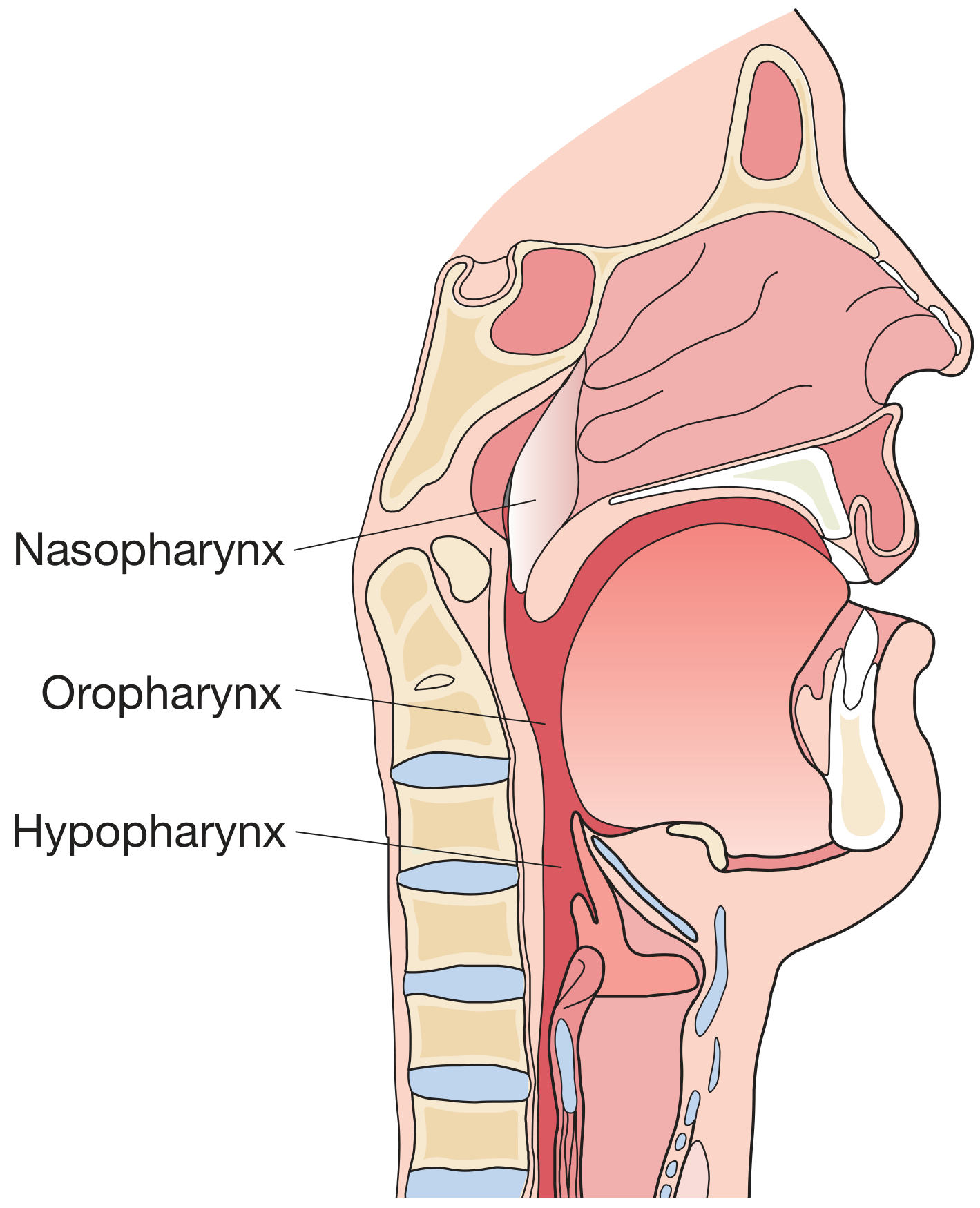
- Nasopharynx – behind the choanae, above the soft palate (the region continuous with the posterior wall of the nose)
- Oropharynx – from the soft palate to the tip of the epiglottis
- Laryngopharynx (hypopharynx) – from the epiglottis to the lower border of the cricoid cartilage, continuing into the oesophagus
Contents
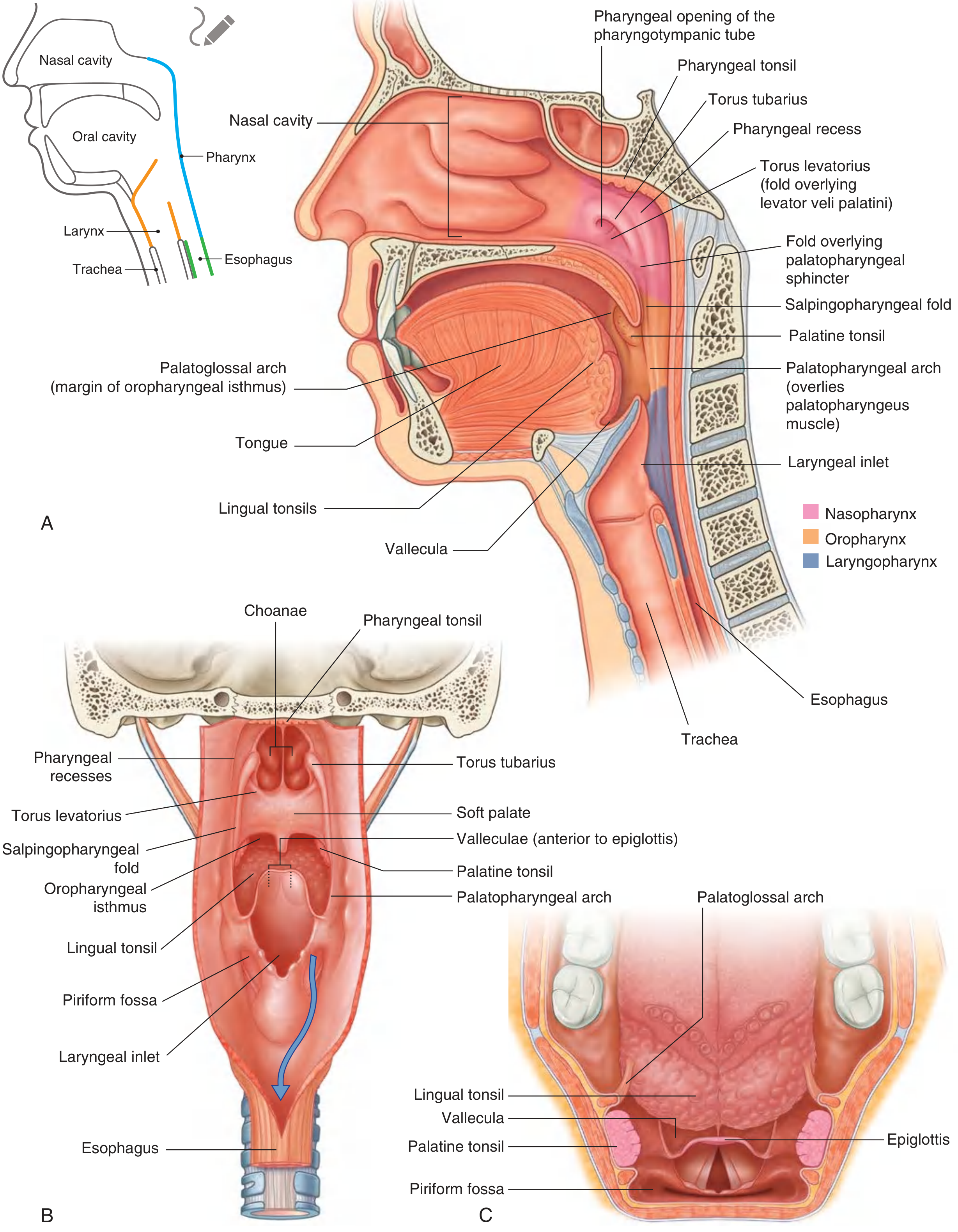
- Adenoids (pharyngeal tonsil) – a mass of lymphoid tissue situated at the junction of the roof and posterior wall of the nasopharynx; forms the superior component of Waldeyer's ring (along with the palatine and lingual tonsils) - Bailey and Love's Short Practice of Surgery, p. 8994.
- Eustachian (pharyngotympanic) tube opening – located on the lateral wall, posterosuperiorly, just above and behind the level of the hard palate.
- Torus tubarius – a mucosal elevation formed by the cartilaginous end of the Eustachian tube, projecting into the nasopharynx from a posterolateral direction.
- Fossa of Rosenmuller (pharyngeal recess) – a slit-like recess situated dorsal and superior to the tubal opening, bounded anteriorly by a ridge raised by the salpingopharyngeus muscle; a common site for development of nasopharyngeal carcinoma - Bailey and Love's Short Practice of Surgery, p. 9021.
- Salpingopharyngeal fold – mucosal fold overlying the salpingopharyngeus muscle running down from the torus tubarius.
- Mucosa here is lined mainly by respiratory (ciliated columnar) epithelium, transitioning to stratified squamous over areas of friction such as the roof.
Applied Anatomy
- Adenoid hypertrophy – physiological enlargement of the pharyngeal tonsil in childhood can obstruct the choanae/nasopharynx, causing mouth breathing, snoring, nasal twang, "adenoid facies," and predisposing to otitis media with effusion (via Eustachian tube blockage).
- Choanal atresia – a congenital failure of canalization of the posterior nasal aperture, presenting with respiratory distress (especially if bilateral) in neonates who are obligate nasal breathers.
- Nasopharyngeal carcinoma (NPC) – classically arises in the fossa of Rosenmuller; can present with unilateral nasal obstruction, epistaxis, unilateral serous otitis media (Eustachian tube involvement), or a neck node with an occult primary, and may invade the skull base.
- Posterior epistaxis – bleeding from the posterior nasal cavity/nasopharyngeal region (Woodruff's plexus) is harder to control than anterior (Little's area) bleeds and may need posterior nasal packing or sphenopalatine artery ligation/cautery.
- Posterior rhinoscopy / nasopharyngoscopy – clinical examination of this region (mirror or endoscope) to assess adenoids, choanal patency, and the fossa of Rosenmuller, important in diagnosing NPC, adenoid hypertrophy, and choanal masses (e.g., antrochoanal polyp presenting through the choana).
- Antrochoanal polyp – arises in the maxillary antrum and prolapses through the choana into the nasopharynx, illustrating the direct communication between the nasal cavity and nasopharynx via this posterior aperture.
I aslo need images related to it
nasopharynx lateral wall Eustachian tube opening fossa of Rosenmuller
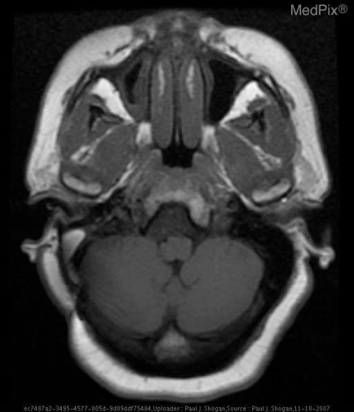
**Imaging Modality:** Magnetic Resonance Imaging (MRI), T1-weighted sequence.
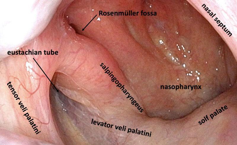
This clinical diagnostic image is a high-definition endoscopic view of the right lateral nasopharynx in a healthy adult. The image clearly demonstrates the anatomical landmarks of the pharyngeal orifice of the Eustachian tube. Key structures labeled include the Eustachian tube opening appearing as a dark slit, the Rosenmüller fossa (pharyngeal recess) located posteriorly, and the torus tubarius. The muscular anatomy is delineated, showing the tensor veli palatini anteriorly and the levator veli palatini inferiorly. The salpingopharyngeus muscle is visible as a vertical fold of mucosa. Other landmarks include the soft palate forming the inferior boundary, the nasopharyngeal wall, and the posterior edge of the nasal septum. The mucosa appears healthy, pink, and well-vascularized without signs of inflammation, lymphoid hyperplasia, or obstruction. This image serves as a reference for normal nasopharyngeal anatomy and the study of peritubal muscle relationships relevant to Middle Ear Myoclonus (MEM) and Eustachian tube dysfunction.
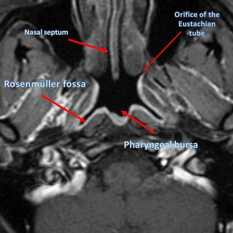
This diagnostic image is an axial (transverse) Magnetic Resonance Imaging (MRI) scan of the human nasopharynx in a healthy subject. The view provides a detailed anatomical study of the upper airway and its surrounding mucosal landmarks. Centrally, the posterior aspect of the nasal septum is visible, exhibiting a slight lateral deviation. The nasopharyngeal lumen is clearly defined, showing key lateral and posterior wall structures. The Rosenmüller fossa (lateral pharyngeal recess) is identified as a deep indentation on the lateral wall, appearing with a heterogeneous signal intensity and ill-defined borders. Anterior to this fossa is the orifice of the Eustachian tube, visualized as a small opening with a dark signal. On the midline of the posterior wall, the pharyngeal bursa is depicted as a small, smooth-contoured protrusion. This imaging modality is essential for evaluating obstructive sleep apnea (OSA), nasopharyngeal carcinoma, or lymphoid hypertrophy, as it allows for precise assessment of spatial relationships and mucosal symmetry within the rhinopharynx.
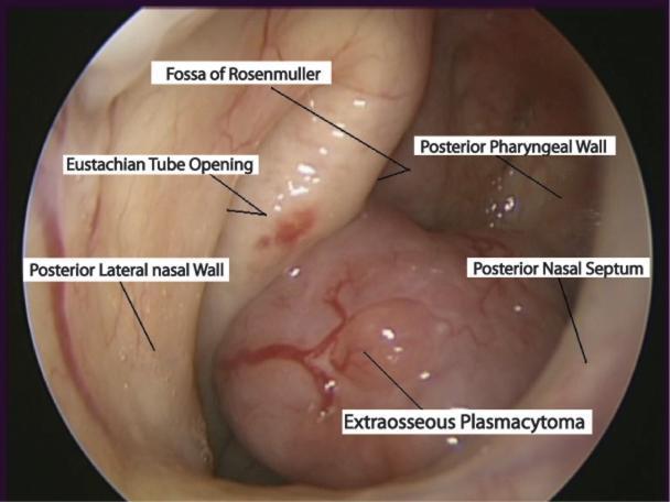
This diagnostic endoscopic image captures the nasopharynx, demonstrating an extraosseous plasmacytoma in a 75-year-old male. The primary finding is a large, smooth, fleshy pink-to-red mass protruding into the nasopharyngeal space. The mass exhibits prominent surface vascularity with branching vessels and is shown effacing the posterior pharyngeal wall. Key anatomical landmarks are annotated, including the Eustachian tube opening and the Fossa of Rosenmuller located superior to the lesion. The mass is positioned adjacent to the posterior nasal septum and the posterior lateral nasal wall. This clinical photograph illustrates the typical presentation of a rare extramedullary plasma cell neoplasm in the upper respiratory tract, which may present with symptoms such as epistaxis. The image serves as an educational resource for Otolaryngology and Oncology, highlighting the importance of endoscopic examination in evaluating nasopharyngeal masses.
posterior nasal aperture choana anatomy skull
adenoids pharyngeal tonsil nasopharynx sagittal section