
How is muscle tone maintained in the body
muscle spindle stretch reflex arc gamma motor neuron diagram
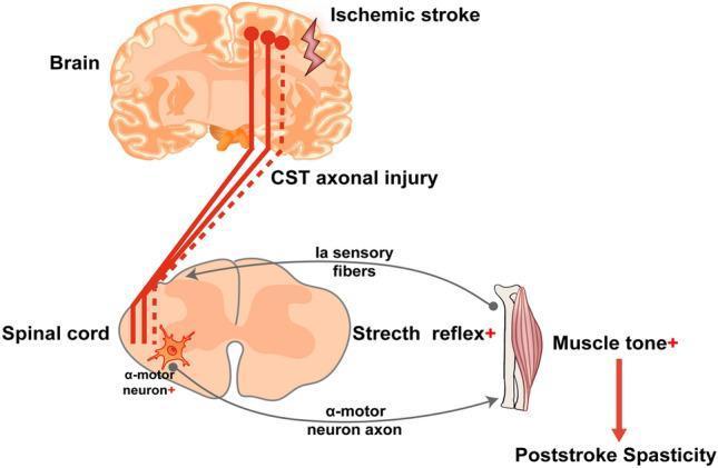
A pathophysiology diagram illustrating the mechanisms of post-stroke spasticity (PSS) involving the corticospinal tract (CST) and the spinal stretch reflex arc. The diagram is divided into three main levels: the brain, the spinal cord, and the effector muscle. In the brain, an ischemic stroke is depicted, leading to CST axonal injury, represented by solid red lines transitioning into a dashed red line to signify disrupted descending signals. At the spinal cord level, the diagram shows the interaction between the damaged CST and the lower motor neurons. The stretch reflex arc is highlighted, featuring Ia sensory fibers from the muscle spindle synapsing onto an alpha-motor neuron in the anterior horn. A '+' sign next to the alpha-motor neuron and the stretch reflex indicates abnormal hyperexcitability and hyperreflexia due to the loss of descending inhibitory control from the CST. The pathway concludes at the skeletal muscle, where increased muscle tone (+) is indicated, leading to the clinical manifestation of post-stroke spasticity. The illustration serves as an educational tool for neurology and physical medicine, demonstrating the upper motor neuron lesion model.
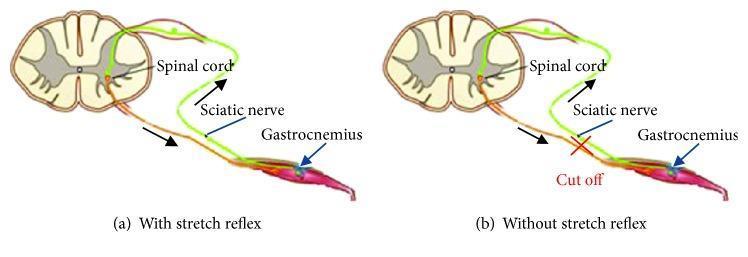
Anatomical diagrams illustrating the components of the monosynaptic stretch reflex arc in two experimental states. Both panels show a transverse section of the spinal cord (white and gray matter) connected to the gastrocnemius muscle via the sciatic nerve. Panel (a), labeled 'With stretch reflex', depicts an intact neural circuit: a sensory (afferent) pathway, colored green, carries signals from the muscle spindle to the dorsal horn, while a motor (efferent) pathway, colored orange/brown, carries impulses from the ventral horn back to the muscle. Black arrows indicate the direction of signal flow. Panel (b), labeled 'Without stretch reflex', demonstrates a disrupted circuit where the sciatic nerve has been severed, indicated by a red 'X' and the text 'Cut off'. This visualization highlights how the interruption of the peripheral nerve abolishes the reflex arc between the spinal cord and the effector muscle, commonly used in physiological studies to compare muscle response forces with and without neural feedback.
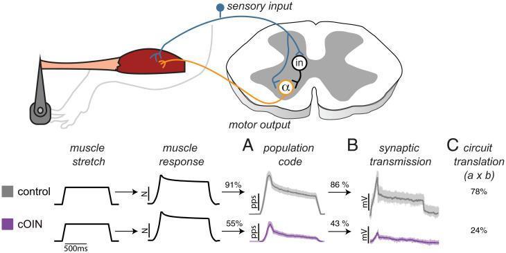
This medical illustration and data plot set depict the pathophysiology of sensorimotor circuit dysfunction, comparing control conditions with chronic oxaliplatin-induced neuropathy (cOIN). The top anatomical diagram illustrates a spinal reflex arc: mechanical muscle stretch triggers sensory input via propriosensory neurons (blue) into the spinal cord, synapsing with interneurons ('in') and alpha-motoneurons (orange, 'α') to produce motor output. Below, comparative waveforms track signal translation through three stages: (A) Population Code, representing peripheral sensory encoding; (B) Synaptic Transmission, representing central processing; and (C) Circuit Translation, the compounded accuracy. Data show that while muscle stretch and physical response remain identical, cOIN specimens exhibit a severe degradation in signal fidelity. Specifically, the sensory population code accuracy drops from 91% to 55%, and central synaptic transmission accuracy falls from 86% to 43%. The total compounded circuit translation accuracy is drastically reduced from 78% in controls to 24% in the cOIN model, illustrating how peripheral and central defects compound to cause sensorimotor disability.
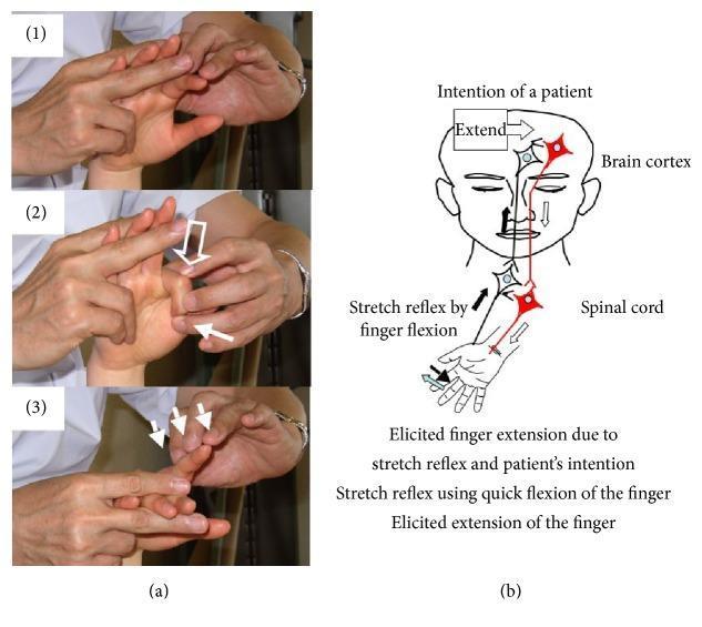
This composite educational graphic illustrates the Repetitive Facilitative Exercise (RFE) technique for hemiplegic fingers, divided into a clinical demonstration and a pathophysiology diagram. Panel (a) shows a three-step manual therapy sequence: (1) quick passive finger flexion to elicit a stretch reflex; (2) simultaneous manual flexion of the metacarpophalangeal (MP) joint with an 'Extend' verbal cue to facilitate voluntary effort; and (3) application of slight resistance during finger extension to maintain the alpha-gamma linkage and muscle contraction. Panel (b) provides a neuroanatomical schematic depicting the hypothesized mechanism. It labels the brain cortex and spinal cord, tracing a dual pathway where the 'intention of the patient' (descending motor tracts) is combined with sensory input from 'stretch reflex by finger flexion' (ascending/local arcs). The integration of these signals at the spinal level is shown to result in 'elicited finger extension.' This visual is intended for rehabilitation specialists and physical therapists focusing on neuroplasticity and motor recovery in stroke or hemiplegic patients.
Maintenance of Muscle Tone
1. The Muscle Spindle - The Peripheral Sensor
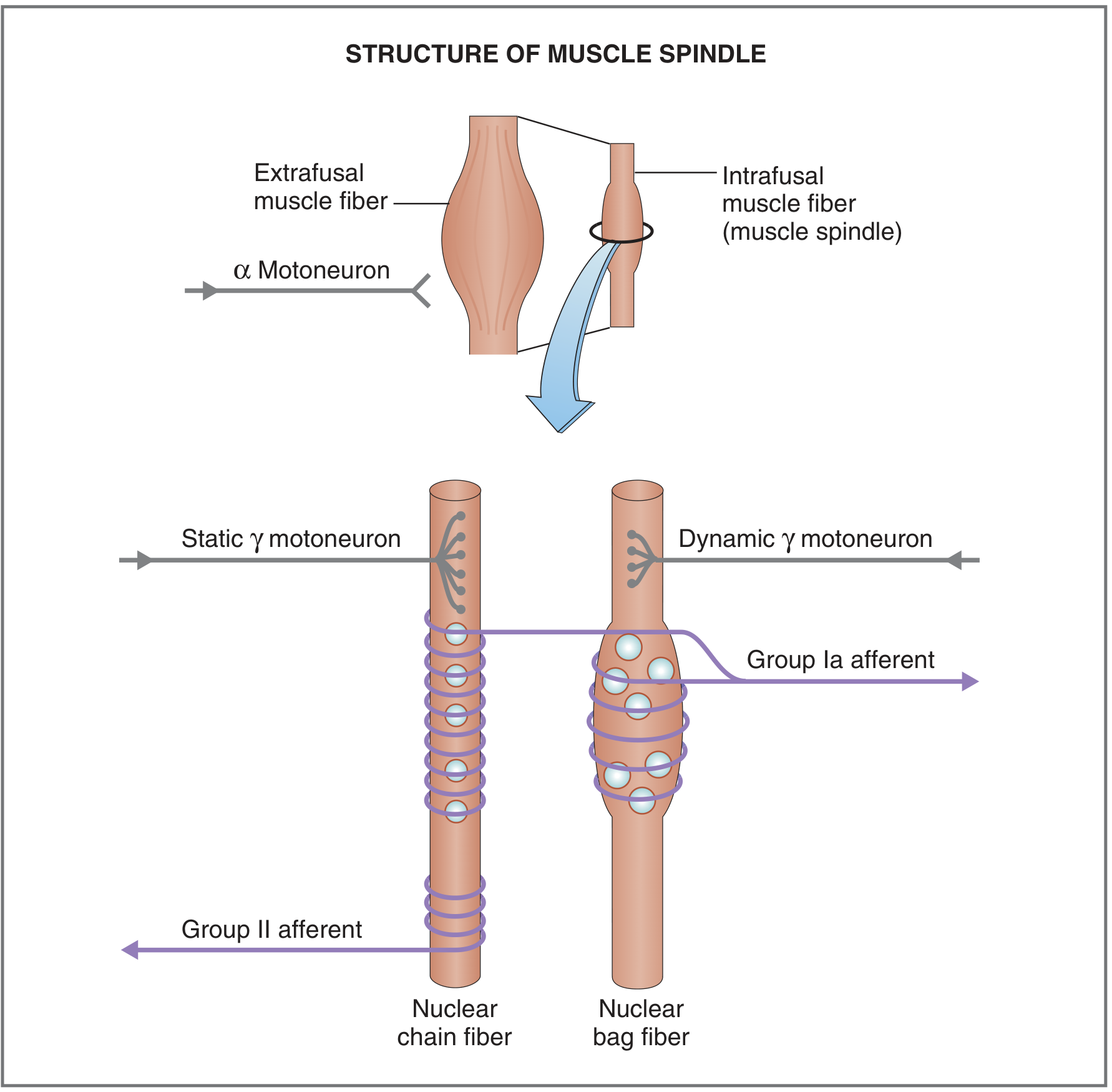
- Nuclear bag fibers (2 per spindle) - larger, with nuclei clustered centrally; include both dynamic and static subtypes
- Nuclear chain fibers (5-6 per spindle) - smaller, nuclei arranged in rows
- Group Ia (primary) afferents - wrap around both nuclear bag and chain fibers; exquisitely sensitive to the velocity of stretch (dynamic response). They provide fast signals about the speed of length change.
- Group II (secondary) afferents - innervate nuclear chain and static bag fibers; signal steady-state length (static response, tonic activity).
2. The Stretch Reflex (Myotatic Reflex)
- Spindle afferents (particularly Ia) fire
- Ia fibers make monosynaptic excitatory connections directly onto the alpha (α) motor neurons in the anterior horn
- α motor neurons fire, causing the stretched muscle to contract - resisting the stretch
- Simultaneously, Ia inhibitory interneurons (Ia inhibitory INs) inhibit the antagonist muscle (reciprocal inhibition)
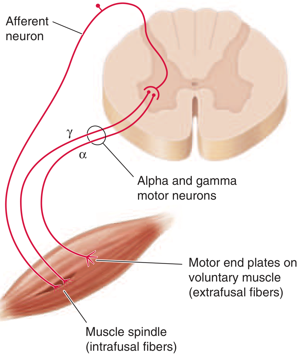
3. The Gamma Motor Neuron System - The Tone Setter
| Type | Target | Effect |
|---|---|---|
| Dynamic γ neurons | Dynamic nuclear bag fibers | Increase velocity sensitivity (dynamic response) of Ia afferents |
| Static γ neurons | Static bag + nuclear chain fibers | Increase tonic (resting) firing of Ia and II afferents; reduce silencing of Ia during sustained stretch |
"The nervous system uses the fusimotor system to fine-tune muscle spindles so that the ensemble output of the spindles provides information most appropriate for a task." - Kandel, Principles of Neural Science
4. The Golgi Tendon Organ - The Protective Brake
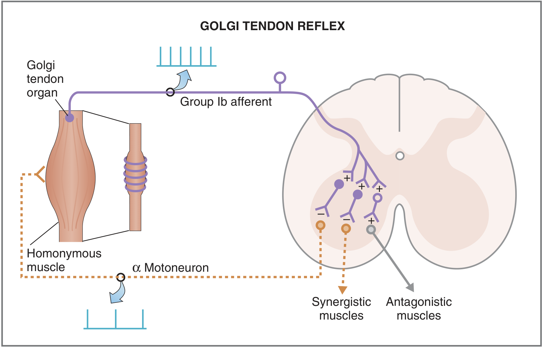
- Muscle contracts → GTO activates → Group Ib afferents fire
- Ib afferents synapse on inhibitory interneurons in the spinal cord
- These inhibitory interneurons suppress α motor neuron firing
- The contracting muscle relaxes; synergistic muscles also relax while antagonists contract
5. Supraspinal (Descending) Control
| Descending Pathway | Effect on Tone |
|---|---|
| Reticulospinal tract (pontine) | Facilitatory - increases tone via excitation of γ motor neurons |
| Reticulospinal tract (medullary) | Inhibitory - decreases tone |
| Vestibulospinal tract | Facilitatory to extensor tone (maintains posture against gravity) |
| Corticospinal tract | Mainly modulates fine voluntary movement; also regulates interneurons that control tone |
- γ motor neuron discharge rate is the dominant determinant of tone level: muscles are generally hypotonic when γ discharge is low, and hypertonic when it is high. - Ganong's Review of Medical Physiology, 26th Ed.
- Cutting the motor nerve → flaccidity (no spindle input, no motor output)
- Upper motor neuron lesion (e.g., stroke) → spasticity (loss of inhibitory descending control → uninhibited stretch reflexes → high γ discharge)
- Acute spinal cord or cerebral lesion → initial hypotonia (spinal shock), transitioning to spasticity
Summary
| Component | Role in Tone |
|---|---|
| Muscle spindle (Ia/II afferents) | Continuous sensory signaling of muscle length → drives tonic α motor neuron activity |
| α motor neurons | Execute the contraction that produces tone |
| γ motor neurons | Tune spindle sensitivity; set the gain of the stretch reflex |
| Alpha-gamma co-activation | Maintains spindle function during active movement |
| Golgi tendon organs (Ib) | Protective inhibition to prevent excessive tension |
| Descending tracts | Modulate γ neuron activity from brainstem/cortex |
| Reciprocal inhibition | Balances tone between agonist and antagonist pairs |