
Cholera microbiology laq
I'll look up the medical library skill first, then pull comprehensive cholera microbiology content.
Reading File
Now let me search the library for cholera microbiology content in parallel:
Finding Sources
Finding Sources
Excellent results. The best microbiology-focused sources are Sherris & Ryan's Medical Microbiology and Medical Microbiology 9e. Let me read all the key sections in parallel:
Reading File
Reading File
Finding Sources
Great content so far. Let me read more of Sherris (rest of the CHOLERA section) and Jawetz (classic microbiology textbook), plus the Medical Microbiology section on pathogenesis, all in parallel:
Reading File
Reading File
Reading File
Excellent content! Let me now fetch the key diagram of cholera toxin action and read the Jawetz pathogenesis section for more detail:
Loading Image
Reading File
I now have comprehensive content from multiple authoritative textbooks. Let me compile the complete LAQ answer.
Cholera - Microbiology (Long Answer Question)
1. Introduction
Cholera is an acute secretory diarrheal illness caused by Vibrio cholerae. It remains a major public health problem in developing countries and has caused seven documented worldwide pandemics since 1817. The disease is a classic model for understanding toxin-mediated secretory diarrhea.
2. Morphology
- Shape: Comma-shaped (curved rod), 2-4 µm long; on prolonged culture may straighten and resemble other Gram-negative enteric rods
- Gram stain: Gram-negative
- Motility: Actively motile by a single polar flagellum ("darting/shooting star" motility on dark-field microscopy)
- Arrangement: Non-capsulate (O1 strains); O139 and non-O1 strains produce an acidic polysaccharide capsule
- Pili: Long filamentous pili, particularly the toxin-coregulated pilus (TCP) - critical for virulence and colonization
3. Cultural Characteristics
| Feature | Details |
|---|---|
| Growth media | Grows on blood agar, MacConkey agar |
| Selective media | TCBS (Thiosulfate-Citrate-Bile Salts-Sucrose) agar - produces yellow colonies (sucrose fermenter) on dark green background |
| Enrichment broth | Alkaline peptone broth/water (pH 8.5-9.5, 1% NaCl) |
| Temperature | 37°C optimal; range 14-40°C |
| pH | Optimal at alkaline pH 8.0-9.5; killed rapidly by acids |
| Atmosphere | Facultative anaerobe |
| Colony morphology | Convex, smooth, round, opaque, granular in transmitted light |
| NaCl requirement | V. cholerae can grow without supplemental NaCl (unlike other halophilic vibrios) |
| Transport medium | Cary-Blair medium for delayed processing |
"V. cholerae produces yellow colonies (sucrose fermented) on TCBS agar that are readily visible against the dark-green background." - Jawetz, Melnick & Adelberg's Medical Microbiology
4. Biochemical Reactions
- Oxidase positive (key preliminary identification test)
- Ferments sucrose and mannose but NOT arabinose
- Voges-Proskauer (VP) test: Positive in El Tor biotype; negative in Classical biotype
- Lysine decarboxylase: positive
- String test: Positive (forms a mucoid "string" when colony touched with loop in 0.5% sodium deoxycholate)
5. Antigenic Structure & Classification
O (Somatic) Antigen
- Based on LPS O polysaccharide; >200 serogroups exist
- Only O1 and O139 cause epidemic/pandemic cholera; all others are non-epidemic
O1 Serogroup - Further Subdivisions
By serotype (based on Inaba and Ogawa antigens):
| Serotype | Antigen |
|---|---|
| Ogawa | A + B antigens |
| Inaba | A + C antigens |
| Hikojima (transitional) | A + B + C antigens |
By biotype:
| Feature | Classical | El Tor |
|---|---|---|
| Hemolysin | Negative | Positive |
| Voges-Proskauer | Negative | Positive |
| Polymyxin B | Sensitive | Resistant |
| Pandemic | 1st-6th | 7th (current) |
O139 (Bengal) Serogroup
- Emerged in 1992 in India/Bangladesh
- Phenotypically resembles O1 El Tor but produces a polysaccharide capsule
- Does NOT carry the O1 LPS gene
H (Flagellar) Antigen
- Single heat-labile flagellar antigen; shared by many vibrios; not protective
6. Virulence Factors
| Virulence Factor | Function |
|---|---|
| Cholera toxin (CT) | Main virulence factor; causes massive secretory diarrhea |
| Toxin-coregulated pilus (TCP) | Mediates colonization of small intestinal mucosa; receptor for bacteriophage CTXφ |
| Neuraminidase | Modifies GM2 → GM1 on cell surface, increasing binding sites for CT |
| Accessory cholera enterotoxin (Ace) | Increases intestinal fluid secretion |
| Zonula occludens toxin (ZOT) | Increases intestinal permeability by affecting tight junctions |
| Chemotaxis protein | Acts as adhesin factor |
| LPS (Lipid A) | Endotoxin activity |
7. Cholera Toxin (CT) - Mechanism of Action
CT is an A-B type ADP-ribosylating toxin (MW ~84,000). This is the single most important concept in cholera microbiology.
Structure
- 5 B subunits: Binding subunits (pentamer ring)
- 1 A subunit: Contains A1 (toxic) and A2 (linker) polypeptides joined by a disulfide bond
Step-by-Step Mechanism
Step 1 - Binding: The 5 B subunits bind to GM1-ganglioside receptors on the apical surface of intestinal epithelial cells (enterocytes). Neuraminidase enhances this by converting GM2 to GM1.
Step 2 - Entry: The A1 subunit is released by reduction of the A1-A2 disulfide bond and translocates into the cell.
Step 3 - ADP-ribosylation: A1 catalyzes ADP-ribosylation (ADPR) of the Gsα (stimulatory G-protein). This locks the Gsα protein in a permanently active state - it cannot hydrolyze GTP and cannot dissociate from adenylate cyclase.
Step 4 - cAMP accumulation: Persistent activation of adenylate cyclase converts ATP → cAMP in massive amounts at the basolateral membrane.
Step 5 - Ion and water secretion: Elevated cAMP activates protein kinase A → phosphorylates CFTR chloride channels → massive efflux of Cl⁻, K⁺, HCO₃⁻, Na⁺, and water into the intestinal lumen → profuse "rice-water" stools.
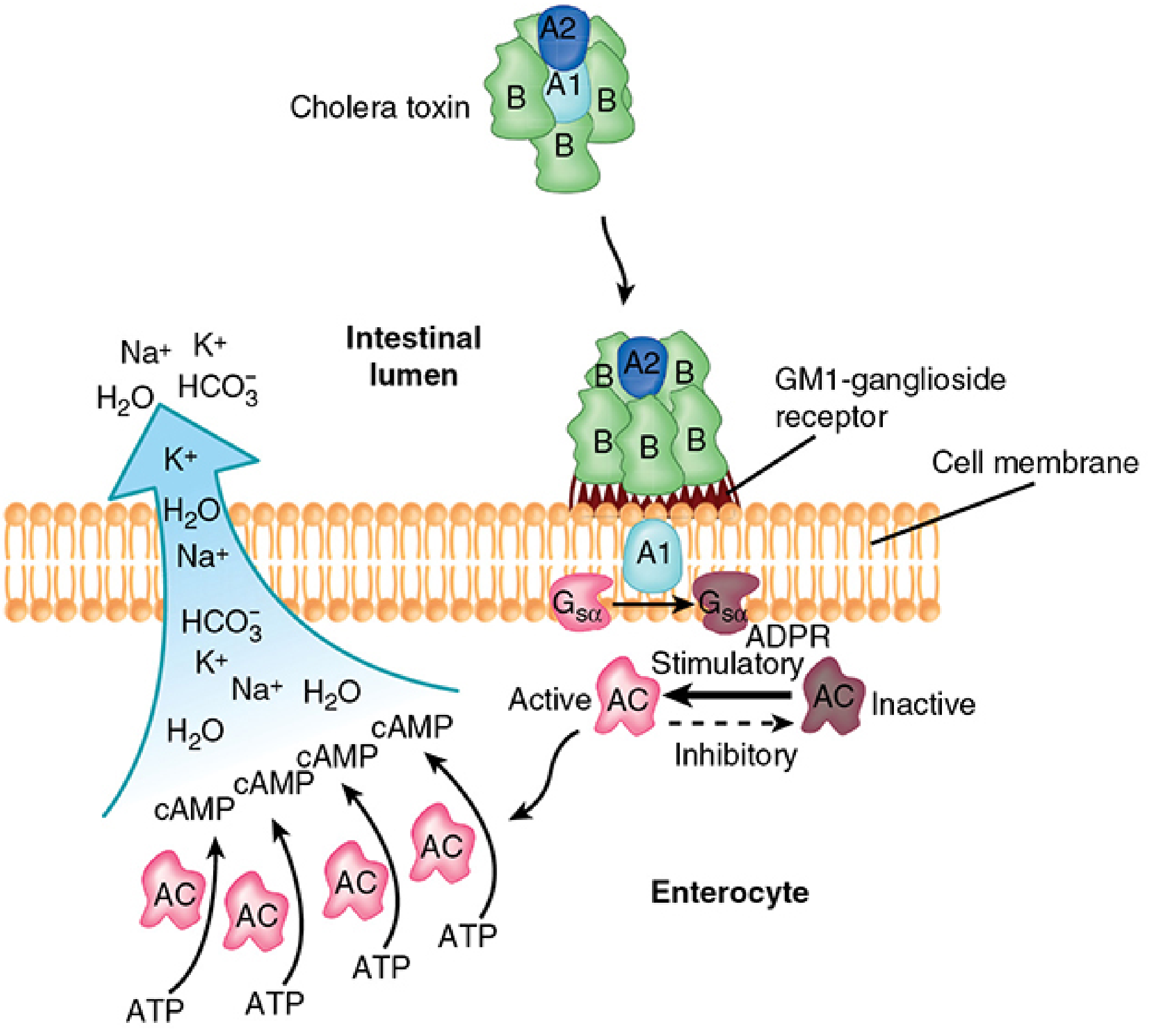
"CT catalyzes the ADPR of the G-protein, rendering it unable to dissociate from the active adenylate cyclase complex... net effect is excessive accumulation of cAMP at the cell membrane causing hypersecretion of chloride, potassium, bicarbonate, and water out of the cell." - Sherris & Ryan's Medical Microbiology, 8th Ed.
8. Pathogenesis
- Ingestion of large inoculum (10⁸ organisms needed; reduced to 10³ if gastric acid neutralized)
- Organisms survive alkaline conditions; killed by gastric acid
- Colonization of small intestinal mucosa via TCP pili and chemotaxis protein
- No mucosal invasion - purely non-invasive, toxin-mediated disease
- CT produced → secretory diarrhea as above
- Fluid loss of 10-20 L/day leading to rapid dehydration, electrolyte imbalance, metabolic acidosis
- No bacteremia in classic O1 cholera (no capsule = no systemic spread)
9. Epidemiology
- Transmission: Feco-oral route; mainly through contaminated water; foodborne (shellfish, raw fish - ceviche)
- Reservoir: Estuarine and marine environments; chitinous shellfish; human carriers
- Incubation period: 6 hours to 5 days (classically ~2 days)
- Infectious dose: High (~10⁸ organisms); much lower if gastric acid reduced
- Person-to-person spread: Rare due to high infectious dose
- 7 Pandemics: 1st-6th caused by Classical biotype; 7th (ongoing, from 1961) by El Tor biotype; O139 (Bengal) emerged 1992
10. Clinical Features
| Severity | Presentation |
|---|---|
| Mild | Self-limiting watery diarrhea |
| Moderate | Profuse watery diarrhea with mild dehydration |
| Severe ("cholera gravis") | "Rice-water" stools (grey, turbid, fishy-smelling); vomiting; rapid dehydration; hypovolemic shock; "washerwoman's hands", sunken eyes, decreased skin turgor; "cholera voice" (aphonia) |
11. Laboratory Diagnosis
| Method | Details |
|---|---|
| Specimen | Fresh stool (early in disease); rectal swab |
| Transport | Cary-Blair medium; alkaline peptone water |
| Direct microscopy | Dark-field/phase-contrast: "shooting star" motility; comma-shaped rods; inhibited by O1 antiserum (immobilization test) |
| Culture | TCBS agar - yellow colonies; alkaline peptone broth (enrichment) |
| Biochemical | Oxidase positive; String test positive; VP test positive (El Tor) |
| Serology | Widal-type agglutination with O1/O139 antisera; immunoassay for CT or LPS |
| Molecular | Multiplex PCR / NAAT - gold standard in resource-rich settings; detects CT gene |
12. Treatment
| Component | Details |
|---|---|
| Rehydration (mainstay) | ORS (oral rehydration solution) for mild-moderate; IV Ringer's lactate for severe |
| Antibiotics | Reduce bacterial burden, shorten disease duration by ~50%; options: Doxycycline (single dose, adults), Azithromycin (children/pregnant), Tetracycline, Ciprofloxacin |
| Zinc | Adjunct in children |
Note: Antibiotics are adjunct only; fluid replacement is the cornerstone of treatment.
13. Prevention and Control
- Safe water supply and sewage treatment
- Proper food hygiene (cooked food, pasteurized water)
- Vaccines:
- Dukoral: Killed whole-cell O1 + recombinant CT-B subunit (oral); ~50-60% efficacy for 2-3 years
- Shanchol/mORCVAX: Bivalent killed whole-cell O1 + O139 (oral); no CT-B component; ~65% efficacy
- Provides limited protection and herd immunity
14. Key Exam Points (Summary Table)
| Feature | Key Fact |
|---|---|
| Organism | Gram-negative curved rod, polar flagellum |
| Selective medium | TCBS - yellow colonies |
| Enrichment | Alkaline peptone broth (pH 8.5-9.5) |
| Pandemic strains | O1 (Classical + El Tor) and O139 |
| Current pandemic biotype | El Tor |
| Toxin type | A-B enterotoxin (ADP-ribosylating) |
| Receptor for B subunit | GM1-ganglioside |
| Target of A1 | Gsα protein (ADP-ribosylation) |
| Final mediator | cAMP (persistent elevation) |
| Stool description | Rice-water stools |
| Disease mechanism | Non-invasive, purely toxin-mediated |
| Oxidase | Positive |
| String test | Positive |
| El Tor vs Classical | El Tor: VP+, hemolysin+, polymyxin B resistant |
Sources:
- Sherris & Ryan's Medical Microbiology, 8th Edition
- Jawetz, Melnick & Adelberg's Medical Microbiology, 28th Edition
- Medical Microbiology, 9th Edition (Murray et al.)
This is a shared conversation. Sign in to Orris to start your own chat.