
Arm anatomy
Reading File
The arm has two main segments: the upper arm (between shoulder and elbow) and the forearm (between elbow and wrist), plus the hand. Each region has distinct compartments, bones, muscles, nerves, and vessels.
Searching sources...
Finding Sources
Finding Sources
Finding Sources
Finding Sources
Reading File
Finding Sources
Finding Sources
Finding Sources
Reading File
Finding Sources
Loading Image
Loading Image
Reading File
Excellent - I have great textbook content and cross-sectional diagrams. Let me now compile the full answer with these rich visual resources.
Arm Anatomy
The "arm" in strict anatomical terminology refers to the upper arm (shoulder to elbow), but clinically it encompasses the entire upper limb: upper arm, forearm, and hand. Here is a region-by-region breakdown.
1. Bones
| Region | Bones |
|---|---|
| Upper arm | Humerus |
| Forearm | Radius (lateral), Ulna (medial) |
| Wrist | 8 carpal bones (scaphoid, lunate, triquetrum, pisiform, trapezium, trapezoid, capitate, hamate) |
| Hand | 5 metacarpals + 14 phalanges |
The radius and ulna are connected by the interosseous membrane, which serves as both a structural stabilizer and an origin for several forearm muscles.
2. Compartments and Muscles
Upper Arm
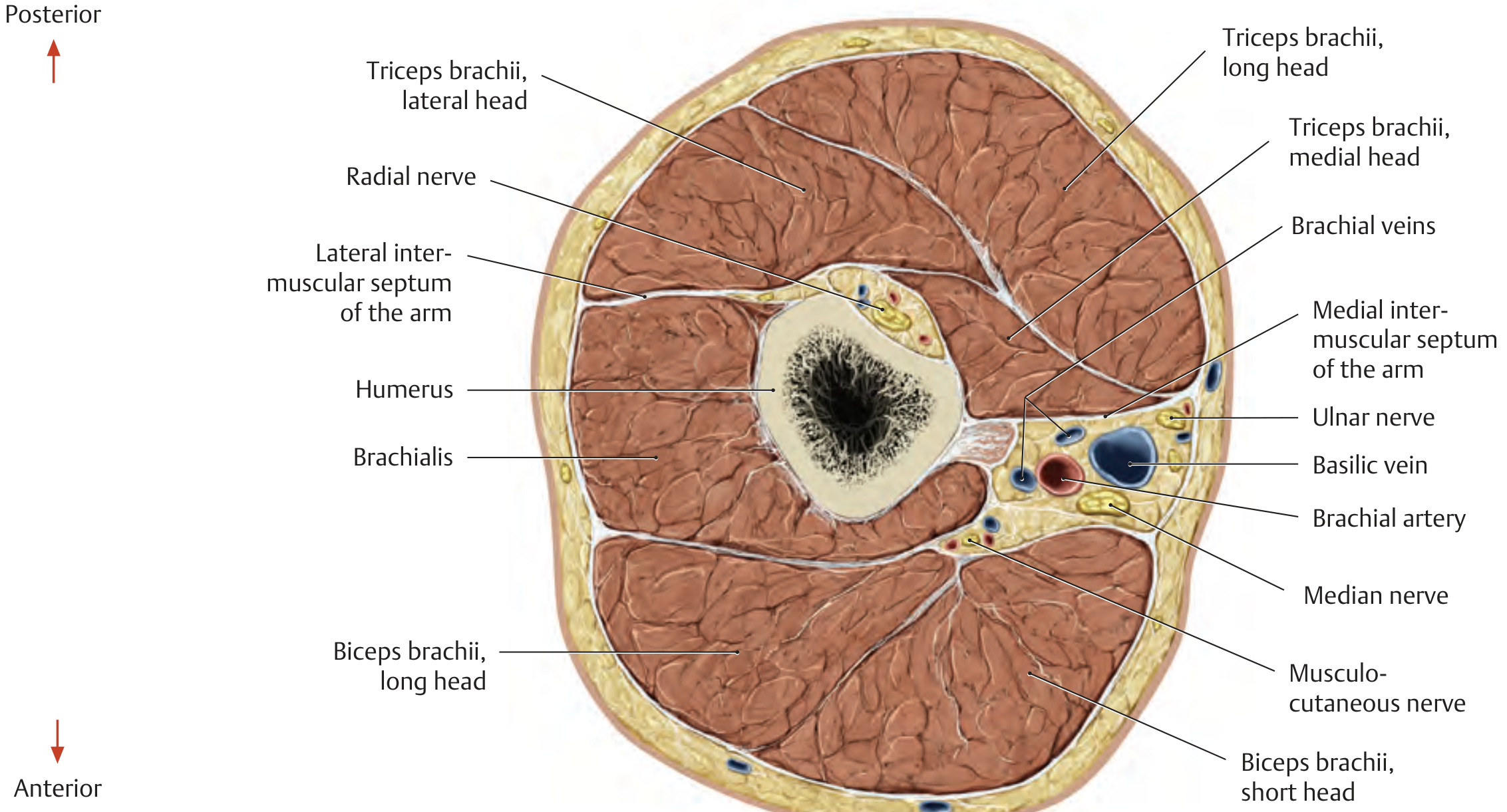
The humerus is surrounded by two fascial compartments divided by the medial and lateral intermuscular septa:
Anterior (flexor) compartment:
- Biceps brachii (long + short heads) - flexes elbow and supinates forearm
- Brachialis - primary elbow flexor
- Coracobrachialis - shoulder flexion/adduction
Posterior (extensor) compartment:
- Triceps brachii (long, lateral, and medial heads) - extends elbow
The cross section below (right arm, proximal view) shows this arrangement clearly:
Forearm
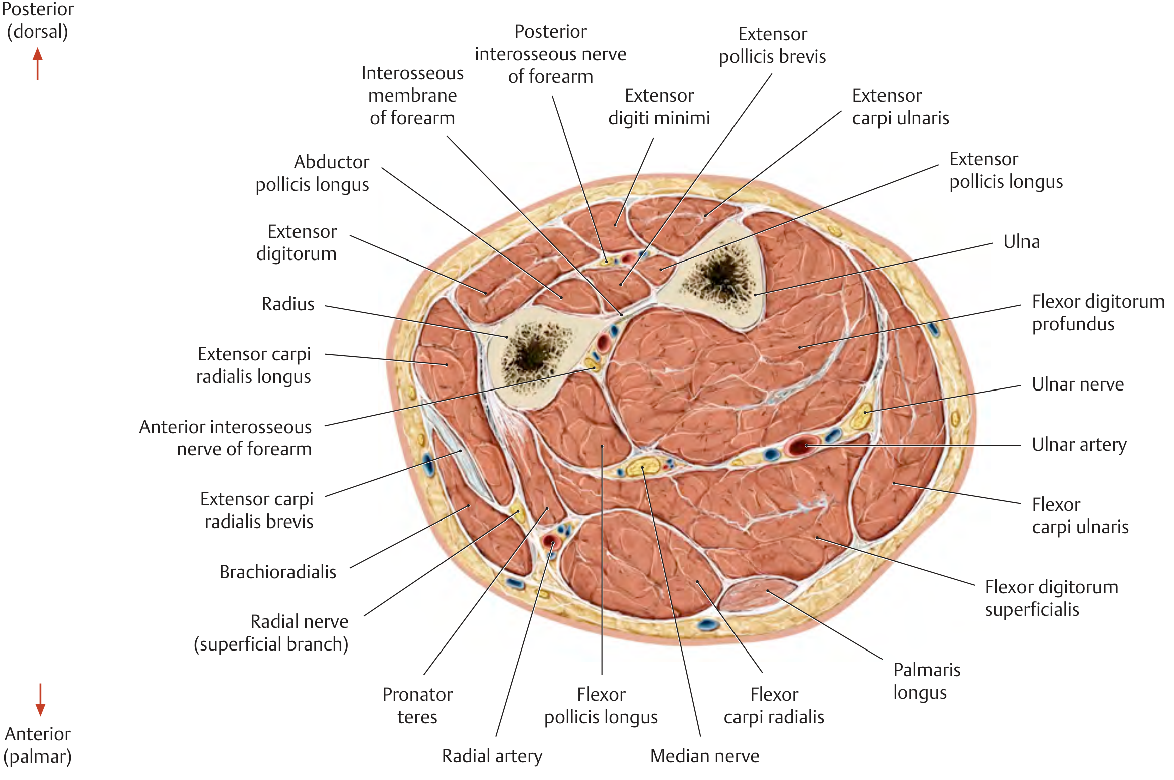
The forearm contains two bones (radius and ulna) and is divided into two compartments:
Anterior (flexor/pronator) compartment - 8 muscles in 3 layers:
- Superficial: Pronator teres, Flexor carpi radialis, Palmaris longus, Flexor carpi ulnaris
- Intermediate: Flexor digitorum superficialis
- Deep: Flexor digitorum profundus, Flexor pollicis longus, Pronator quadratus
Posterior (extensor/supinator) compartment - 12 muscles:
- Superficial: Brachioradialis, ECRL, ECRB, Extensor digitorum, Extensor digiti minimi, Extensor carpi ulnaris
- Deep: Supinator, Abductor pollicis longus, Extensor pollicis brevis, Extensor pollicis longus, Extensor indicis
The cross-section below shows the full forearm arrangement with both radius and ulna, the interosseous membrane, and all muscles + neurovascular bundles:
3. Innervation (Brachial Plexus - C5 to T1)
Almost all arm muscles are innervated by the brachial plexus (C5-T1). The key principle: extensor muscles are supplied by posterior divisions (radial, axillary nerves), while flexor muscles are supplied by anterior divisions (musculocutaneous, median, ulnar nerves).
| Nerve | Key muscles innervated |
|---|---|
| Musculocutaneous (C5-C7) | Coracobrachialis, Biceps brachii, Brachialis |
| Axillary (C5-C6) | Deltoid, Teres minor |
| Radial (C5-T1) | Triceps brachii, Anconeus, Brachioradialis, all wrist/finger extensors, Abductor pollicis longus |
| Median (C6-T1) | Pronator teres, Pronator quadratus, FCR, Palmaris longus, FDS, FDP (lateral half), FPL, thenar muscles, 1st & 2nd lumbricals |
| Ulnar (C8-T1) | FCU, FDP (medial half), hypothenar muscles, interossei, 3rd & 4th lumbricals, Adductor pollicis |
Source: General Anatomy and Musculoskeletal System (THIEME Atlas of Anatomy) - Classification of upper limb muscles by innervation
4. Vasculature
Arterial supply:
- Axillary artery continues as the brachial artery at the lower border of teres major
- The brachial artery runs in the anteromedial arm, adjacent to the median nerve
- At the elbow (cubital fossa), it divides into the radial artery (lateral) and ulnar artery (medial)
- The radial and ulnar arteries anastomose at the superficial and deep palmar arches in the hand
Venous drainage:
- Deep veins follow the arteries (brachial veins paired with the brachial artery)
- Superficial: Cephalic vein (lateral, drains into axillary), Basilic vein (medial, pierces fascia at mid-arm), Median cubital vein (connects the two at the elbow - used for venipuncture)
5. Key Anatomical Spaces
| Space | Location | Contents |
|---|---|---|
| Axilla | Between arm and chest wall | Brachial plexus, axillary artery/vein, lymph nodes |
| Cubital fossa | Anterior elbow | Brachial artery, median nerve, biceps tendon, radial nerve (lateral) |
| Carpal tunnel | Wrist, deep to flexor retinaculum | Median nerve + 9 flexor tendons |
| Guyon's canal | Medial wrist | Ulnar nerve + ulnar artery |
6. Quick Clinical Memory Aids
- "Saturday night palsy" = radial nerve compression (wrist drop, loss of finger extension)
- Carpal tunnel syndrome = median nerve compression (thenar wasting, loss of thumb opposition, 1st-3rd finger numbness)
- Cubital tunnel syndrome = ulnar nerve compression at elbow ("ring and little finger" numbness, interosseous wasting)
- "Carrying angle" = normal valgus angle at elbow (~5-15° in males, up to 20° in females)
- Volkmann's contracture = compartment syndrome of forearm leading to ischemic contracture of flexors
Source: THIEME Atlas of Anatomy - General Anatomy and Musculoskeletal System, Sections 2 and 3.7
This is a shared conversation. Sign in to Orris to start your own chat.