Secretary stages of Amylogenesis
Reading File
Finding Sources
Finding Sources
Finding Sources
Reading File
Finding Sources
Reading File
Loading Image
Loading Image
Loading Image
Reading File
I now have comprehensive information on the secretory (and maturation) stages of amelogenesis. Let me compile the full answer.
Note on terminology: "Amylogenesis" is not a standard term. You likely mean amelogenesis - the process of dental enamel formation. Here is a detailed account.
Secretory Stage of Amelogenesis
Amelogenesis is the matrix-mediated biomineralization process by which dental enamel is formed. It proceeds through two major stages:
- Matrix production (Secretory stage)
- Matrix maturation (Maturation stage)
Stage 1: Matrix Production - Secretory Stage
Overview
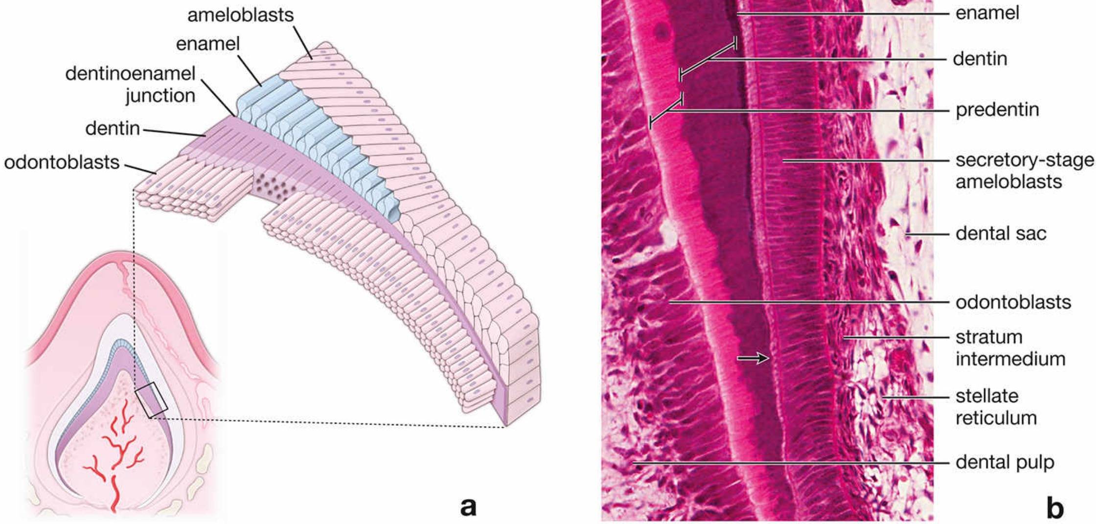
Dentin is produced first by odontoblasts. Then, a partially mineralized enamel matrix is deposited directly onto the surface of the previously formed dentin. The cells responsible for this are secretory-stage ameloblasts, derived from the inner enamel epithelium of the enamel organ.
Morphology of Secretory-Stage Ameloblasts
Secretory-stage ameloblasts are tall, polarized columnar cells lying directly adjacent to the developing enamel. Key structural features include:
| Feature | Detail |
|---|---|
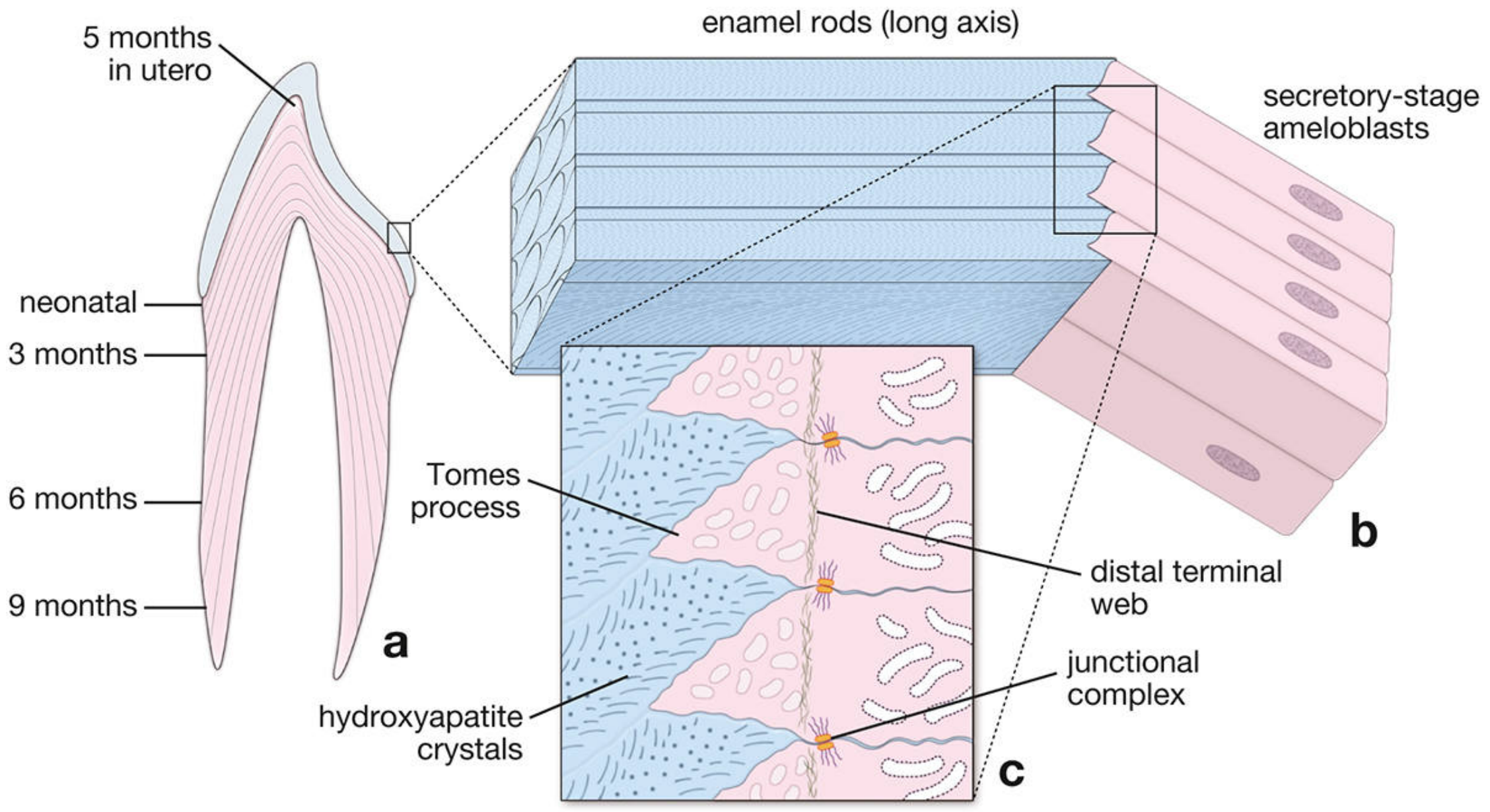
| Tomes process | Apical cytoplasmic extension projecting into the developing enamel; packed with secretory vesicles containing enamel matrix proteins |
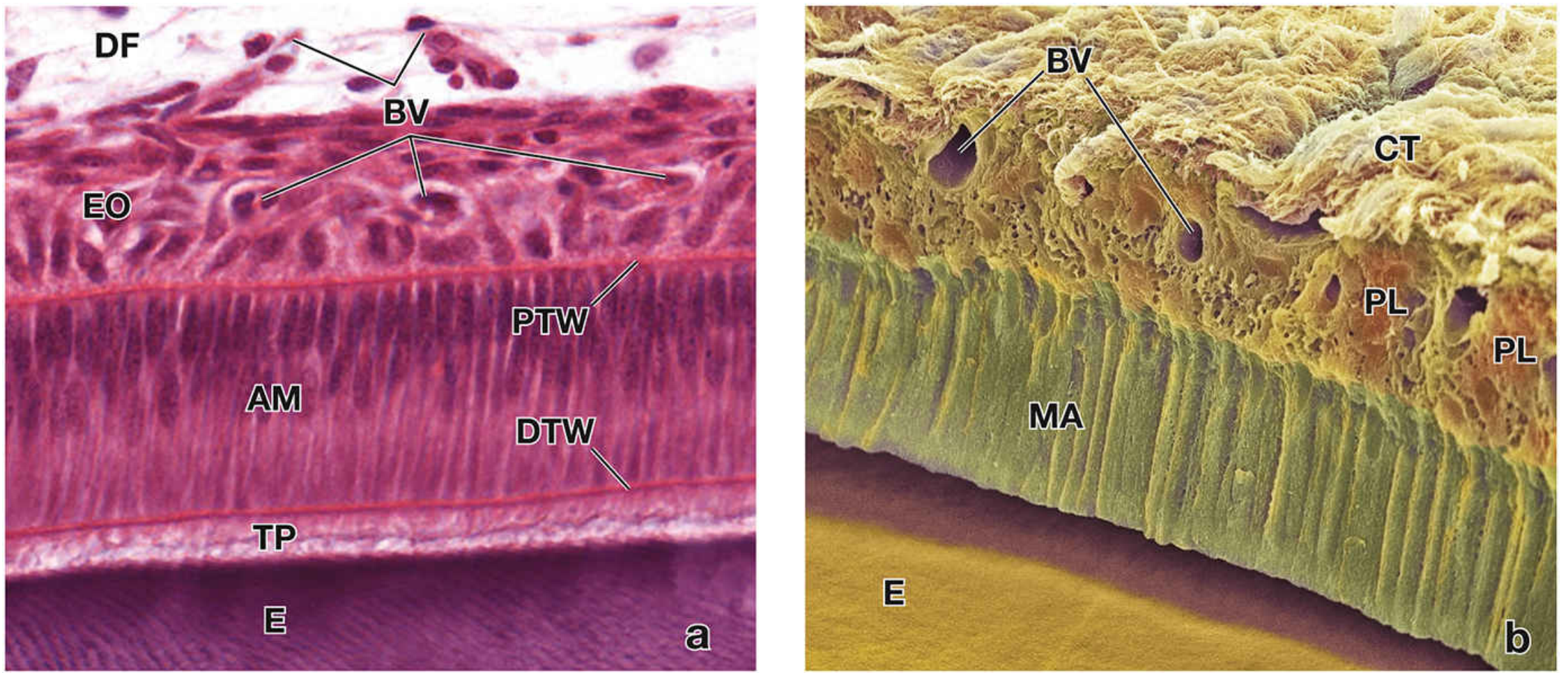
| Distal terminal web (DTW) | Band of actin filaments at the junction between the cell body and Tomes process |
| Proximal terminal web (PTW) | Second actin band at the base (proximal end) of the ameloblast |
| Junctional complexes | Located at the apical pole; seal adjacent ameloblasts to regulate passage of ions and molecules |
| Organelles | Abundant rER, prominent Golgi apparatus, and secretory granules - all reflecting high secretory activity |
The enamel rod produced by each ameloblast forms in the wake of the cell as it moves away from the dentinoenamel junction. The direction of the enamel rod is thus a permanent record of the path taken by the secretory-stage ameloblast.
Matrix Proteins Secreted
- Amelogenin - the dominant structural protein (~90%); initiates and guides growth of hydroxyapatite crystals; encoded on the X chromosome
- Enamelin - non-amelogenin matrix protein
- Ameloblastin (sheathlin) - associated with Tomes processes
These proteins are produced via rER → Golgi → secretory granules → exocytosis, analogous to osteoblast matrix secretion.
Supporting Cells - Stratum Intermedium
At the base (proximal pole) of secretory-stage ameloblasts lies the stratum intermedium - a layer of enamel organ cells whose plasma membrane contains alkaline phosphatase, an enzyme active in calcification. Beyond the stratum intermedium are the stellate reticulum cells, separated from adjacent blood vessels by a basal lamina.
Stage 2: Matrix Maturation Stage
Once the full thickness of enamel matrix is deposited, the secretory-stage ameloblasts differentiate into maturation-stage ameloblasts.
Key Events in Maturation
-
Removal of organic matrix - enzymes degrade and reabsorb the organic proteins
-
Continued influx of Ca²⁺ and phosphate - enamel becomes progressively more mineralized (~96% mineral in mature enamel)
-
Cyclic modulation - maturation-stage ameloblasts alternate between:
- Striated (ruffled) border (~70% of time): Secretes HCO₃⁻ ions and pumps Ca²⁺ via plasma membrane Ca²⁺-ATPase (PMCA) into enamel
- Smooth-ended border (~30% of time): Secretes enzymes (e.g., kallikrein-4) to degrade and remove residual matrix
-
The stratum intermedium disappears - replaced by a reorganized papillary layer containing stellate papillary cells with abundant mitochondria (reflecting high energy demand for ion transport)
Summary Table
| Parameter | Secretory Stage | Maturation Stage |
|---|---|---|
| Cell type | Secretory-stage ameloblast | Maturation-stage ameloblast |
| Apical feature | Tomes process | Striated/smooth-ended border |
| Function | Deposits organic enamel matrix | Removes organic matrix; mineralizes enamel |
| Key proteins | Amelogenin, enamelin, ameloblastin | MMP-20, kallikrein-4 (degrade matrix) |
| Adjacent layer | Stratum intermedium | Papillary layer |
| Mineralization | Partial (~30%) | Complete (~96%) |
Sources: Histology: A Text and Atlas with Correlated Cell and Molecular Biology (9781975181512), pp. 1445-1450; Junqueira's Basic Histology, 17e (9781264930395)
Give a short note on secretory stage of amelogenesis for three marks, point-wise answer. Don't give too much long answer, don't give too much short answer. It is of three marks, so make answer according to that.
Secretory Stage of Amelogenesis
-
Definition: It is the first stage of amelogenesis in which secretory-stage ameloblasts deposit a partially mineralized organic enamel matrix. Dentin is always formed first, and enamel matrix is then deposited directly onto the dentin surface.
-
Cell morphology: Secretory-stage ameloblasts are tall, polarized columnar cells. Each cell has an apical cytoplasmic extension called the Tomes process, which is packed with secretory granules containing enamel matrix proteins.
-
Matrix proteins secreted: The main proteins produced are amelogenin (~90%), enamelin, and ameloblastin. These are synthesized via rER → Golgi → secretory granules and guide hydroxyapatite crystal growth.
-
Direction of enamel rod formation: As each ameloblast moves away from the dentinoenamel junction, the enamel rod forms in its wake. The direction of the rod is thus a permanent record of the ameloblast's path.
-
Supporting cells: At the base of secretory-stage ameloblasts lies the stratum intermedium, whose plasma membrane contains alkaline phosphatase - an enzyme active in calcification.
-
End point: The secretory stage continues until the full thickness of the future enamel layer is achieved, after which ameloblasts differentiate into maturation-stage ameloblasts.
Point wise
Secretory Stage of Amelogenesis
-
First stage of amelogenesis; dentin is formed first, then enamel matrix is deposited onto the dentin surface.
-
Performed by secretory-stage ameloblasts - tall, polarized columnar cells derived from the inner enamel epithelium.
-
Each ameloblast has an apical Tomes process packed with secretory granules containing enamel matrix proteins.
-
Main proteins secreted: amelogenin (~90%), enamelin, and ameloblastin - synthesized via rER → Golgi → secretory granules.
-
These proteins guide hydroxyapatite crystal nucleation and growth, producing a partially mineralized matrix.
-
Enamel rods form in the wake of the moving ameloblast; rod direction reflects the path of the ameloblast.
-
Stratum intermedium lies at the base of ameloblasts and provides alkaline phosphatase for calcification support.
-
Stage ends when the full thickness of enamel is achieved; cells then differentiate into maturation-stage ameloblasts.
Why don't you understand my thing? First of all, write definition and tell me in where does it occur, what is its function, location, significance. According to that, please give me the answer and in point wise, make a subheading and give point wise answer. Similarly, do that answer.
Secretory Stage of Amelogenesis
Definition
- It is the first and major stage of amelogenesis in which ameloblasts actively secrete organic enamel matrix proteins to form the full thickness of enamel.
Where It Occurs
- Occurs in the developing tooth germ, specifically at the dentinoenamel junction (DEJ)
- Takes place after dentin formation by odontoblasts
- Enamel matrix is laid down directly on the surface of dentin
Cells Involved
- Performed by secretory-stage ameloblasts - tall, polarized columnar cells derived from the inner enamel epithelium
- Each ameloblast has an apical extension called the Tomes process, packed with secretory granules
Function
- Synthesizes and secretes enamel matrix proteins via rER → Golgi → secretory granules
- Main proteins: Amelogenin (~90%), Enamelin, Ameloblastin
- These proteins guide hydroxyapatite crystal nucleation and growth
- Continues until the full thickness of future enamel is achieved
Location of Supporting Structures
- Stratum intermedium lies at the base of ameloblasts - provides alkaline phosphatase for calcification
- Stellate reticulum lies external to stratum intermedium
- Blood vessels supply nutrients through the enamel organ
Significance
- Establishes the complete thickness and rod pattern of enamel
- Direction of each enamel rod permanently records the path taken by each ameloblast
- Lays the organic framework that will be fully mineralized in the next (maturation) stage
- Disruption during this stage leads to Amelogenesis Imperfecta (hypoplastic type)


