
Altered PT,APTT,INR approach
Approach to Altered PT, aPTT, and INR
Understanding What Each Test Measures
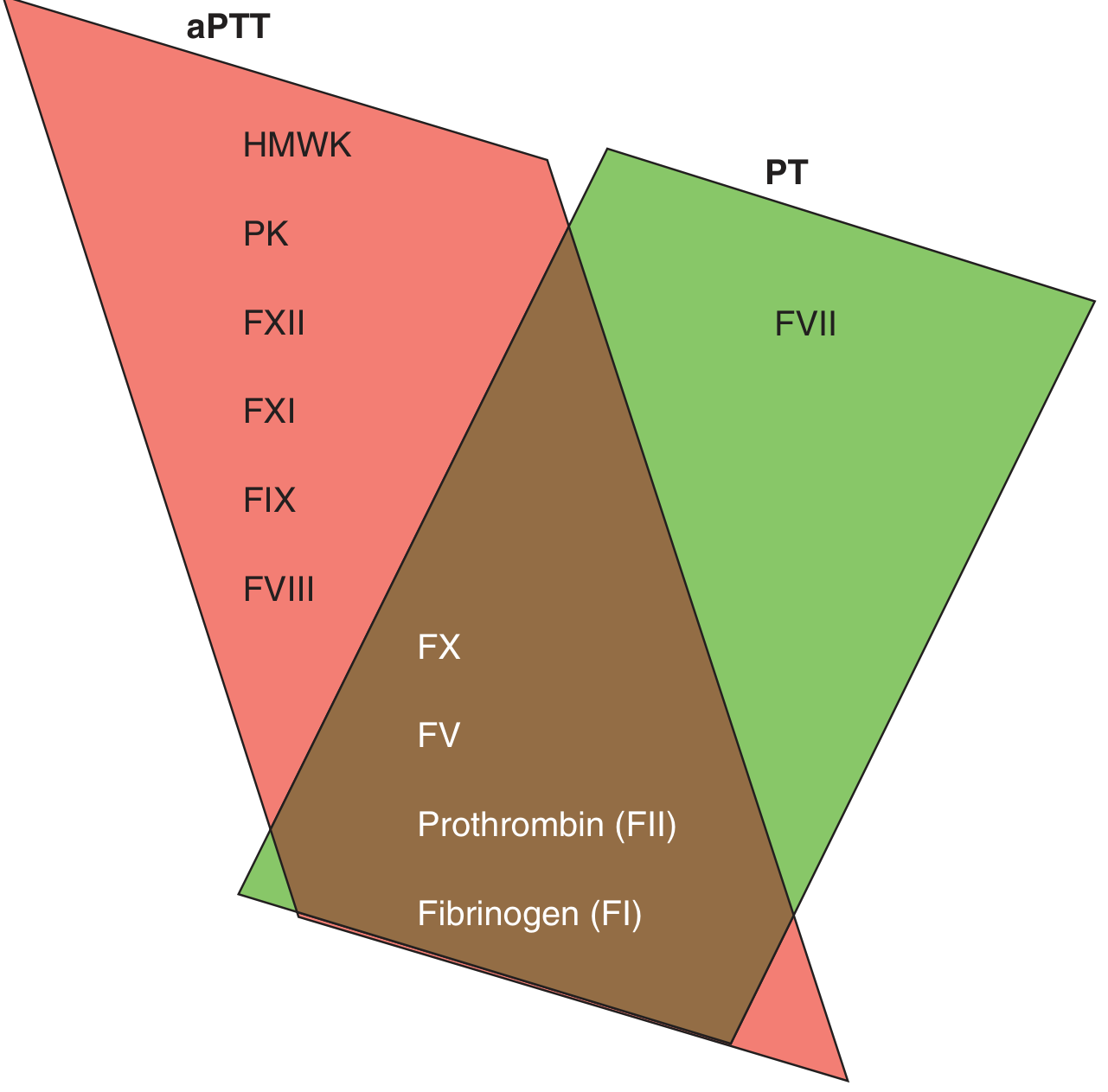
| Test | Pathway Tested | Factors Assessed |
|---|---|---|
| PT / INR | Extrinsic + Common pathway | I (fibrinogen), II, V, VII, X |
| aPTT | Intrinsic + Common pathway | I, II, V, VIII, IX, X, XI, XII, HMWK, Prekallikrein |
| Both prolonged | Common pathway | I, II, V, X (fibrinogen, prothrombin) |
The INR = (PT / PT normal mean)^ISI. It was developed to standardize vitamin K antagonist monitoring and is used in liver disease assessment, though it may overestimate bleeding risk in mild liver dysfunction. PT reagents have variable sensitivity to direct Xa inhibitors; PT is usually normal with apixaban.
- Harrison's Principles of Internal Medicine 22E, p. 518
Step 1 - Pre-Analytical Checks Before Interpreting Results
- Underfilled tube (must be >90% fill) - gives falsely prolonged PT/aPTT
- High hematocrit (>55%) - decreases plasma:anticoagulant ratio, gives false values
- Poor mixing or delayed processing
- Harrison's Principles of Internal Medicine 22E, p. 518
Step 2 - Pattern Recognition (The Core Framework)
Pattern 1: Isolated Prolonged PT (aPTT normal)
| Cause | Notes |
|---|---|
| Factor VII deficiency (congenital) | TT normal, aPTT normal, PT elevated |
| Early vitamin K deficiency | Factor VII has the shortest half-life (~6h); first to fall |
| Warfarin/vitamin K antagonists | Most common cause |
| Direct Xa inhibitors (rivaroxaban) | Prolongs PT, but PT may be normal with apixaban |
| Liver disease (mild-early) | Factor VII also synthesized in liver |
Pattern 2: Isolated Prolonged aPTT (PT normal)
| aPTT pattern | Factors | Clinical bleeding |
|---|---|---|
| No clinical bleeding | ↓ Factor XII, HMWK, Prekallikrein | None (contact activation factors) |
| Mild/variable bleeding | Mild ↓ Factor VIII or IX | Variable |
| Severe bleeding | Severe ↓ Factor VIII (Hemophilia A) or IX (Hemophilia B) | Yes, severe |
| No bleeding + thrombosis | Lupus anticoagulant | Paradoxical thrombosis |
| Anticoagulant use | Heparin, direct thrombin inhibitors (dabigatran) | Drug effect |
- Harrison's Principles of Internal Medicine 22E, Table 69-3
Pattern 3: Both PT and aPTT Prolonged
| Cause | Key Feature |
|---|---|
| Liver disease | Decreased synthesis of factors II, V, VII, X + fibrinogen |
| DIC (Disseminated Intravascular Coagulation) | + thrombocytopenia, elevated D-dimer, low fibrinogen |
| Vitamin K deficiency (severe/late) | Affects II, VII, IX, X |
| Massive transfusion / dilutional coagulopathy | History of large volume transfusion |
| Factor V, X, II, or fibrinogen deficiency | Rare congenital deficiencies |
| Warfarin overdose | INR markedly elevated |
| Supratherapeutic heparin | Also prolongs aPTT more |
-
Fibrinogen deficiency: TT ↑↑, aPTT ↑↑, PT ↑↑
-
Prothrombin (FII) or FV deficiency: TT normal, aPTT ↑, PT ↑
-
Factor X deficiency: TT normal, aPTT ↑, PT ↑
-
Vitamin K-dependent factor deficiency: TT normal, aPTT ↑, PT ↑↑
-
Goldman-Cecil Medicine, Table 157-2
Step 3 - The Mixing Study (Distinguishing Factor Deficiency vs. Inhibitor)
Corrects immediately AND stays corrected on incubation at 37°C
→ FACTOR DEFICIENCY (normal plasma supplied the missing factor)

Corrects immediately but PROLONGS on incubation at 37°C
→ ACQUIRED FACTOR INHIBITOR (most commonly acquired factor VIII inhibitor)

Does NOT correct at any time point
→ INHIBITOR present (lupus anticoagulant, heparin, paraproteins, fibrin split products)
- Harrison's Principles of Internal Medicine 22E, p. 518
Step 4 - Specific Second-Line Tests
| Test | When to Use | What It Detects |
|---|---|---|
| Specific factor assays (VIII, IX, XI, VII, etc.) | After mixing study guides you | Exact factor level |
| Fibrinogen level (Clauss) | Both PT + aPTT prolonged | Hypo/afibrinogenemia, DIC |
| Thrombin time (TT) | Suspected fibrinogen problem, heparin | Fibrinogen conversion to fibrin |
| Reptilase time | TT prolonged - is it heparin? | Normal with heparin (distinguishes heparin from fibrinogen defect) |
| D-dimer + fibrinogen | Suspected DIC | Consumption coagulopathy |
| Anti-Xa level | LMWH monitoring, apixaban/rivaroxaban activity | Direct factor Xa inhibitor levels |
| dRVVT | Suspected lupus anticoagulant | Phospholipid-dependent test |
| vWF antigen + activity | aPTT prolonged, mild bleeding | von Willebrand disease (vWD) |
Step 5 - Clinical Context Integration
| Bleeding site | Suggests |
|---|---|
| Mucosal, petechiae, immediate after trauma | Platelet/vascular defect (PT/aPTT may be normal) |
| Deep hematomas, hemarthroses, delayed after trauma | Coagulation factor deficiency |
| Mixed picture | DIC, liver disease |
- Liver disease: PT/INR prolonged earliest (factor VII), then aPTT; thrombocytopenia often co-exists. Note: mild INR elevation may overestimate true bleeding risk (thrombin generation may be preserved).
- DIC: PT ↑ + aPTT ↑ + platelets ↓ + fibrinogen ↓ + D-dimer ↑ - treat the underlying cause.
- Warfarin: Isolated PT/INR prolongation; aPTT can also prolong at supratherapeutic levels.
- Heparin (UFH): Predominantly prolongs aPTT; TT also prolonged; reptilase time normal.
- Dabigatran: Prolongs TT markedly; aPTT sensitive but not linear.
- Apixaban: PT often normal; anti-Xa assay needed to assess drug levels.
- Hemophilia A/B: Isolated aPTT prolongation, corrects on mixing, factor VIII or IX assay confirms.
- Lupus anticoagulant: aPTT prolonged, does NOT correct on mixing, dRVVT confirmatory, associated with thrombosis not bleeding.
Quick Algorithm Summary
ABNORMAL PT / aPTT / INR
         ↓
1. Exclude pre-analytical error (repeat if needed)
         ↓
2. Pattern:
   PT only ↑ → Extrinsic (FVII, early VitK deficiency, warfarin, Xa inhibitors)
   aPTT only ↑ → Intrinsic (FVIII/IX/XI deficiency, heparin, lupus AC, XII/HMWK/PK)
   Both ↑ → Common pathway / multiple (liver disease, DIC, VitK deficiency, FV/FX/FII/Fibrinogen)
         ↓
3. Mixing study (1:1 patient:normal plasma):
   Corrects → Factor deficiency → specific factor assay
   No correction → Inhibitor → lupus AC workup (dRVVT), factor inhibitor assay
         ↓
4. Correlate with: clinical bleeding, family history, medications, liver function, DIC screen
Generate a diagnostic flowchart as a visual decision tree for PT/aPTT patterns
A detailed clinical diagnostic flowchart / decision tree for interpreting abnormal PT, aPTT, and INR coagulation test results. The flowchart starts at the top with a box labeled "ABNORMAL PT / aPTT / INR" with an arrow pointing down to "Exclude Pre-Analytical Error (underfilled tube, high hematocrit, repeat test)". Then branches into three main pattern boxes side by side: 1. LEFT: "Isolated PT ↑ (aPTT Normal)" - colored orange/amber 2. CENTER: "Both PT ↑ AND aPTT ↑" - colored red 3. RIGHT: "Isolated aPTT ↑ (PT Normal)" - colored blue Under LEFT branch (PT only elevated), list causes in a box: • Factor VII deficiency • Early Vitamin K deficiency • Warfarin / VKA therapy • Rivaroxaban (direct Xa inhibitor) • Mild liver disease Under CENTER branch (both elevated), arrow goes down to a box labeled "MIXING STUDY" then branches: - LEFT sub-branch: "Corrects → Factor Deficiency" listing: DIC (+ low fibrinogen, high D-dimer, low platelets), Liver disease (II, V, VII, X, fibrinogen ↓), Severe Vitamin K deficiency, Factor V / X / II / Fibrinogen deficiency, Massive transfusion - RIGHT sub-branch: "No Correction → Inhibitor": Heparin supratherapeutic, Paraproteins, Fibrin split products Under RIGHT branch (aPTT only elevated), arrow goes down to "MIXING STUDY (1:1 patient + normal plasma)" which then branches into TWO paths: - LEFT: "Corrects → Factor Deficiency" with sub-boxes: "Factor VIII ↓ → Hemophilia A", "Factor IX ↓ → Hemophilia B", "Factor XI ↓ → Hemophilia C", "Factor XII / HMWK / PK ↓ → No bleeding", "vWD (check vWF antigen + activity)" - RIGHT: "No Correction → Inhibitor" further branching into: "Immediate no correction → Lupus Anticoagulant (confirm with dRVVT, associated with THROMBOSIS not bleeding)" and "Corrects then prolongs on incubation → Acquired Factor VIII Inhibitor (check factor VIII inhibitor assay)" At the bottom, a footer box labeled "KEY ADDITIONAL TESTS: Thrombin Time (TT) • Reptilase Time • D-dimer • Fibrinogen level • Anti-Xa level • Factor-specific assays • dRVVT for lupus anticoagulant" Clean white background, medical textbook style, professional clinical flowchart, clear arrows, color-coded branches (orange for PT-only, blue for aPTT-only, red for both elevated, green for corrects, purple for no correction), bold headings, neat sans-serif font, high resolution.
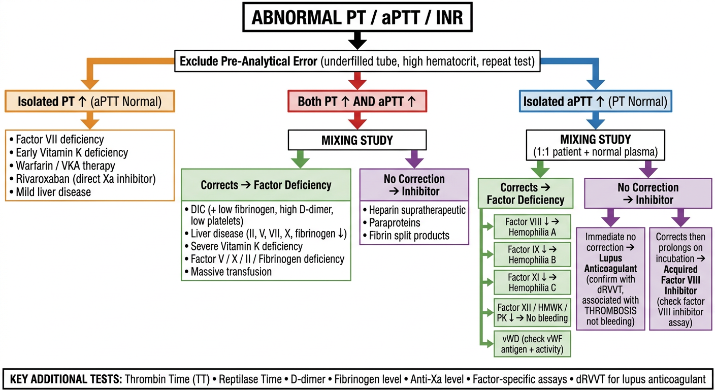
- Orange (left) - Isolated PT elevation - points to extrinsic pathway problems (Factor VII, early vitamin K deficiency, warfarin, rivaroxaban)
- Red (center) - Both PT and aPTT elevated - common/multiple pathway involvement (DIC, liver disease, severe vitamin K deficiency, supratherapeutic heparin)
- Blue (right) - Isolated aPTT elevation - intrinsic pathway, leads to the mixing study
- Corrects (green) - factor deficiency - proceed to specific factor assays
- No correction (purple) - inhibitor present
- Immediate non-correction - Lupus Anticoagulant (confirm with dRVVT; causes thrombosis, not bleeding)
- Corrects then prolongs on 37°C incubation - Acquired Factor VIII inhibitor
Search for recent guidelines on perioperative coagulation management
perioperative coagulation management guidelines
perioperative coagulation management guidelines 2024 2025
PMID: 39029742
PMID: 38992466
PMID: 41654142
perioperative anticoagulation reversal bleeding management
PMID: 40448969
PMID: 38383680
viscoelastic hemostatic assay TEG ROTEM perioperative transfusion
Recent Guidelines on Perioperative Coagulation Management (2024-2026)
1. ISTH SSC - Surgical & Procedural Bleed Risk Stratification (2026)
- Provides a practical procedure-specific bleeding risk stratification framework for use in adults on anticoagulation undergoing elective procedures
- Addresses when to interrupt anticoagulation, for how long, and when to safely restart
- Recognizes that discordant perioperative management is largely driven by the lack of harmonized bleed risk schemas
- Intended to work alongside thromboembolic risk assessment tools
2. ISTH SSC - Reversal of Direct Oral Anticoagulants (2024)
| DOAC | Specific Reversal Agent |
|---|---|
| Apixaban, Rivaroxaban, Edoxaban (Factor Xa inhibitors) | Andexanet alfa |
| Dabigatran (direct thrombin inhibitor) | Idarucizumab |
| When specific agents unavailable | Prothrombin complex concentrates (PCCs) |
- Also covers reversal of oral FXIa inhibitors (a new class currently in clinical development)
- Non-specific agents (4-factor PCC, activated PCC) remain a fallback option when specific reversal agents are unavailable
3. 2025 DOAC Guidelines - Practical Perioperative & Bleeding Management
- No routine "bridging" with LMWH when interrupting DOACs for planned procedures - this is now firmly consensus across guidelines
- DOACs do not require routine laboratory testing for monitoring
- For planned procedures: interrupt and resume without bridging
- Coagulation test guidance for urgent surgery:
- aPTT for dabigatran (qualitative guidance)
- PT ratio for rivaroxaban (ratio ≤1.2 with high probability rules out significant activity)
- PT is not reliable for apixaban or edoxaban - use anti-Xa assay
- Plasma DOAC levels <30 ng/mL considered acceptable for surgery with standard bleeding risk
4. 2025 Multisociety Consensus - Perioperative Antithrombotic Management (Spain/Europe)
- For UFH: discontinue 4-6 hours before the procedure; 12 hours for prophylactic doses
- For VKAs (warfarin): restart as early as possible postoperatively (requires 36-48 hours to achieve therapeutic effect)
- When bridging is needed: restart heparin (LMWH or UFH) at therapeutic doses within the first 24 hours postoperatively
- Neuraxial/regional anesthesia with DOACs (per 2025 ASRA 5th edition guidelines):
- High-dose DOACs: 72-hour hold before neuraxial procedures
- Acceptable residual DOAC: levels <30 ng/mL or anti-Xa activity ≤0.1 IU/mL
- Reversal agents (andexanet alfa, idarucizumab, PCC) are not recommended for routine reversal prior to neuraxial procedures
5. French National Authority for Health (HAS) - Perioperative Patient Blood Management (2024)
| Perioperative Phase | Key Recommendations |
|---|---|
| Preoperative | Detect and treat anemia/iron deficiency well in advance of surgery with moderate-high bleeding risk |
| Intraoperative | Tranexamic acid (TXA) for high-bleeding-risk surgery; cell salvage for surgeries with major transfusion risk |
| Postoperative | Limit unnecessary blood sampling; monitor for postoperative anemia; IV iron where indicated |
| Transfusion threshold | "Restrictive" threshold adjusted for comorbidities and patient tolerance |
6. Perioperative Management of Antithrombotics in Intracranial Surgery (2024)
- Best methodological quality: European Society of Anaesthesiology (ESA) guidelines (two sets) and American College of Chest Physicians (ACCP)
- Highlights ongoing uncertainty due to lack of high-quality RCT data in neurosurgical patients specifically
- Key tension: risk of thromboembolism vs. perioperative hemorrhage in a closed intracranial space
7. HIT in Cardiac Surgery - ISTH Guidance (2026)
Summary of Cross-Guideline Consensus Points (2024-2026)
| Topic | Current Consensus |
|---|---|
| Bridging with LMWH for DOACs | Not recommended for most elective procedures |
| DOAC reversal for life-threatening bleeding | Andexanet alfa (Xa inhibitors), Idarucizumab (dabigatran); PCC if unavailable |
| Monitoring DOACs pre-urgent surgery | Anti-Xa assay for apixaban/edoxaban; PT ratio for rivaroxaban; aPTT for dabigatran |
| Acceptable DOAC level for surgery | <30 ng/mL plasma level |
| Neuraxial anesthesia DOAC hold | 72 hours for high-dose DOACs (ASRA 2025) |
| Tranexamic acid | Recommended for high-bleeding-risk surgery (strong evidence) |
| Preoperative anemia | Detect and treat iron deficiency well before elective surgery |
| Viscoelastic tests (TEG/ROTEM) | Increasingly supported for goal-directed transfusion in cardiac/major surgery |
Factor XII / HMWK / PK deficiency approach
Factor XII HMWK prekallikrein deficiency clinical approach prolonged aPTT no bleeding
factor XII deficiency contact activation thrombosis aPTT
Factor XII / HMWK / Prekallikrein Deficiency - Clinical Approach
The Paradox in One Sentence
The Contact Activation System - What It Is
| Protein | Role | Alternate Name |
|---|---|---|
| Factor XII | Initiating serine protease; activated by negatively charged surfaces | Hageman factor |
| Prekallikrein (PK) | Reciprocal activator of FXII; converted to kallikrein by FXIIa | Fletcher factor |
| High-molecular-weight kininogen (HMWK) | Non-enzymatic cofactor; anchors PK and FXII to activating surfaces | Fitzgerald factor |
| Factor XI | Downstream target of FXIIa; bridges contact pathway to intrinsic pathway | Note: FXI deficiency DOES cause mild bleeding - separate entity |
Negatively charged surface (glass, kaolin, celite, ellagic acid)
        ↓
FXII → FXIIa  [HMWK as cofactor]
        ↓
Prekallikrein → Kallikrein  [amplification loop back to FXII]
        ↓
FXI → FXIa → FIX → FX → Thrombin → Fibrin
- Goldman-Cecil Medicine, p. 1798 | Braunwald's Heart Disease, Chapter 95
Why the aPTT is Markedly Prolonged
A clue: an aPTT >100-120 seconds with completely no bleeding history strongly suggests a contact factor deficiency rather than hemophilia A or B.
Key Clinical Features - Comparison Table
| Feature | FXII Deficiency | Prekallikrein Deficiency | HMWK Deficiency |
|---|---|---|---|
| aPTT | Markedly prolonged | Markedly prolonged | Markedly prolonged |
| PT | Normal | Normal | Normal |
| Clinical bleeding | None | None | None |
| Thrombotic risk | Increased (8-10% VTE, occasionally fatal) | Possibly increased | Possibly increased |
| Inheritance | Autosomal recessive | Autosomal recessive | Autosomal recessive |
| Prevalence | Rare; carrier rate ~1:1000 | Very rare | Very rare |
| Famous case | John Hageman (original patient) died of PE | - | - |
Hageman (FXIIa) deficiency: the original patient after whom factor XII was named died of a pulmonary embolism - highlighting the paradox.
Diagnostic Approach
Step 1 - Recognize the Pattern
- Isolated markedly prolonged aPTT + PT normal + no bleeding history = contact factor deficiency until proven otherwise
Step 2 - Exclude Heparin Contamination
- Clinical history (heparin flushes in lines)
- Heparin neutralization (Hepzyme procedure)
- Thrombin time: prolonged in heparin; reptilase time: normal in heparin
Step 3 - Mixing Study (1:1 patient + normal plasma)
- Contact factor deficiency: corrects on mixing
- Rosner Index: (mix aPTT - normal aPTT) / patient aPTT × 100
- <12% = correction (factor deficiency)
- 12-15% = gray zone
- >15% = no correction (inhibitor)
A Special Clue: Incubation Time Effect
- Kaolin/celite can slowly activate residual FXII even at low levels
- Prekallikrein deficiency in particular may "normalize" aPTT on longer incubation
If aPTT shortens or normalizes with prolonged incubation time → strongly suggests prekallikrein deficiency
- Goldman-Cecil Medicine, p. 2142 | Quick Compendium of Clinical Pathology 5th ed.
Step 4 - Specific Factor Assays
- Factor XII activity assay
- Prekallikrein (PK) activity assay
- HMWK activity assay
- These are available at specialized coagulation labs
Management
No treatment is required for contact activation factor deficiencies, regardless of how prolonged the aPTT is.
| Scenario | Management |
|---|---|
| Incidental finding pre-surgery | Reassure; proceed with surgery without factor replacement or FFP |
| Major/elective surgery | No special preparation; standard perioperative care |
| Acute bleeding during surgery | Bleeding is NOT due to the contact deficiency; investigate other causes |
| Venous thromboembolism | Treat with standard anticoagulation (LMWH, DOACs) - no contraindication |
| aPTT monitoring during heparin therapy | Use anti-Xa assay instead, as aPTT is unreliable in these patients |
"Deficiencies of the contact activation factors, however severe they are and however prolonged the associated aPTT may be, do not cause clinical bleeding problems, even in response to surgery or trauma. Therefore, no therapy is indicated." - Goldman-Cecil Medicine, p. 2147
The Thrombosis Paradox - Emerging Biology
- Medical devices (stents, mechanical valves, CPB circuits, ECMO) activate FXII on foreign surfaces → device-related thrombosis
- Neutrophil extracellular traps (NETs) activate the contact pathway → thromboinflammation
- Polyphosphates released from platelet dense granules activate FXII in vivo
- Animal studies: FXII-deficient mice form small, unstable thrombi at sites of arterial/venous injury
- In humans: Factor XI antisense oligonucleotides and FXIa inhibitors reduce postoperative VTE better than enoxaparin in knee arthroplasty
- Braunwald's Heart Disease, Chapter 95 | Henry's Clinical Diagnosis and Management by Laboratory Methods
Quick Summary Algorithm
Markedly prolonged aPTT + PT normal + NO bleeding history
                    ↓
         Exclude heparin contamination
                    ↓
              Mixing study → Corrects
                    ↓
     Does aPTT normalize on prolonged incubation?
         YES → Prekallikrein deficiency likely
         NO  → Factor XII or HMWK deficiency likely
                    ↓
     Specific factor assays: FXII, PK, HMWK
                    ↓
     DIAGNOSIS CONFIRMED
     ↓                        ↓
NO TREATMENT NEEDED     Screen for VTE risk
Proceed with surgery    Standard anticoagulation if thrombosis