
What is the blood supply of brain?
blood supply of brain circle of Willis cerebral arteries diagram
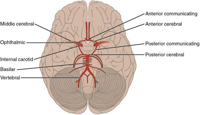
An anatomical diagram illustrating the arterial supply of the brain from an inferior (basal) perspective, specifically highlighting the Circle of Willis and its contributing vessels. The diagram shows the bilateral internal carotid arteries branching into the middle cerebral (MCA) and ophthalmic arteries. Centrally, the anterior cerebral arteries (ACA) are seen originating and coursing medially into the longitudinal fissure, connected by the anterior communicating artery (ACoA). The posterior circulation is depicted with the vertebral arteries merging into the basilar artery, which subsequently divides into the posterior cerebral arteries (PCA). Posterior communicating arteries (PCoA) are shown forming the critical anastomotic link between the internal carotid and posterior cerebral systems. The diagram is superimposed on the ventral surface of the brain, demonstrating the spatial relationship of these vessels to the frontal, temporal, and occipital lobes, as well as the cerebellum and brainstem. This illustration is an educational resource for understanding neurovascular anatomy and the territories susceptible to ischemic stroke.
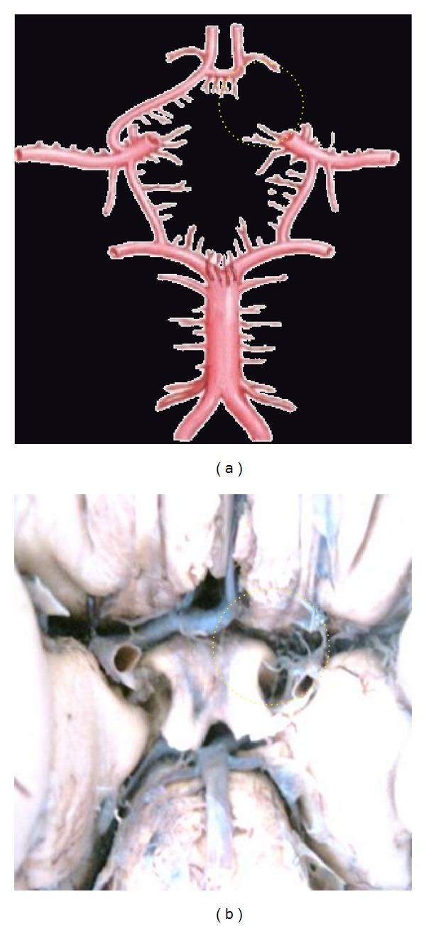
This medical visual content consists of an anatomical diagram (a) and a corresponding clinical photograph of a cadaveric dissection (b) illustrating neurovascular anatomy. The focus is on the Circle of Willis, specifically demonstrating a congenital vascular variation known as aplasia of the A1 segment of the anterior cerebral artery (ACA). In both panels, a yellow dotted circle highlights the area where the right A1 segment should connect the internal carotid artery to the anterior communicating artery (ACoA). The absence of this segment results in an incomplete Circle of Willis, which has clinical significance for collateral cerebral blood flow. The diagram (a) clearly depicts the major components of the cerebral arterial circle, including the basilar artery, posterior cerebral arteries, internal carotid arteries, and the intact left A1 and A2 segments. The clinical photograph (b) provides a real-world view of the same anatomical deficiency at the base of the brain. This material is designed for advanced medical education in neuroanatomy and radiology, emphasizing common vascular anomalies.
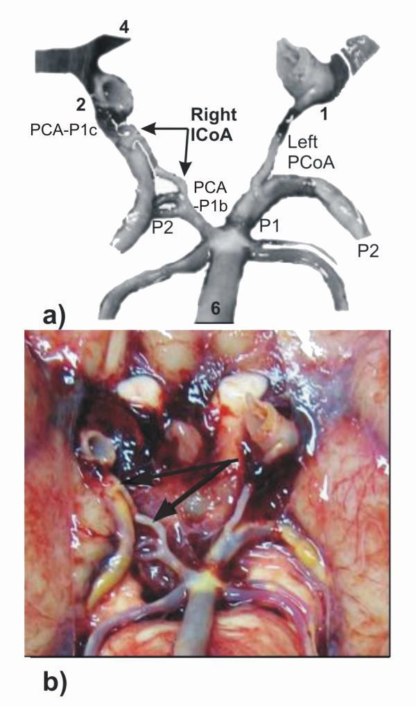
This medical figure consists of an anatomical diagram (a) and a matching intraoperative/cadaveric photograph (b) illustrating an anatomical variant in the posterior Circle of Willis. The visual focus is the 'intermediate communicating artery' (ICoA) on the right side. In the diagram, the right posterior cerebral artery (PCA) is shown with dual pre-communicating origins: a carotid origin segment (PCA-P1c) and a basilar origin segment (PCA-P1b), linked by the right ICoA (indicated by arrows). The left side shows a conventional posterior communicating artery (PCoA) connecting the internal carotid artery (1) to the PCA. Other labeled structures include the basilar artery (6), internal carotid arteries (1, 2), and the post-communicating PCA segments (P2). The clinical photograph (b) provides a real-world correlate of this vascular configuration on the basal surface of the brain, highlighting the small caliber and anatomical path of the accessory ICoA. This material is designed for advanced neuroanatomical education, demonstrating rare vascular complexities and variations in cerebral blood supply.
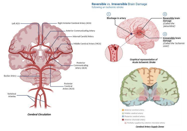
Educational medical infographic containing three panels illustrating cerebral neurovasculature and acute ischemic stroke pathophysiology. The first panel, 'Cerebral Circulation', features a detailed anatomical diagram of the Circle of Willis, labeling the vertebral arteries, basilar artery, and its bifurcation into the posterior cerebral arteries (PCA). It further illustrates the internal carotid arteries, middle cerebral arteries (MCA), and the anterior cerebral arteries (ACA) connected by the anterior communicating artery. The second panel, 'Graphical representation of Acute Ischaemic Stroke', depicts a sagittal view of the brain showing an arterial blockage. It differentiates between the 'ischemic core' (irreversible damage) and the surrounding 'penumbra' (reversible/salvageable tissue). The third panel, 'Cerebral Artery Supply Zones', is a coronal brain cross-section color-coded to show perfusion territories, specifically highlighting regions supplied by the ACA (orange), MCA (green), PCA (blue), and anterior choroidal artery (red). This resource is designed for students learning about stroke mechanisms, vascular anatomy, and clinical neuro-perfusion.
Blood Supply of the Brain
- Gray's Anatomy for Students, p. 1017
- Neuroanatomy through Clinical Cases, 3rd Edition, p. 70
1. Arterial Supply - Two Systems
A. Anterior Circulation (Internal Carotid Arteries)
- The internal carotid arteries (ICA) arise from the common carotid arteries, ascend through the neck, enter the skull via the carotid canals, and give off the following branches inside the cranial cavity:
- Ophthalmic artery - supplies the eye and orbit
- Posterior communicating artery - connects ICA to the posterior cerebral artery (links anterior and posterior circulations)
- Middle cerebral artery (MCA) - the largest branch; supplies the lateral surface of the cerebral hemispheres, deep structures of the basal forebrain (lenticulostriate arteries), and internal capsule
- Anterior cerebral artery (ACA) - supplies the medial surface of the frontal and parietal lobes
B. Posterior Circulation (Vertebrobasilar System)
- The vertebral arteries arise from the subclavian arteries, pass upward through the foramina transversaria of the upper 6 cervical vertebrae, and enter the skull via the foramen magnum.
- Inside the skull, each vertebral artery gives off:
- Posterior inferior cerebellar artery (PICA) - supplies the posterior inferior cerebellum and lateral medulla
- Posterior spinal artery - descends on the posterior spinal cord
- Anterior spinal artery (formed by contributions from both vertebrals) - descends in the anterior median fissure of the spinal cord
- The two vertebral arteries fuse at the base of the pons to form the single basilar artery
- The basilar artery runs rostrally along the anterior pons and gives off:
- Anterior inferior cerebellar artery (AICA) - supplies anterior inferior cerebellum
- Pontine arteries - multiple small branches to the pons
- Superior cerebellar artery (SCA) - supplies the superior cerebellum
- Posterior cerebral arteries (PCA) - terminal branches; supply the occipital lobe (primary visual cortex), inferior temporal lobe, thalamus, and midbrain
2. The Circle of Willis (Cerebral Arterial Circle)
| Component | Connection |
|---|---|
| Anterior communicating artery (ACoA) | Connects left and right anterior cerebral arteries |
| Posterior communicating artery (PCoA) x2 | Each connects the internal carotid artery to the ipsilateral posterior cerebral artery |

3. Cortical Territory Distribution
| Artery | Territory Supplied |
|---|---|
| ACA | Medial frontal lobe, medial parietal lobe (motor/sensory for the leg) |
| MCA | Lateral cortex (frontal, parietal, temporal), internal capsule, basal ganglia |
| PCA | Occipital lobe (visual cortex), inferior/medial temporal lobe, thalamus |
| PICA | Lateral medulla, posterior inferior cerebellum |
| AICA | Anterior inferior cerebellum, lateral pons |
| SCA | Superior cerebellum, lateral midbrain/pons |
4. Venous Drainage
- Cerebral veins (superficial and deep) drain into the dural venous sinuses (superior sagittal sinus, inferior sagittal sinus, straight sinus, transverse sinuses, sigmoid sinuses)
- The sigmoid sinuses empty into the internal jugular veins at the jugular foramina
Clinical Significance
- MCA occlusion: Contralateral hemiparesis (arm > leg), hemisensory loss, homonymous hemianopia, aphasia (dominant hemisphere)
- ACA occlusion: Contralateral leg weakness and sensory loss
- PCA occlusion: Contralateral homonymous hemianopia
- Lateral medullary (Wallenberg) syndrome: PICA occlusion - ipsilateral facial, contralateral body sensory loss, dysphagia, Horner's syndrome