
Anatomy of the upper limb
Reading File
The query is broad - upper limb anatomy covers bones, joints, muscles, nerves, vessels, and more. Let me clarify the focus before diving in.
Asking for Preferences
Which aspect of upper limb anatomy are you most interested in? · What level is this for?
I'll deliver a comprehensive overview covering all major aspects. Let me search the library in parallel.
Finding Sources
Finding Sources
Finding Sources
Reading File
Finding Sources
Finding Sources
Reading File
Finding Sources
Finding Sources
Reading File
Finding Sources
Reading File
Reading File
Reading File
I now have rich content from Gray's Anatomy for Students. Let me fetch the key brachial plexus diagram and the nerve innervation diagram to include in the answer.
Loading Image
Loading Image
Loading Image
Excellent - I now have comprehensive content and diagrams. Let me compile the full answer.
Anatomy of the Upper Limb
Source: Gray's Anatomy for Students; Rosen's Emergency Medicine
1. Overview and Regions
The upper limb is divided into:
- Shoulder / pectoral girdle (clavicle + scapula connecting the limb to the axial skeleton)
- Arm (shoulder to elbow)
- Forearm (elbow to wrist)
- Hand (carpals, metacarpals, phalanges)
- Axilla (armpit - a pyramid-shaped space that serves as the neurovascular highway)
2. Bones
| Region | Bones |
|---|---|
| Pectoral girdle | Clavicle, Scapula |
| Arm | Humerus |
| Forearm | Radius (lateral), Ulna (medial) |
| Wrist | 8 Carpal bones (proximal row: Scaphoid, Lunate, Triquetrum, Pisiform; distal row: Trapezium, Trapezoid, Capitate, Hamate) |
| Hand | 5 Metacarpals, 14 Phalanges (proximal, middle, distal - except thumb which has 2) |
Key landmarks:
- Humerus: greater and lesser tubercles, intertubercular sulcus (biceps tendon), surgical neck (axillary nerve), radial groove (radial nerve), medial and lateral epicondyles
- Ulna: olecranon, coronoid process, trochlear notch
- Radius: head, radial tuberosity (biceps insertion), styloid process
3. Joints
| Joint | Type | Key Features |
|---|---|---|
| Sternoclavicular | Synovial, saddle | Only bony articulation of upper limb with axial skeleton |
| Acromioclavicular | Synovial, plane | Site of AC joint dislocation |
| Glenohumeral (shoulder) | Synovial, ball-and-socket | Most mobile but least stable joint; reinforced by rotator cuff |
| Elbow (humeroulnar + humeroradial) | Synovial, hinge | Flexion/extension; carrying angle ~170° |
| Proximal + Distal Radioulnar | Synovial, pivot | Pronation/supination |
| Wrist (radiocarpal) | Synovial, condyloid | Flexion, extension, abduction, adduction |
| Carpometacarpal of thumb | Saddle | Enables thumb opposition |
Rotator Cuff - four muscles that stabilize the glenohumeral joint (mnemonic SITS):
- Supraspinatus (abduction, first 15°)
- Infraspinatus (lateral rotation)
- Teres minor (lateral rotation)
- Subscapularis (medial rotation)
4. Brachial Plexus
The brachial plexus is the network of nerves supplying virtually all of the upper limb. It is formed by the anterior rami of C5-T1.
Structure: Roots → Trunks → Divisions → Cords → Branches
(Mnemonic: Real Texans Drink Cold Beer)


Trunks
| Trunk | Roots |
|---|---|
| Superior | C5 + C6 |
| Middle | C7 |
| Inferior | C8 + T1 |
Cords (named by relation to axillary artery)
| Cord | Origin | Terminal nerves |
|---|---|---|
| Lateral (C5-C7) | Anterior div. of Superior + Middle | Musculocutaneous, lateral root of Median |
| Medial (C8-T1) | Anterior div. of Inferior | Ulnar, medial root of Median |
| Posterior (C5-T1) | All three posterior divs. | Radial, Axillary |
Major Terminal Nerves
| Nerve | Root | Motor | Sensory | Injury |
|---|---|---|---|---|
| Musculocutaneous | C5-C7 | Biceps, brachialis, coracobrachialis | Lateral forearm (as lateral cutaneous n.) | Weak elbow flexion/supination |
| Median | C5-T1 | Forearm flexors (except FCU + medial FDP), thenar muscles, lumbricals 1&2 | Lateral 3.5 digits (palm) | Carpal tunnel; "ape hand", loss of thumb opposition |
| Ulnar | C8-T1 | FCU, medial FDP, hypothenar, interossei, lumbricals 3&4, adductor pollicis | Medial 1.5 digits | "Claw hand" (ring/little fingers); at medial epicondyle |
| Radial | C5-T1 | All extensors (triceps, wrist/finger extensors), supinator | Posterior arm/forearm, dorsal lateral 3.5 digits | "Wrist drop" (posterior cord/spiral groove injury) |
| Axillary | C5-C6 | Deltoid, teres minor | Lateral shoulder (regimental badge area) | Loss of shoulder abduction; surgical neck fracture |
5. Muscles
Arm
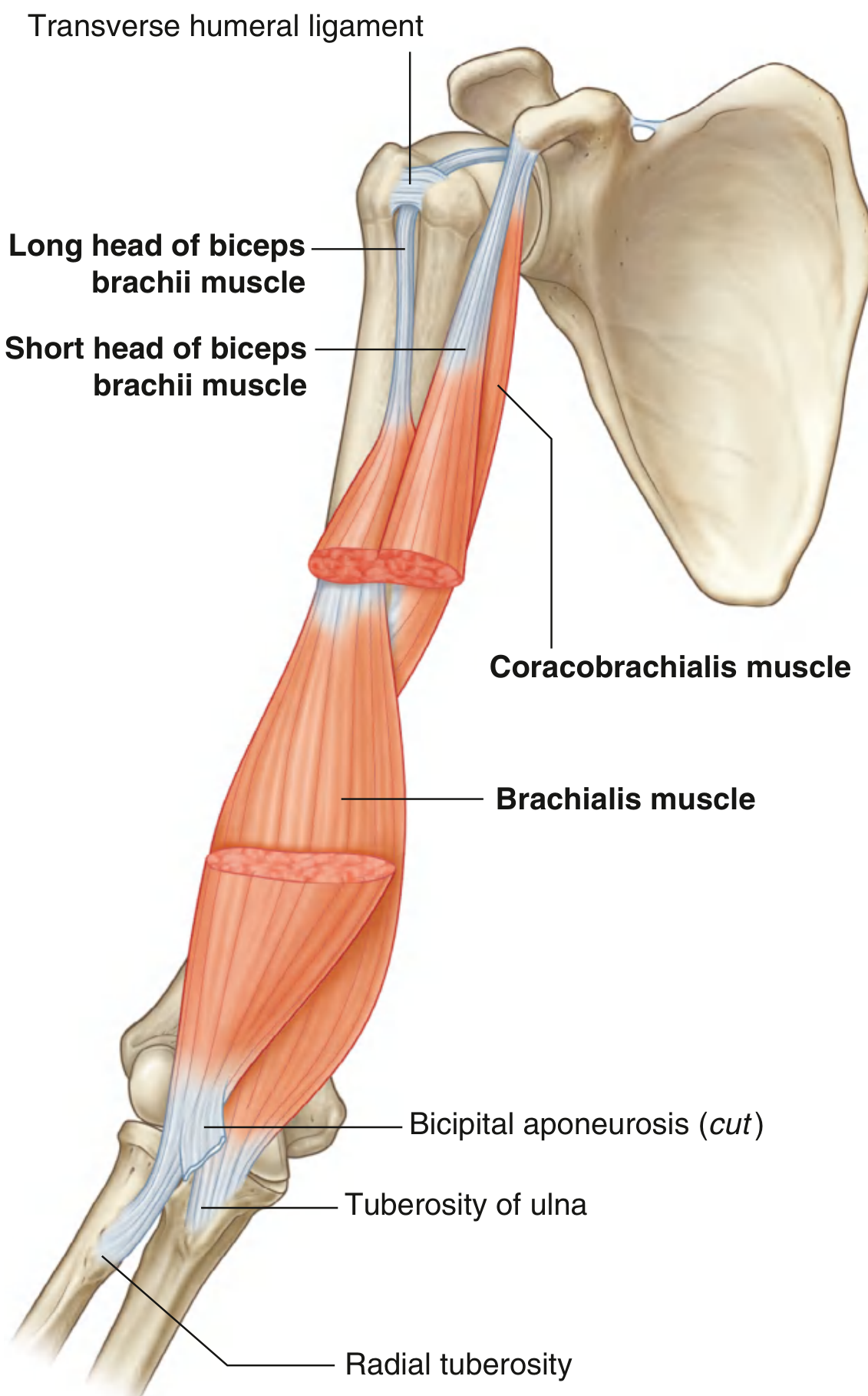
Anterior compartment (innervated by musculocutaneous nerve, C5-C7):
- Biceps brachii - flexes elbow, supinates forearm (long head from supraglenoid tubercle; short head from coracoid; inserts on radial tuberosity)
- Brachialis - prime mover of elbow flexion (inserts on ulnar tuberosity)
- Coracobrachialis - flexes arm (from coracoid to medial humerus)
Posterior compartment (innervated by radial nerve, C6-C8):
- Triceps brachii - extends elbow; long head originates from infraglenoid tubercle; inserts on olecranon
Forearm
Anterior compartment (flexors, generally median and ulnar nerves):
Superficial layer: Pronator teres, Flexor carpi radialis (FCR), Palmaris longus, Flexor carpi ulnaris (FCU - ulnar n.), Flexor digitorum superficialis (FDS)
Deep layer: Flexor digitorum profundus (FDP - lateral half median, medial half ulnar), Flexor pollicis longus (FPL - AIN/median), Pronator quadratus (AIN/median)
Posterior compartment (extensors, radial nerve):
Superficial: Brachioradialis, ECRL, ECRB, Extensor digitorum, Extensor digiti minimi, Extensor carpi ulnaris, Anconeus
Deep: Supinator, APL, EPL, EPB, Extensor indicis
Hand
- Thenar muscles (median nerve): Abductor pollicis brevis, Flexor pollicis brevis (superficial head), Opponens pollicis
- Hypothenar muscles (ulnar nerve): Abductor digiti minimi, Flexor digiti minimi, Opponens digiti minimi
- Lumbricals: flex MCP, extend IP joints (L1&2 = median; L3&4 = ulnar)
- Interossei: Dorsal = abduct fingers (DAB); Palmar = adduct fingers (PAD) - all ulnar nerve
- Adductor pollicis (ulnar nerve)
6. Blood Supply
| Vessel | Origin | Territory |
|---|---|---|
| Subclavian artery | Brachiocephalic trunk (R) / Aortic arch (L) | Becomes axillary artery at lateral border of 1st rib |
| Axillary artery (3 parts, divided by pectoralis minor) | From subclavian | Branches: thoracoacromial, lateral thoracic, subscapular, anterior/posterior circumflex humeral |
| Brachial artery | Continues from axillary at lower border of teres major | Entire arm; divides at cubital fossa |
| Radial artery | Terminal branch of brachial | Lateral forearm, anatomical snuffbox, deep palmar arch |
| Ulnar artery | Terminal branch of brachial | Medial forearm, superficial palmar arch |
Palmar arches:
- Superficial palmar arch = primarily ulnar artery + small contribution from radial
- Deep palmar arch = primarily radial artery + deep branch of ulnar
7. Venous Drainage
Superficial veins (clinically important):
- Cephalic vein - lateral; runs in deltopectoral groove; drains to axillary vein
- Basilic vein - medial; pierces deep fascia in arm; drains to axillary vein
- Median cubital vein - connects cephalic and basilic in the cubital fossa; preferred site for venipuncture
Deep veins accompany the arteries (venae comitantes).
8. Lymphatics
Axillary lymph nodes (drain the upper limb and breast) are arranged in 5 groups:
- Pectoral (anterior) - drain anterior chest wall and breast
- Humeral (lateral) - drain most of the upper limb
- Subscapular (posterior) - drain posterior wall, shoulder, neck
- Central - receive from the above three groups
- Apical - drain all other groups; efferent vessels form the subclavian trunk
Axillary node clearance (e.g., in breast cancer surgery) or axillary radiotherapy can damage lymphatic drainage, causing lymphoedema of the upper limb.
9. Key Clinical Correlates
| Injury | Mechanism | Deficit |
|---|---|---|
| Erb's palsy (upper brachial plexus, C5-C6) | Forced neck-shoulder separation | "Waiter's tip" - arm adducted, medially rotated, elbow extended |
| Klumpke's palsy (lower, C8-T1) | Forced arm abduction | Intrinsic hand weakness; "claw hand" ± Horner syndrome |
| Axillary nerve injury | Shoulder dislocation / surgical neck fracture | Loss of deltoid contraction; loss of regimental badge sensation |
| Radial nerve in spiral groove | Mid-shaft humeral fracture | Wrist drop; loss of finger extension |
| Median nerve at carpal tunnel | CTS | Thenar wasting; loss of thumb opposition; first 3.5 digit numbness |
| Ulnar nerve at medial epicondyle | Cubitus valgus fracture | Ring/little finger claw; loss of interossei; Froment's sign |
| Anterior interosseous nerve | Compressive injury | Loss of FDP (index/middle) + FPL; "pinch" defect; no sensory loss |
Sources: Gray's Anatomy for Students (9780323934237), Rosen's Emergency Medicine (9780323757898)
This is a shared conversation. Sign in to Orris to start your own chat.