
Organising Pneumonia workup
I'll look up the medical library skill first, then research Organising Pneumonia workup comprehensively.
Reading File
Now I'll run parallel searches across the medical library, PubMed, and web simultaneously.
Finding Sources
Finding Sources
Searching PubMed
"organizing pneumonia" AND diagnosis
Searching Images
organising pneumonia CT scan chest radiology
Good - I found dedicated sections in Grainger & Allison's and Murray & Nadel's. Let me read the key sections now in parallel.
Reading File
Finding Sources
Finding Sources
Good. Now let me read the rest of the detailed OP section from Grainger & Allison's, and also the Murray & Nadel's section on OP, in parallel.
Reading File
Finding Sources
Excellent - I have multiple authoritative sources. Let me read the key clinical sections in parallel now.
Reading File
Reading File
Reading File
Loading Image
Excellent - I now have very rich textbook data. Let me also read the Murray & Nadel's clinical features continuation and the Goldman-Cecil diagnosis section, plus fetch the COP CT image from Murray & Nadel's.
Reading File
Reading File
Loading Image
I now have comprehensive data from four major textbooks. Let me compile the full, structured workup answer.
Organising Pneumonia (OP) - Diagnostic Workup
Organising pneumonia (OP) is a clinico-radiological-pathological entity characterised by excessive proliferation of granulation tissue (Masson bodies) within alveolar ducts, alveoli, and small airways, with surrounding chronic inflammation. The term COP (Cryptogenic OP) is used only when all secondary causes are excluded.
1. Classification First - Is it COP or Secondary OP?
Before the workup, frame the question:
| Type | Definition |
|---|---|
| COP | Idiopathic - no cause found after thorough investigation |
| Secondary OP | Caused by: infections (bacterial/viral/COVID-19), drugs, connective tissue disease (polymyositis, RA, Sjogren's), vasculitis (EGPA, GPA), radiation, transplant (GVHD), malignancy, aspiration, toxic fume exposure |
The two types have no major clinical differences - workup aims to exclude secondary causes. - Murray & Nadel's Textbook of Respiratory Medicine
2. Clinical Presentation
- Demographics: Mean age 50-60 years, equal sex distribution, no strong smoking association
- Onset: Subacute (usually <2 months), often misdiagnosed as community-acquired pneumonia
- Symptoms: Flu-like illness - nonproductive cough, progressive exertional dyspnoea, fever, malaise, fatigue, weight loss, night sweats
- Examination: Focal inspiratory crackles; finger clubbing is rare
- Key clue: Failure to respond to antibiotics in a patient treated for "pneumonia" - Harrison's Principles of Internal Medicine 22E
3. Laboratory Investigations
Bloods
| Test | Finding in OP |
|---|---|
| FBC | Leukocytosis (no eosinophilia) in ~50% |
| ESR / CRP | Frequently elevated (nonspecific) |
| LDH | May be mildly elevated |
| ANA, anti-dsDNA, anti-Ro/La, anti-Scl-70, anti-Jo-1 | Screen for CTD (polymyositis, RA, Sjogren's, SSc) |
| RF, anti-CCP | Screen for RA |
| ANCA (c-ANCA / p-ANCA) | Screen for GPA, EGPA |
| Myositis panel (anti-Jo-1, anti-MDA5) | Especially if inflammatory myopathy suspected |
| HIV serology | Immunocompromised screening |
| Serum precipitins (HP panel) | Exclude hypersensitivity pneumonitis |
These serologies are clinically important - CTD-associated OP often requires prolonged therapy, distinguishing it from isolated COP. - Harrison's 22E
Microbiology
- Sputum culture + sensitivities
- Blood cultures if febrile
- Urinary Legionella and pneumococcal antigen
- Respiratory viral panel (including COVID-19, influenza)
- Consider Pneumocystis jirovecii (PCP) PCR if immunocompromised
4. Pulmonary Function Tests
| Parameter | Typical Finding |
|---|---|
| Pattern | Restrictive (most common) |
| FEV1/FVC | Obstructive in ~20% (mostly current/ex-smokers with COPD) |
| DLCO | Reduced (<80% predicted) in ~75% of patients |
| Hypoxaemia | Present at rest and with exercise (very common) |
Murray & Nadel's Textbook of Respiratory Medicine
5. Imaging
Chest X-Ray
- Patchy unilateral or bilateral alveolar opacities, often peripheral
- Normal lung volumes (distinguishes from pulmonary fibrosis)
- Small nodular opacities in 10-50% of cases
- Pleural effusion and cavities are uncommon
HRCT Chest (KEY investigation)
The CT findings in OP are distinctive:
| Finding | Significance |
|---|---|
| Peripheral, subpleural or peribronchial consolidation | Classic - present in ~90%; lower zone predominance |
| Ground-glass opacification | Common, often surrounding consolidation |
| Migratory opacities | Highly characteristic - change in size/location over time even without treatment |
| Reverse halo / Atoll sign | Central GGO surrounded by a ring of consolidation; seen in ~20% of COP; highly specific |
| Linear opacities (Type I: radial bands; Type II: subpleural curvilinear) | Less common pattern |
| Nodules (up to 1 cm) | Can be sole manifestation; if solitary mass, can mimic lung cancer |
| Bronchial wall thickening and dilation | Common CT finding |
CT findings are often far more extensive than expected from the chest X-ray. Grainger & Allison's Diagnostic Radiology
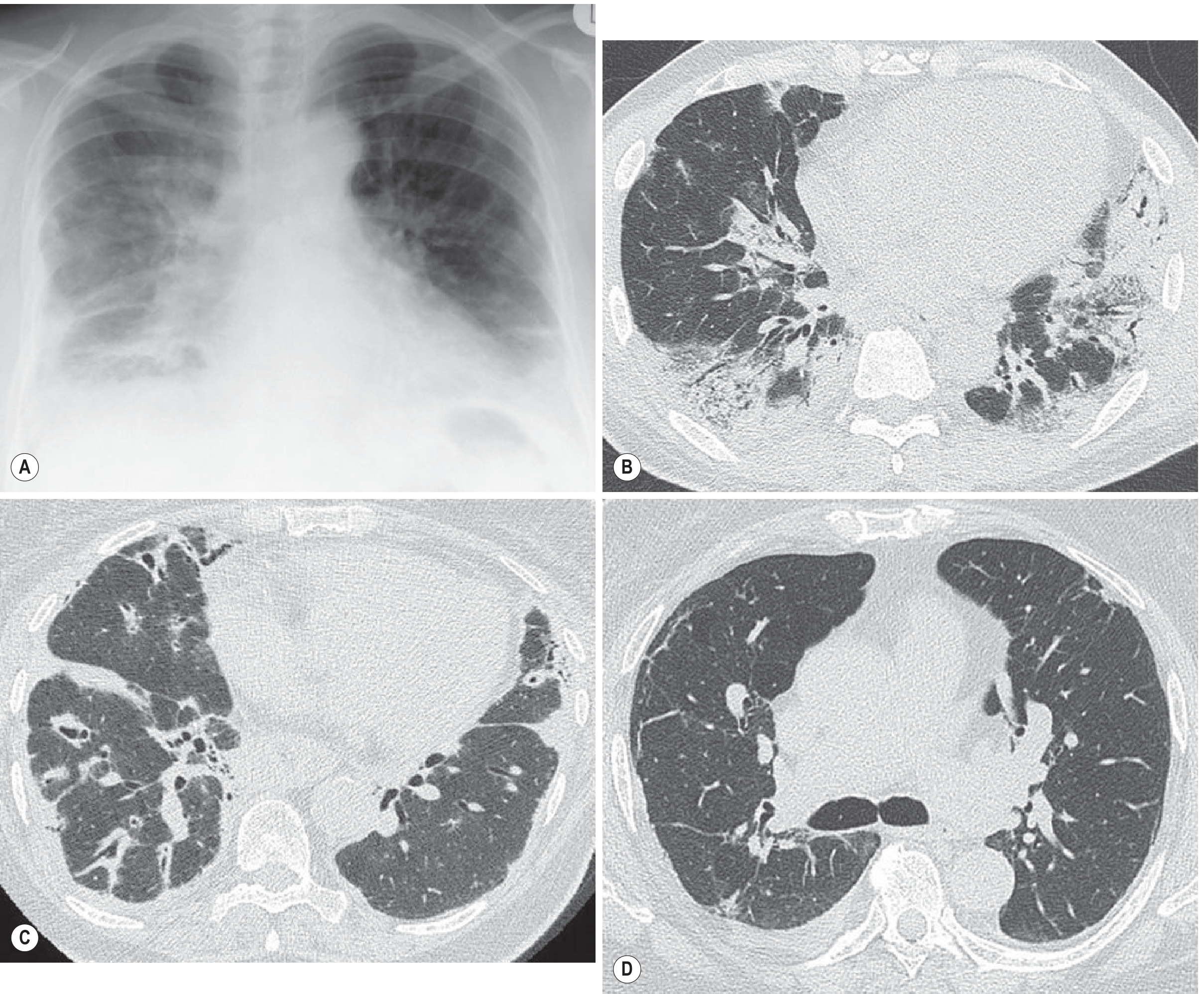
Fig: Variable CXR and CT patterns of OP. (A) CXR with typical mid/lower zone consolidation. (B) Classical peripheral lower zone consolidation on CT. (C) Bronchocentric consolidation (drug-induced OP - nitrofurantoin). (D) Radial (Type I) and subpleural curvilinear (Type II) linear bands. - Grainger & Allison's Diagnostic Radiology
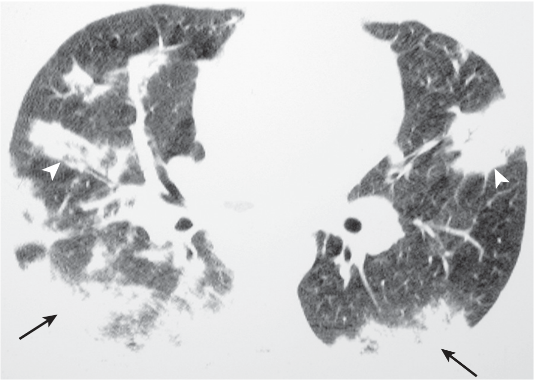
Fig: Axial CT in COP showing areas of peripheral (arrows) and peribronchial (arrowheads) consolidation. - Murray & Nadel's Textbook of Respiratory Medicine
6. Bronchoalveolar Lavage (BAL)
BAL is nonspecific but supports the diagnosis and helps exclude infection and other ILDs:
| Cell | Typical finding in OP |
|---|---|
| Lymphocytes | Increased (20-40%) - dominant finding |
| Neutrophils | Mildly increased (~10%) |
| Eosinophils | Mildly increased (~5%) |
| CD4/CD8 ratio | Usually decreased (in contrast to sarcoidosis, where it is increased) |
| Plasma cells / mast cells | May also be increased |
The BAL pattern of increased lymphocytes > eosinophils is characteristic. It also helps exclude infection, malignancy (cytology), and other diagnoses. - Murray & Nadel's Textbook of Respiratory Medicine
7. Lung Biopsy
When is biopsy needed?
- Atypical clinical/radiological features
- Poor response to corticosteroids
- Diagnostic uncertainty (e.g., single mass lesion mimicking malignancy)
- In immunocompromised hosts (e.g., post-HSCT), biopsy may be required earlier
Biopsy approaches (in order of invasiveness)
| Approach | Notes |
|---|---|
| Transbronchial biopsy (TBBx) | Sufficient in some cases with typical CT; yield lower than surgical biopsy |
| CT-guided transthoracic biopsy | Useful for focal/solitary OP lesion |
| Video-assisted thoracoscopic surgery (VATS) / surgical lung biopsy | Gold standard for definitive histology when non-invasive workup inconclusive |
Histopathological features (Masson bodies)
- Intraluminal fibroblastic buds (Masson bodies) in respiratory bronchioles, alveolar ducts, and alveoli - the hallmark
- Foamy macrophages in alveolar spaces
- Type 2 pneumocyte hyperplasia
- Interstitial mononuclear inflammatory infiltrate
- Notably: the lung architecture is preserved (no fibrosis/honeycombing distinguishes from UIP/IPF)
Murray & Nadel's Textbook of Respiratory Medicine, Harrison's 22E
8. Additional Investigations to Exclude Secondary Causes
| Suspected cause | Investigation |
|---|---|
| Drug-induced OP | Thorough medication history (antibiotics, amiodarone, chemotherapy, immunotherapy, NSAIDs); check WHO VigiAccess / pneumotox.com |
| CTD | ANA, ENA panel, myositis panel, RF, anti-CCP, complement |
| Vasculitis | ANCA, skin/renal biopsy if indicated |
| Post-radiation | RT field history; CT shows geographic distribution matching radiation port |
| Post-infectious | Paired serology, nasopharyngeal swabs, COVID-19 history |
| Malignancy-associated OP | CT-PET; bronchoscopy + BAL cytology |
| Post-transplant (GVHD) | Transplant history; GVHD workup |
| Hypersensitivity pneumonitis | Antigen exposure history, serum precipitins, BAL lymphocytosis |
9. Diagnostic Algorithm Summary
Subacute flu-like illness + consolidation not responding to antibiotics
         ↓
HRCT chest (first key investigation)
         ↓
Classical CT pattern (peripheral/subpleural consolidation ± reverse halo)?
    YES ──→ BAL + serology screen (CTD, ANCA, serum precipitins)
               ↓
            Exclude secondary causes → if none found → COP likely
               ↓
            Consider TBBx; if inconclusive → VATS biopsy for histology
    NO  ──→ Consider alternative ILD / malignancy / infection workup
10. Treatment (Brief)
- Oral corticosteroids (prednisolone 0.75-1 mg/kg/day) for 6 months: rapid, dramatic response is characteristic and itself supports the diagnosis
- Relapse rate is high (~50%) on steroid taper - often needs prolonged treatment
- Steroid-sparing agents for relapsing disease: azathioprine, mycophenolate mofetil, cyclophosphamide
- Clarithromycin 250-500 mg/day: an alternative for mild disease (FVC >80% predicted)
- Rituximab: emerging evidence in steroid-refractory OP (especially CTD-associated)
- Spontaneous remission occurs in a minority
Goldman-Cecil Medicine; Harrison's 22E; Murray & Nadel's
Key sources: Grainger & Allison's Diagnostic Radiology | Murray & Nadel's Textbook of Respiratory Medicine | Harrison's Principles of Internal Medicine 22E | Goldman-Cecil Medicine | Algorithmic approach review: Cherian et al., Chest 2022 (PMID 35038455)
This is a shared conversation. Sign in to Orris to start your own chat.