
Urea breath test
"urea breath test" AND "Helicobacter pylori"
urea breath test Helicobacter pylori mechanism diagram
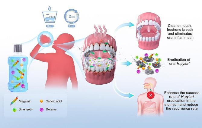
This medical infographic illustrates the mechanism and application of MAXPOWER Biological Antibacterial Liquid, a mouthwash designed to eradicate oral Helicobacter pylori. The diagram outlines a two-step clinical protocol: Step 1 involves measuring 20ml of the solution, and Step 2 specifies a 2-minute gargle duration. The formulation's active components are visualized as molecular icons: Magainin (spiral structure), Sinensetin (green hexagonal shape), Caffeic acid (orange circle), and Betaine (purple circle). The central illustration depicts the mouthwash acting within the oral cavity to disrupt H. pylori bacteria. The educational outcomes are highlighted through three pathways: clinical oral hygiene (cleansing, breath freshening, and inflammation reduction), microbiological eradication (direct killing of oral H. pylori), and gastroenterological benefit (enhancing the success rate of gastric H. pylori eradication and reducing recurrence by eliminating oral reservoirs). The graphic serves as a schematic for oral-gastric healthcare protocols targeting bacterial colonization.
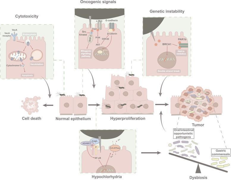
This pathophysiology diagram illustrates the 'hit and run' mechanism of Helicobacter pylori-induced gastric carcinogenesis. The central flowchart depicts the progression from normal epithelium to hyperproliferation and eventual tumor formation, accompanied by gastric dysbiosis. Detailed callouts outline four specific mechanisms: 1) Cytotoxicity: VacA binding and endocytosis leading to mitochondrial cytochrome C release, vacuolation, and apoptosis. 2) Oncogenic Signaling: CagA translocation via Type IV Secretion System (T4SS), involving Src kinase-mediated mitogenic signaling, c-Met/PI3K/AKT activation, and E-cadherin/beta-catenin disruption (Wnt signaling). 3) Genetic Instability: CagA-mediated PAR1b inhibition and BRCA1 interference leading to DNA double-strand breaks. 4) Hypochlorhydria: CagL interaction with integrins and ADAM17, inhibiting H,K-ATPase via NF-kB to cause gastric acid hyposecretion. These molecular pathways contribute to cellular transformation, genetic instability, and an altered microbial environment (dysbiosis) where opportunistic pathogens outcompete gastric commensals, promoting a persistent tumor phenotype.
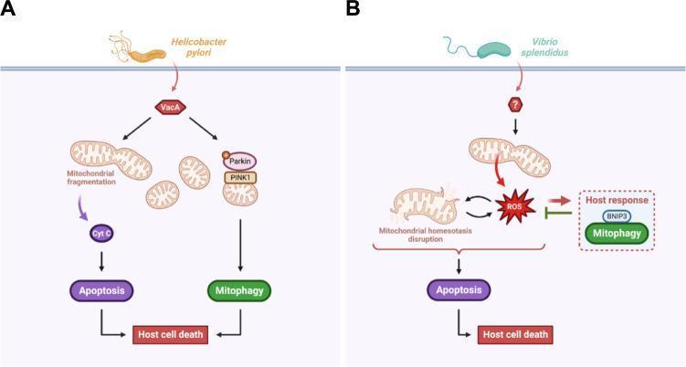
This pathophysiology diagram illustrates the signaling pathways through which extracellular pathogens, Helicobacter pylori and Vibrio splendidus, manipulate host mitochondrial homeostasis to induce cell death. Panel A details the H. pylori mechanism: the virulence effector VacA is introduced into the host cell, where it bifurcates into two pathways. One pathway causes mitochondrial fragmentation and cytochrome c (Cyt C) release, triggering apoptosis. The other activates the PINK1/Parkin-mediated mitophagy pathway. Both lead to host cell death. Panel B illustrates the V. splendidus pathway: infection triggers mitochondrial homeostasis disruption and the accumulation of mitochondrial reactive oxygen species (mtROS). This oxidative stress leads to host cell apoptosis. In response, a defensive host mechanism is shown where BNIP3-mediated mitophagy is initiated to eliminate damaged mitochondria and limit mtROS-induced cell death. The diagram highlights key educational concepts in medical microbiology and cellular pathology, specifically the intersection of mitochondrial stress, mitophagy, and programmed cell death during bacterial infection.
Urea Breath Test (UBT)
Overview
Principle / Mechanism
- The fasting patient ingests a solution of radiolabeled urea (¹³C-urea or ¹⁴C-urea)
- If H. pylori urease is present, the urea is split into labeled CO₂ and ammonia
- Labeled ¹³CO₂ (or ¹⁴CO₂) is exhaled and measured in breath samples collected in a balloon or specialized container
- Detection of labeled CO₂ confirms active H. pylori infection
"The patient drinks a solution of urea labeled with the nonradioactive isotope ¹³C and then blows into a tube. If H. pylori urease is present, the urea is hydrolyzed, and labeled carbon dioxide is detected in breath samples." - Harrison's Principles of Internal Medicine 22E
Two Forms: ¹³C vs ¹⁴C
| Feature | ¹³C-UBT | ¹⁴C-UBT |
|---|---|---|
| Isotope | Stable, nonradioactive | Radioactive |
| Detection method | Mass spectrometry | Scintillation counter (low-level radioactivity) |
| Safety | Preferred; safe in children, pregnant women | Low-level radiation; avoid in pregnancy/children |
| Availability | Less widely available locally | More widely available |
| Special utility | Pediatric patients | Cost-effective in adults |
Diagnostic Accuracy
Indications
- Diagnosis of active H. pylori infection (non-endoscopic "test and treat" strategy for young, dyspeptic patients without alarm symptoms)
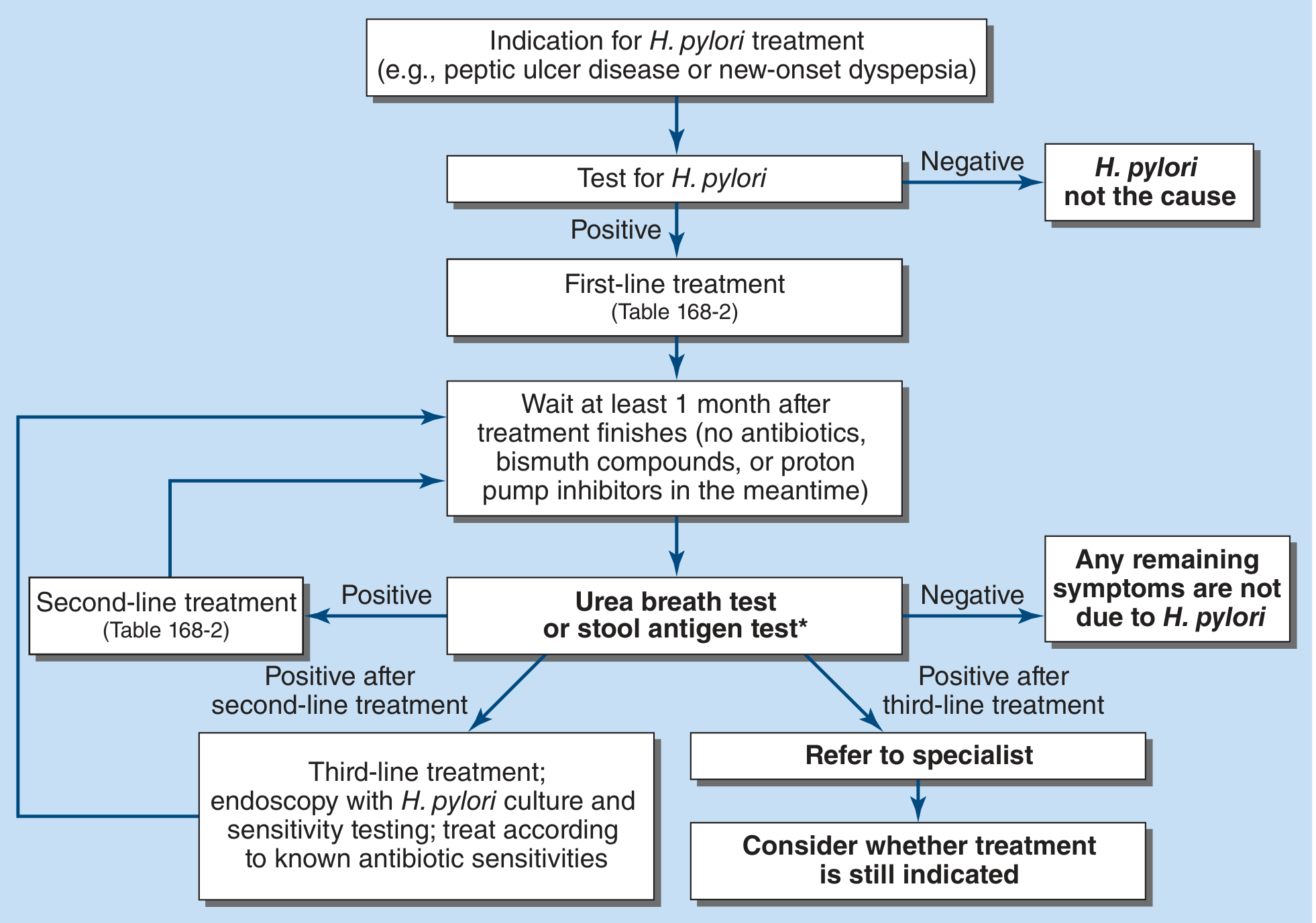
- Confirming eradication after antibiotic treatment (preferred over serology for this purpose)
- Patients with peptic ulcer disease, functional dyspepsia, MALT lymphoma workup
- Follow-up in gastric ulcer (to confirm healing and clearance)
- Surveillance in areas with high H. pylori prevalence (>20% community prevalence)
Comparison with Other H. pylori Tests
| Test | Sensitivity | Specificity | Notes |
|---|---|---|---|
| ¹³C Urea Breath Test | >95% | >95% | Best for confirmation and post-treatment follow-up |
| Stool antigen test | >95% | >95% | Monoclonal antibody-based; convenient; less popular due to stool handling |
| Serology (IgG) | 57-100% | 58-96% | Cannot confirm eradication; remains elevated >1 year after cure |
| Biopsy urease (CLO test) | 84-95% | 95-100% | Requires endoscopy; rapid (~minutes to 24 h) |
| Histology | ~95% | ~99% | Most accurate biopsy test; detects gastritis severity |
| Culture | ~80% | 100% | Allows antibiotic susceptibility testing |
Pre-test Conditions (Critical for Accuracy)
- Proton pump inhibitors (PPIs) - suppress H. pylori, causing false-negatives
- Discontinue PPIs 2 weeks before testing
- Antibiotics - kill H. pylori, causing false-negatives
- Discontinue antibiotics 4 weeks before testing
- H2-receptor antagonists - less effect, but best to discontinue
- Acute GI bleeding - reduces sensitivity
Post-treatment Confirmation
- Serology remains positive for >1 year and cannot assess treatment response
- UBT should be performed at least 4 weeks after completing therapy to avoid false-negative results
- Either the UBT or stool antigen test is acceptable for post-treatment assessment
Key Points Summary
- Principle: H. pylori urease cleaves labeled urea → labeled CO₂ detected in breath
- Isotopes: ¹³C (nonradioactive, preferred) or ¹⁴C (radioactive, widely available)
- Accuracy: Sensitivity and specificity both >95%
- Stop PPIs 2 weeks and antibiotics 4 weeks before testing
- Test of choice for confirming eradication (performed ≥4 weeks post-therapy)
- Preferred over serology for active infection diagnosis
- ¹³C-UBT particularly useful in children and pregnant women