
Hernia anatomy
inguinal hernia anatomy diagram Hesselbach triangle
inguinal canal anatomy
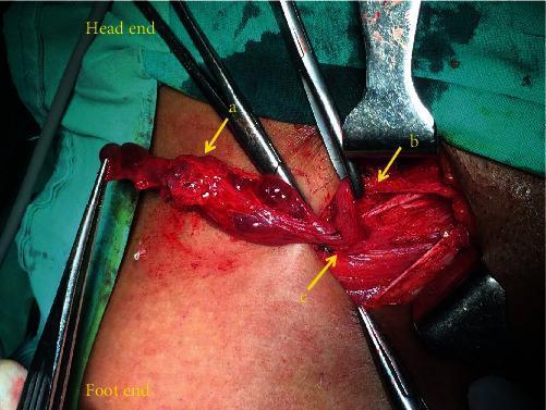
This intraoperative clinical photograph displays an exploration of the right inguinal canal in a female patient. The primary pathology is a multiloculated cystic mass (labeled 'a'), characterized by several interconnected, reddish-brown, semi-translucent sacs with thin walls and an irregular surface. The mass is positioned anterior to the round ligament of the uterus (labeled 'b'), which appears as a distinct fibrous cord-like structure. The deep inguinal ring (labeled 'c') is visible as the anatomical gateway where these structures emerge from the abdominal cavity. Surgical instruments including retractors and forceps are used to expose the operative field. The visual findings are consistent with Nuck's canal endometriosis or a multilocular hydrocele of the canal of Nuck. This image serves as an educational resource for general surgeons and gynecologists regarding the differential diagnosis of inguinal masses in women and the surgical anatomy of the round ligament within the inguinal canal.
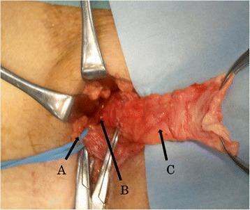
This intraoperative clinical photograph captures a surgical repair of a right internal inguinal hernia (supravesical hernia). The image shows the open surgical field with various anatomical structures labeled for educational identification. The spermatic cord (A) is visible as a reddish-brown bundle of tubular structures, isolated and retracted with a blue vessel loop and surgical instruments. Centrally, the hernia orifice (B) is identified as a dark, recessed opening on the posterior wall of the inguinal canal near the vesical side. A distended, pale pink membranous structure represents the hernia sac (C), which has been dissected and is being held outward by surgical clamps. Metal retractors are positioned to maintain visualization of the inguinal canal anatomy. The image demonstrates the spatial relationships between the cord structures and the hernia defect during a Lichtenstein-type repair, highlighting the surgical anatomy of the inguinal region in a patient with a supravesical-type hernia.
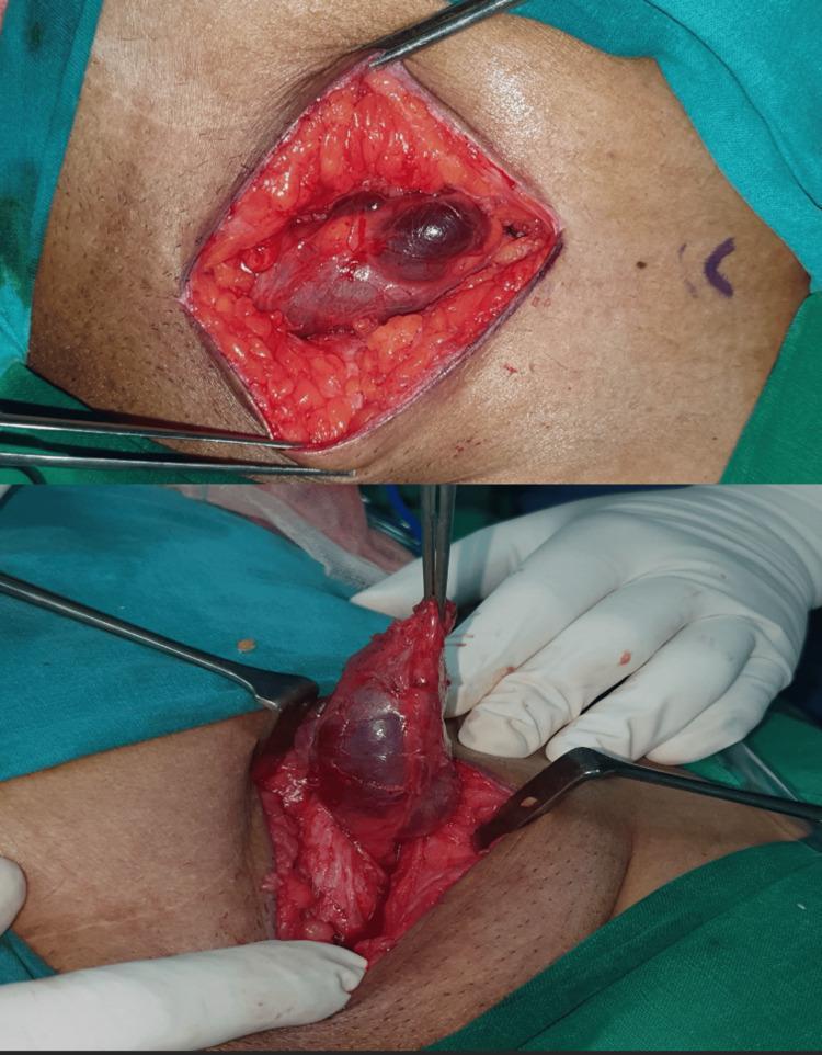
Two intraoperative clinical photographs demonstrate a surgical exploration of the inguinal region for a hydrocele of the canal of Nuck. The top panel shows an open surgical field with skin and subcutaneous fat retracted, revealing an elongated, bluish, fluid-filled cystic sac lying within the inguinal canal. The bottom panel shows the surgical dissection and mobilization of the same sac, which is being held by forceps and pulled upward to demonstrate its attachment to the round ligament. The lesion is smooth-walled, tense, and translucent, characteristic of a hydrocele. The surrounding anatomy includes the dissected inguinal canal and layers of the abdominal wall. These images illustrate the surgical presentation and operative approach for managing Nuck's cyst in a female patient, emphasizing the need to trace the sac to the deep inguinal ring to ensure complete excision.
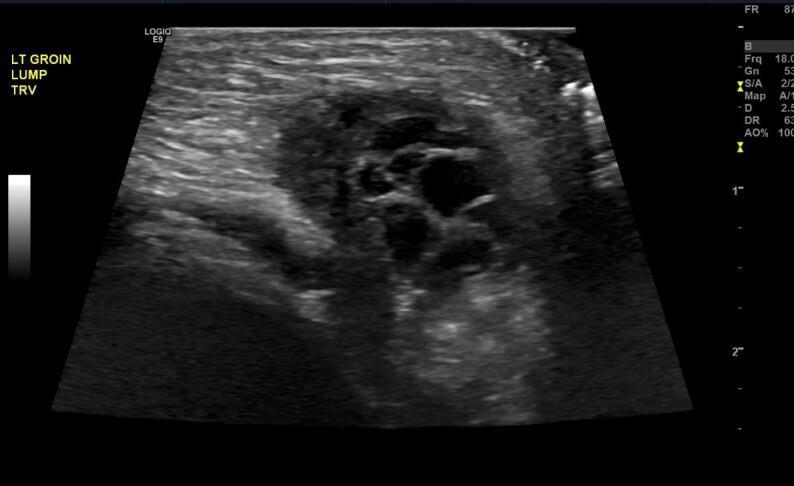
This diagnostic image is a gray-scale ultrasonography of the left groin area in a transverse (TRV) plane. The scan reveals a well-defined mass of mixed echogenicity within the inguinal canal, measuring approximately 2.1 x 1.1 cm. Central to the mass are multiple small, anechoic, circular structures consistent with ovarian follicles. The visualization of these follicles within the inguinal region is diagnostic of an incarcerated ovary within an inguinal hernia. The surrounding stromal tissue of the herniated ovary appears slightly hypoechoic compared to the adjacent hyperechoic subcutaneous fat. This image demonstrates a critical pediatric surgical emergency where normal pelvic anatomy is displaced into the inguinal canal, requiring prompt identification and reduction to prevent ovarian torsion or ischemia. Educational focus is on the sonographic recognition of ovarian tissue by its follicular pattern when evaluating a palpable inguinal lump.
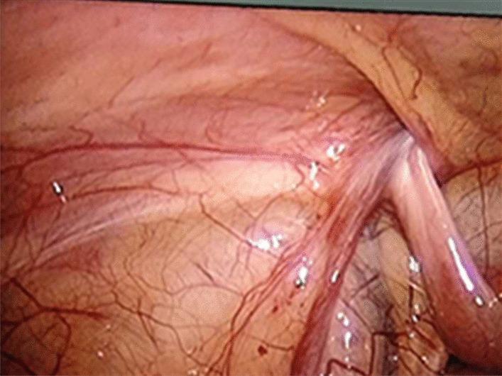
This laparoscopic diagnostic image depicts a view of the internal abdominal cavity, specifically focusing on the pelvic anatomy near the inguinal region. The primary visual element is the oviduct (fallopian tube), which is seen extending into an unclosed internal inguinal canal orifice, characteristic of an indirect inguinal hernia or incarcerated adnexa. The oviduct appears tubular, slightly thickened, and exhibits signs of mild edema and hyperaemia, evidenced by a swollen morphology and a reddish-pink hue due to increased vascularity. The surrounding parietal peritoneum shows a prominent network of small, congested blood vessels. Key anatomical landmarks visible include the internal ring of the inguinal canal and the adjacent abdominal wall lining. This image serves as an educational tool for identifying adnexal involvement in pediatric or adult female inguinal hernias and demonstrates the visual manifestations of early inflammatory changes (edema and hyperemia) in incarcerated tissue during laparoscopic exploration.
Hernia Anatomy — Topic Map
- General abdominal wall layers & the myopectineal orifice of Fruchaud - the shared anatomic "weak zone" that inguinal and femoral hernias both pass through
- Inguinal hernia - inguinal canal, deep/superficial rings, Hesselbach's triangle, direct vs. indirect pathways
- Femoral hernia - femoral canal, femoral ring boundaries (relation to inguinal ligament, femoral vein, lacunar ligament)
- Umbilical / paraumbilical hernia - umbilical ring, linea alba defect
- Epigastric and Spigelian hernia - linea alba above umbilicus; semilunar line defects
- Incisional / ventral hernia - defects through surgical scars in the abdominal wall layers
- Hiatal / diaphragmatic hernia - esophageal hiatus, sliding vs. paraesophageal types
- Rare hernias - obturator, lumbar (Petit's/Grynfeltt's), sciatic, perineal
Which hernia anatomy would you like to go deep on?
Hernia Anatomy — Topic Map
- General abdominal wall layers & the myopectineal orifice of Fruchaud - the shared anatomic "weak zone" that inguinal and femoral hernias both pass through
- Inguinal hernia - inguinal canal, deep/superficial rings, Hesselbach's triangle, direct vs. indirect pathways
- Femoral hernia - femoral canal, femoral ring boundaries (relation to inguinal ligament, femoral vein, lacunar ligament)
- Umbilical / paraumbilical hernia - umbilical ring, linea alba defect
- Epigastric and Spigelian hernia - linea alba above umbilicus; semilunar line defects
- Incisional / ventral hernia - defects through surgical scars in the abdominal wall layers
- Hiatal / diaphragmatic hernia - esophageal hiatus, sliding vs. paraesophageal types
- Rare hernias - obturator, lumbar (Petit's/Grynfeltt's), sciatic, perineal
anatomical diagram of abdominal wall hernia sites epigastric umbilical Spigelian
Hernia Anatomy — Overview by Region
1. The shared "weak zone" — myopectineal orifice of Fruchaud
2. Inguinal hernia
- Inguinal canal: ~4 cm long, running from the deep (internal) inguinal ring (an opening in the transversalis fascia, lateral to the inferior epigastric vessels) to the superficial (external) inguinal ring (an opening in the external oblique aponeurosis, above the pubic tubercle). It transmits the spermatic cord in men / round ligament in women, plus the ilioinguinal and genital branch of the genitofemoral nerve (Gray's Anatomy for Students).
- Hesselbach's triangle: bounded medially by the lateral border of rectus abdominis, superolaterally by the inferior epigastric vessels, and inferiorly by the inguinal ligament. A weakness here (thinned/ballooned transversalis fascia) is the site of direct inguinal hernias.
- Direct vs indirect:
- Indirect: sac enters the deep ring lateral to the epigastric vessels, follows the spermatic cord through the canal, and may exit the superficial ring into the scrotum - congenital, from a patent processus vaginalis.
- Direct: sac pushes straight through Hesselbach's triangle, medial to the epigastric vessels - acquired, from fascial weakness.
- Both directions are above the inguinal ligament, which is the key landmark distinguishing them from femoral hernias (Goldman-Cecil Medicine; Fischer's Mastery of Surgery).
3. Femoral hernia
- The femoral sheath contains (lateral to medial) the femoral artery, femoral vein, and femoral canal.
- The femoral ring (internal opening) is bounded by the inguinal ligament anteriorly, the pectineal ligament/superior pubic ramus posteriorly, the femoral vein laterally, and the lacunar ligament medially.
- A femoral hernia passes through this ring, below and lateral to the pubic tubercle (versus inguinal hernias, which are above/medial to it) - the classic bedside distinguishing point. Femoral hernias are more common in women due to a wider pelvis and larger femoral ring, and they carry higher strangulation risk because the ring is rigid and narrow (Gray's Anatomy for Students; Fischer's Mastery of Surgery).
4. Umbilical / paraumbilical hernia
5. Epigastric and Spigelian hernia
- Epigastric hernia: defect in the linea alba between the xiphoid and umbilicus, where it is narrower and weaker.
- Spigelian hernia: occurs at the semilunar line (the lateral border of the rectus sheath, where internal oblique and transversus abdominis aponeuroses fuse) - the "Spigelian fascia" here is variably thin, widest near the umbilicus. These are often intraparietal (sac dissects between muscle layers under an intact external oblique), which can make them clinically occult (Maingot's Abdominal Operations; Sabiston Textbook of Surgery).
6. Incisional / ventral hernia
7. Hiatal / diaphragmatic hernia
- Type I (sliding) - ~90-95%; the gastroesophageal junction itself slides upward into the thorax.
- Type II (paraesophageal, pure) - GE junction stays in place, but the fundus herniates alongside it.
- Type III (mixed) - combination of sliding and paraesophageal components; most "paraesophageal" hernias are actually this type.
- Type IV - large defect with herniation of other organs (colon, spleen, small bowel) alongside the stomach.
Quick landmark summary
| Hernia | Key structure | Position relative to inguinal ligament / pubic tubercle |
|---|---|---|
| Indirect inguinal | Deep ring, lateral to epigastric vessels | Above |
| Direct inguinal | Hesselbach's triangle, medial to epigastric vessels | Above |
| Femoral | Femoral ring/canal | Below and lateral |
| Umbilical | Umbilical ring / linea alba | Midline, at umbilicus |
| Spigelian | Semilunar line | Lateral rectus border |
| Hiatal | Esophageal hiatus of diaphragm | Not abdominal wall - thoracoabdominal |
Explain surgical repair approaches (open vs. laparoscopic)
"inguinal hernia" AND laparoscopic vs open repair
Surgical Repair Approaches for Hernia: Open vs. Laparoscopic
Open repair
- Bassini repair - the original tension repair; approximates the transversus abdominis aponeurosis and transversalis fascia to the inguinal ligament.
- Shouldice repair - a refined Bassini modification; the transversalis fascia is opened and the inguinal floor is reconstructed in four continuous suture layers. Expert centers report recurrence rates under 2%, but it is technically demanding and results elsewhere often resemble a plain Bassini (Bailey and Love's Short Practice of Surgery).
- McVay (Cooper's ligament) repair - sutures the conjoint tendon to Cooper's ligament rather than the inguinal ligament, which also closes the femoral ring - the preferred tissue repair for femoral hernias or when mesh cannot be used (Schwartz's Principles of Surgery).
- Lichtenstein repair - a flat polypropylene mesh is laid over the posterior inguinal floor and sutured to the conjoint tendon medially and inguinal ligament inferiorly, with a slit for the spermatic cord. This became the benchmark technique because it eliminates suture-line tension, the main driver of recurrence (Sabiston Textbook of Surgery).
- Mesh plug and patch, Prolene Hernia System (PHS) - variations using a plug or bilayer device combining anterior and preperitoneal mesh; PHS relies on a posterior mesh component similar in principle to Lichtenstein. Current guidelines favor flat mesh (Lichtenstein) over 3-dimensional plug devices (Mulholland and Greenfield's Surgery).
- Open preperitoneal repair (Stoppa, transinguinal preperitoneal) - large mesh placed behind the transversalis fascia covering the whole myopectineal orifice; useful for bilateral or recurrent hernias.
Laparoscopic / minimally invasive repair
- TAPP (transabdominal preperitoneal) - the peritoneal cavity is entered first, a peritoneal flap is raised, mesh is placed in the preperitoneal space, and the peritoneum is closed over it. Easier to learn and gives a full view of anatomy, but carries a higher risk of intra-abdominal injury (bowel, vessels) since the peritoneal cavity is opened.
- TEP (totally extraperitoneal) - the preperitoneal space is accessed and developed (often with a balloon dissector) without ever entering the peritoneal cavity. Avoids intraperitoneal complications and adhesions but has a steeper learning curve.
- The HerniaSurge Group's 2018 International Guidelines consider TAPP and TEP largely equivalent in operative time, cost, complications, and recurrence; choice depends on surgeon experience and hernia complexity (large inguinoscrotal or recurrent hernias may favor TAPP for its wider working view) (Current Surgical Therapy).
- Robot-assisted repair is an emerging variant following the same TAPP/TEP anatomic principles with robotic instrumentation.
Open vs. laparoscopic: how they compare
| Factor | Open (Lichtenstein) | Laparoscopic (TEP/TAPP) |
|---|---|---|
| Postoperative pain | More | Less |
| Recovery / return to activity | Slower | Faster |
| Operative time | Shorter (especially unilateral) | Longer, especially early in learning curve |
| Recurrence | Comparable in expert hands; some meta-analyses show a trend toward slightly higher recurrence with laparoscopic in earlier trials | Comparable overall; low with adequate mesh overlap and experience |
| Chronic groin pain / nerve injury | Somewhat higher | Lower (nerves less handled) |
| Bilateral or recurrent hernia | Requires separate/larger incision | Well suited - same ports treat both sides |
| Learning curve | Shorter | Longer, requires significant case volume for TEP/TAPP proficiency |
| Cost | Lower (no laparoscopic equipment) | Higher |
| Anesthesia | Can be done under local/regional | Requires general anesthesia + pneumoperitoneum |
Practical selection points
- Bilateral, recurrent (after prior open repair), or need for rapid return to activity → favor laparoscopic.
- Contaminated/strangulated hernia, local anesthesia needed, low-volume laparoscopic center, or femoral hernia → favor open (Lichtenstein or McVay).
- Ventral/incisional hernia: similar logic applies - laparoscopic (or robotic) IPOM/retromuscular repair gives lower wound infection rates and faster recovery with comparable recurrence versus open repair, per a meta-analysis of 11 studies cited in Schwartz's Principles of Surgery.
- Paraesophageal/hiatal hernia: laparoscopic repair is now the standard approach for elective repair, with outcomes comparable to open transabdominal or transthoracic repair but faster recovery, per comparative series in Maingot's Abdominal Operations.
Recent evidence check
- Learning curve analysis for laparoscopic inguinal hernia repair (PMID: 36416945) - confirms a meaningful proficiency threshold, reinforcing why outcomes vary by surgeon experience.
- Sexual dysfunction after laparoscopic vs open inguinal hernia repair (PMID: 37450061) - suggests laparoscopic repair may have a favorable profile for this specific complication.
- Laparoscopic vs open repair for incarcerated inguinal hernia in children (PMID: 36220132) - relevant if pediatric incarcerated hernia is a concern, an area not detailed in the general textbook comparison above.