
Triple negative breast cancer and molecular classification, prognostic markers, inc marker/tests for breast carcinoma
"triple-negative breast cancer"[MeSH Terms] AND "molecular classification"
triple negative breast cancer molecular subtypes classification
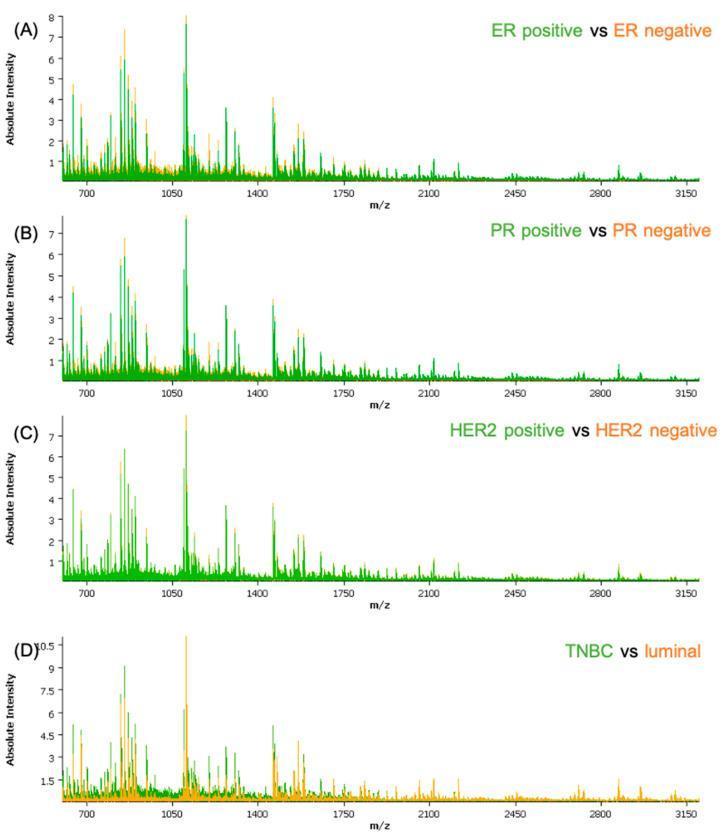
This diagnostic comparison chart displays MALDI-TOF mass spectrometry average spectra for breast cancer classification. The figure contains four subplots (A-D) showing 'Absolute Intensity' on the y-axis against 'm/z' (mass-to-charge ratio) on the x-axis, ranging from 700 to 3150. Subplot (A) compares Estrogen Receptor (ER) positive (green) and negative (orange) samples; (B) compares Progesterone Receptor (PR) positive (green) and negative (orange); (C) shows HER2 positive (green) and negative (orange); and (D) compares Triple-Negative Breast Cancer (TNBC) (green) and luminal subtypes (orange). Visually, the spectra are characterized by dense peptide peaks in the lower m/z range (700-1750), with intensity diminishing at higher mass ranges. Notable differential peak intensities are observed in the ER-positive and luminal groups compared to their counterparts, particularly around 1050 m/z. This educational material demonstrates the use of molecular profiling to identify distinctive peptide signatures associated with breast cancer receptor status and molecular subtypes.
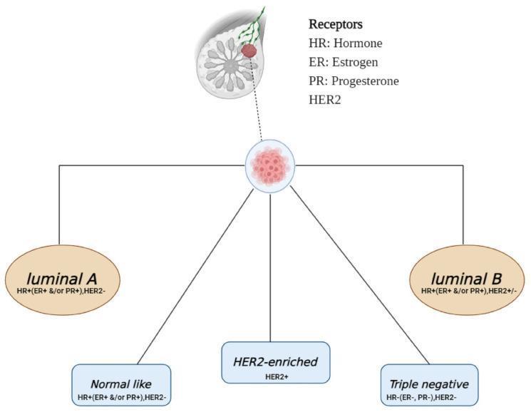
This educational diagram illustrates the intrinsic molecular subtypes of breast cancer categorized by their hormone receptor and HER2 status. At the top, a sagittal anatomical schematic of the breast shows a localized mass, which leads to a central node representing tumor classification. From this node, five distinct pathways branch to define specific subtypes: 1) Luminal A (HR+, ER+ and/or PR+, HER2-); 2) Luminal B (HR+, ER+ and/or PR+, HER2+/-); 3) Normal-like (HR+, ER+ and/or PR+, HER2-); 4) HER2-enriched (HER2+); and 5) Triple negative (HR-, ER-, PR-, HER2-). A legend defines the abbreviations used: HR (Hormone Receptor), ER (Estrogen Receptor), PR (Progesterone Receptor), and HER2 (Human Epidermal Growth Factor Receptor 2). This infographic is designed for medical education, aiding in the understanding of oncology classification, prognostic factors, and the immunohistochemical markers that guide clinical management in breast pathology.
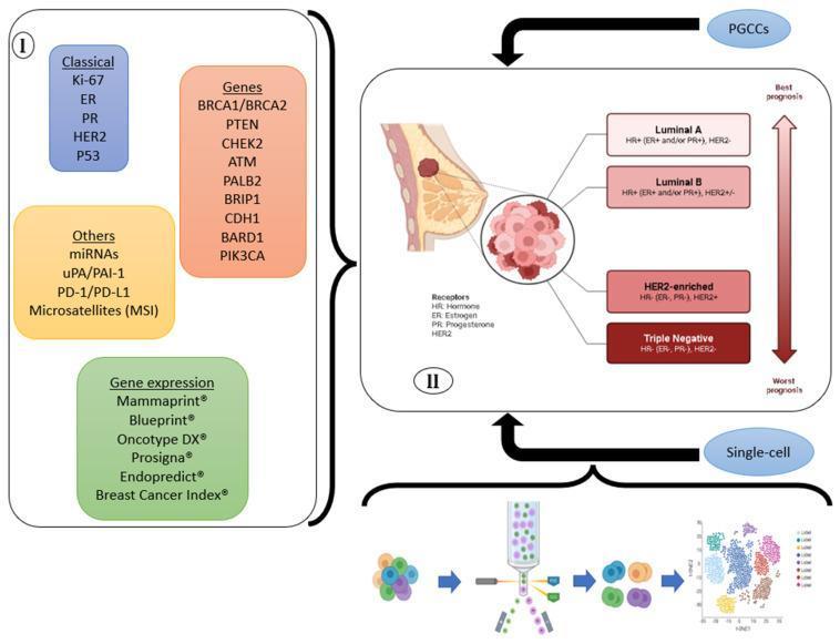
Educational infographic illustrating the classification of breast cancer through various biomarkers and diagnostic approaches. The left panel (I) categorizes breast cancer biomarkers into four domains: 'Classical' (Ki-67, ER, PR, HER2, P53), 'Genes' (including BRCA1/2, PIK3CA, and ATM), 'Gene expression' genomic tests (MammaPrint, Oncotype DX, etc.), and 'Others' (miRNAs, PD-1/PD-L1, MSI). The right panel (II) correlates these markers with the four molecular subtypes: Luminal A (HR+/HER2-), Luminal B (HR+/HER2+/-), HER2-enriched (HR-/HER2+), and Triple Negative (HR-/HER2-), displayed alongside a prognostic arrow ranging from 'Best prognosis' to 'Worst prognosis'. The diagram also includes anatomical cross-sections of the breast and tumor, and introduces emerging technologies like single-cell analysis and PGCCs (Polyploid Giant Cancer Cells) for future classification strategies. This resource is designed for advanced oncology students and clinicians to understand tumor heterogeneity and personalized diagnostic pathways.
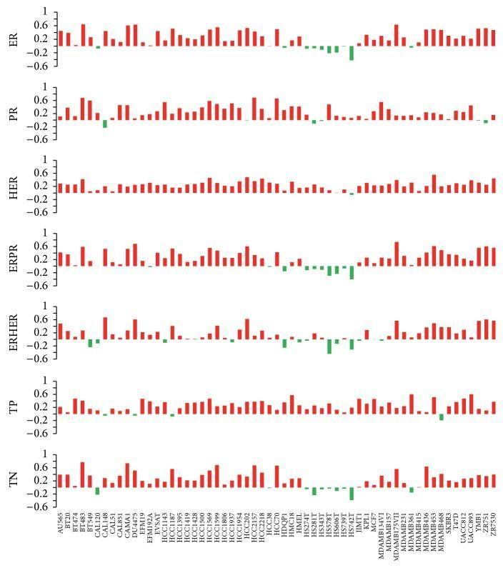
This Comparison Chart illustrates the Spearman correlation coefficients between 59 breast cancer cell lines and primary tumor groups based on copy number variation (CNV) data for 2,426 genes. The visual is organized into seven horizontally stacked bar plots, each representing a specific tumor classification or molecular marker group: ER (estrogen receptor), PR (progesterone receptor), HER (human epidermal growth factor receptor 2), ERPR, ERHER, TP (triple positive), and TN (triple negative). The x-axis lists the 59 individual cell lines (e.g., MCF7, BT20, MDAMB231). The y-axis measures the correlation coefficient ranging from -0.6 to 1.0. Positive correlations are indicated by red vertical bars, while negative correlations are shown in green. The chart serves as a genomic benchmarking tool in oncology research, highlighting how well established cell models reflect the copy number profiles of clinical tumor subtypes. Notably, it demonstrates variability among common models like MCF7 and reveals consistently low correlation across most groups for specific lines such as HS742T, suggesting a more metastatic or drifted genotypic profile.
breast cancer prognostic markers ER PR HER2 Ki67 immunohistochemistry
Triple Negative Breast Cancer (TNBC): Molecular Classification, Prognostic Markers, and IHC/Laboratory Tests
1. Overview of Breast Cancer Molecular Classification
The Three Major Clinical Groups
| Feature | Luminal (ER+/HER2-) | HER2 (±ER) | Triple Negative (TNBC) |
|---|---|---|---|
| Overall frequency | 50-65% | ~20% | 10-20% |
| Typical patients | Older women; BRCA2 mutation | Younger women; germline TP53 mutation | Young women; BRCA1 carriers; African American women |
| Grade | Mainly Grade 1-2 | Mainly Grade 2-3 | Mainly Grade 3 |
| Complete response to chemo | ~10% | 15-60% | ~30% |
| Timing of relapse | Low rate over many years; late recurrence >10 years possible | Bimodal (early and late peaks) | Early peak <8 years; late recurrence rare |
| Metastatic sites | Bone (70-80%), viscera (25-30%), brain (~10%) | Bone (70%), viscera (45%), brain (30%) | Bone (40%), viscera (35%), brain (25%) |
| Common somatic mutations | PIK3CA (29-45%), TP53 (12-29%) | TP53 (70-80%), PIK3CA (~40%) | TP53 (70-80%), PIK3CA (9%) |
2. Intrinsic ("Six Major") Molecular Subtypes by Gene Expression Profiling
| Intrinsic Subtype | IHC Profile | Key Features |
|---|---|---|
| Luminal A | ER+, PR+, HER2-, Ki-67 low | 40-60% of cases; best prognosis; slow growing |
| Luminal B (HER2-) | ER+, PR low/-, HER2-, Ki-67 high | Higher grade, higher proliferation rate |
| Luminal B (HER2+) | ER+, PR low/-, HER2+ | Any Ki-67; benefits from HER2 therapy |
| HER2-enriched | ER-, PR-, HER2+ | 10-20%; aggressive; responds to HER2-targeted drugs |
| Basal-like | ER-, PR-, HER2-; CK5/6+, EGFR+ | Overlaps greatly with TNBC (~80% overlap) |
| Normal-like / Claudin-low | Variable | Stem-cell features; very poor prognosis |
"A useful molecular classification system that better reflects the spectrum of breast cancer biology and behavior separates breast cancers into six major 'intrinsic' subtypes based on gene expression profiling; luminal A, luminal B, HER2 enriched, basal-like, normal-like, and claudin-low."
- Robbins, Cotran & Kumar Pathologic Basis of Disease
3. Triple Negative Breast Cancer (TNBC) - In Depth
Definition
Key Epidemiological Features
- More common in younger women and African American women
- Strongly associated with germline BRCA1 mutations (~20% of TNBC patients have a pathogenic BRCA variant)
- BRCA1/2 mutation carriers should be tested (germline sequencing) because they may be eligible for PARP inhibitors
TNBC and Basal-like Subtype
- TNBC and the "basal-like" intrinsic subtype strongly overlap but are not identical (~80% of TNBC are basal-like; ~85% of basal-like are TNBC)
- Basal-like tumors additionally express basal cytokeratins (CK5/6, CK14, CK17) and EGFR
- The term "basal-like" comes from the resemblance to myoepithelial/basal cells of the normal breast
Lehmann Subtypes (Further TNBC Subclassification)
| Subtype | Abbreviation | Key Features |
|---|---|---|
| Basal-like 1 (BL1) | BL1 | High cell cycle/DNA damage response genes; highest pCR to chemo |
| Basal-like 2 (BL2) | BL2 | Growth factor signaling genes; lower pCR |
| Mesenchymal (M) | M | EMT/stem cell features; may respond to PI3K/mTOR inhibitors |
| Luminal androgen receptor (LAR) | LAR | AR expression; may respond to androgen receptor antagonists |
Prognosis of TNBC
- Worst overall prognosis among the three groups
- Highest risk of developing early metastases and death despite treatment
- No endocrine therapy option (ER-negative)
- No HER2-targeted therapy applicable
- Key advances: pembrolizumab (PD-1 checkpoint inhibitor) + chemotherapy showed markedly improved outcomes in early TNBC (KEYNOTE-522 trial, 2022)
- PARP inhibitors (olaparib, talazoparib) for BRCA1/2-mutated metastatic TNBC
4. Prognostic and Predictive Markers in Breast Cancer
A. Standard IHC Panel (Mandatory for All Invasive Breast Cancers)
| Marker | What It Tests | Clinical Significance |
|---|---|---|
| ER (Estrogen Receptor) | Nuclear IHC (Allred score or % positive cells) | Predicts response to endocrine therapy; favorable prognosis when positive |
| PR (Progesterone Receptor) | Nuclear IHC | Confirms ER-positive phenotype; low PR with positive ER suggests Luminal B |
| HER2 (ERBB2) | IHC (0-3+) ± FISH/ISH for 2+ cases | Predicts response to trastuzumab, pertuzumab, T-DM1, T-DXd |
| Ki-67 | Nuclear IHC (proliferation index) | Distinguishes Luminal A (<14%) from Luminal B (>20%); higher = worse prognosis |
B. Histological Grade (Nottingham Grade / Elston-Ellis System)
- Tubule formation (gland formation)
- Nuclear pleomorphism
- Mitotic count
C. Staging Markers (Anatomic + Biologic - AJCC 8th Edition)
- Tumor size (T): pT1-4
- Node status (N): pN0-3 (sentinel lymph node biopsy / ALND)
- Distant metastasis (M)
- The 8th AJCC edition now incorporates biomarker groups (ER, PR, HER2, grade) into the prognostic stage groups
D. Multi-Gene Expression Assays (Genomic Tests)
| Assay | Genes Analyzed | Clinical Use | Applicable Subtype |
|---|---|---|---|
| Oncotype DX (Genomic Health) | 21 genes → Recurrence Score (RS) 0-100 | Predict benefit from chemo in ER+/HER2-/N0-N1 (TAILORx, RxPONDER) | Luminal only |
| MammaPrint (Agendia) | 70-gene signature | Binary low/high risk; guides chemo decision (MINDACT trial) | ER+/HER2- |
| Prosigna / PAM50 (NanoString) | 50-gene signature | Intrinsic subtype + Risk of Recurrence (RoR) score | ER+/HER2- |
| EndoPredict | 12-gene signature | Predict late recurrence risk (years 5-15) | ER+/HER2- |
| IHC4 score | ER + PR + HER2 + Ki-67 (IHC) | Provides prognostic info similar to Oncotype DX | ER+/HER2- |
"Ki-67 was included with three other widely measured breast cancer markers (ER, PR, and HER2) into a panel of four immunohistochemical makers (IHC4), which together provided similar prognostic information."
- Schwartz's Principles of Surgery, 11th ed.
"Prosigna outperformed other assays in direct comparison of six prognostic signatures." - Fischer's Mastery of Surgery, 8th ed.
5. Additional/Emerging Markers in Breast Carcinoma
Genetic/Hereditary Markers
| Gene | Relevance |
|---|---|
| BRCA1 (17q21) | High-penetrance mutation; strongly associated with TNBC/basal-like; PARP inhibitor eligibility |
| BRCA2 (13q12.3) | Mostly Luminal-type cancers; PARP inhibitor eligibility |
| PALB2 | Partner of BRCA2; high-risk gene |
| ATM, CHEK2, RAD51C/D | Moderate-penetrance genes; homologous recombination deficiency (HRD) |
| TP53 | Somatic mutation rate ~70-80% in TNBC/HER2 cancers |
| PIK3CA | Somatic; most common in Luminal (29-45%); alpelisib target in HR+/HER2-/PIK3CA-mutated disease |
Immunotherapy Markers
| Marker | Relevance |
|---|---|
| PD-L1 (CPS score) | Combined Positive Score (CPS) ≥10 predicts benefit from pembrolizumab in early/metastatic TNBC |
| TILs (Tumor-Infiltrating Lymphocytes) | High TILs in TNBC correlate with better prognosis and response to immunotherapy/chemotherapy |
| TMB (Tumor Mutational Burden) | Emerging marker for checkpoint inhibitor response |
| MSI (Microsatellite Instability) | Rare in breast cancer; predicts response to pembrolizumab |
Other Markers
| Marker | Relevance |
|---|---|
| p53 | Mutated in high-grade/TNBC; adverse prognostic indicator |
| E-cadherin | Lost in lobular carcinoma (ILC); used to distinguish ILC from IDC |
| AR (Androgen Receptor) | Positive in ~10-35% TNBC (LAR subtype); target of enzalutamide |
| EGFR | Often overexpressed in basal-like/TNBC; not yet an established therapeutic target in breast |
| Circulating Tumor DNA (ctDNA) | Liquid biopsy; predicts recurrence in TNBC with residual disease after neoadjuvant chemo |
| CK5/6, CK14, CK17 | Basal cytokeratins; confirm basal-like phenotype |
6. Summary: TNBC vs Other Subtypes at a Glance
Prognosis (best → worst):
Luminal A  →  Luminal B  →  HER2  →  TNBC
- TNBC has no targeted therapy from standard ER/HER2 axis
- Treatment backbone = neoadjuvant chemotherapy (anthracycline + taxane) ± carboplatin
- Pembrolizumab (anti-PD-1) is now standard in early-stage TNBC (CPS-unselected)
- PARP inhibitors for BRCA1/2-mutated TNBC
- Sacituzumab govitecan (anti-Trop-2 ADC) for metastatic TNBC
- Robbins & Kumar Basic Pathology (Table 17.7, p. 709-710) - major subtype comparison table
- Robbins, Cotran & Kumar Pathologic Basis of Disease - six intrinsic subtypes; BRCA genetics; breast cancer evolution model
- Fischer's Mastery of Surgery, 8th ed. (p. 1780-1781) - clinical classification table with Ki-67
- Sabiston Textbook of Surgery - molecular markers, Oncotype DX, MammaPrint, PAM50
- Chen et al., 2025 - "Classifications of TNBC: insights and current therapeutic approaches" (PMID: 39893480)
- Varzaru et al., 2024 - "TNBC: Molecular Particularities Still a Challenge" (PMID: 39272660)
Include clinical features too prepare for 10 marks question
breast cancer clinical signs peau d'orange nipple retraction skin dimpling
breast cancer skin changes peau orange inflammatory breast carcinoma
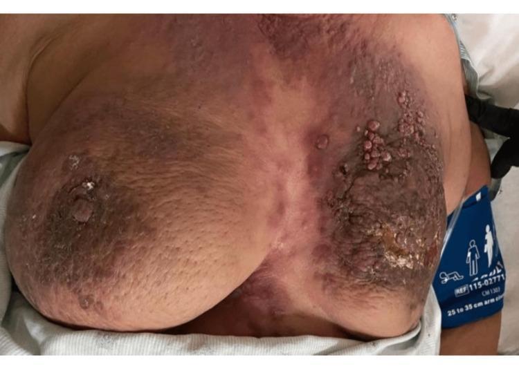
This clinical photograph displays a frontal view of the anterior chest wall, showcasing advanced dermatological manifestations of bilateral breast pathology. Both breasts exhibit significant erythema, diffuse hyperpigmentation, and the characteristic 'peau d'orange' texture, which is indicative of dermal lymphatic invasion. The left breast demonstrates more severe involvement, including extensive nodular skin thickening, verrucous (wart-like) skin nodules, and a retracted nipple. Additionally, a focal area of frank skin ulceration and crusting is visible on the left breast, associated with an underlying tumor mass. The right breast shows similar but less advanced findings with prominent nipple retraction and peau d'orange changes. These visual signs are classic clinical indicators of inflammatory breast cancer or advanced metastatic breast carcinoma with cutaneous involvement.
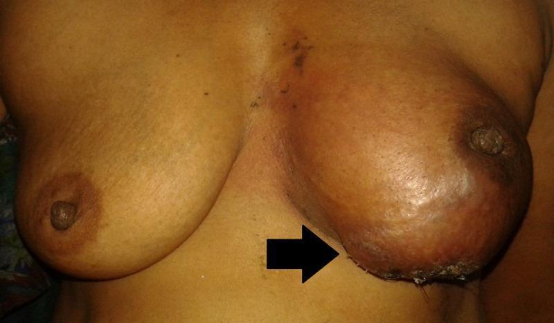
This clinical photograph shows a comparative frontal view of the female chest, highlighting significant pathology in the left breast. The left breast is markedly enlarged and asymmetric compared to the right. The overlying skin of the left breast demonstrates diffuse erythema, peau d'orange-like dimpling, and textural changes indicative of underlying malignancy. The nipple of the left breast is noticeably retracted and inverted. A black arrow points to the inferior aspect of the left breast where there is evidence of skin ulceration and crusting. These clinical findings, including the inflammatory appearance and structural distortion, are highly suggestive of advanced breast cancer, specifically metaplastic carcinoma or inflammatory breast cancer. This image is used in medical education to demonstrate classic physical examination signs of locally advanced breast malignancy, such as nipple retraction, skin dimpling, and inflammatory changes.
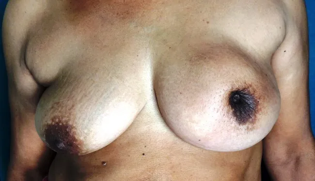
Clinical photograph of a female patient in her mid‑fifties presenting with a left breast mass. The image demonstrates an enlarged left breast with conspicuous nipple retraction and diffuse peau d’orange skin texture consistent with lymphatic obstruction and dermal edema. A firm, irregular 6 cm palpable mass is evident in the upper outer quadrant, correlating with invasive disease. The skin over the lesion shows tethering and dimpling owing to invasion of mammary ducts and stromal fibrosis. This presentation aligns with locally advanced breast cancer and is typical of invasive ductal carcinoma, which commonly presents as a hard, non-mobile lesion with skin involvement and nipple changes. In population-based screening, such cancers are often small and asymptomatic, yet clinically detected cases show palpable masses or skin changes; metastasis to axillary nodes is a consideration. Radiologic correlation (mammography/ultrasound) would typically reveal a spiculated density or irregular mass with posterior acoustic shadowing, and histopathology would confirm infiltrating ductal carcinoma. Clinically, nipple inversion and peau d’orange reflect tumor infiltration and dermal lymphatic obstruction. Early detection improves prognosis; however, skin involvement indicates higher stage. Differential considerations include inflammatory breast cancer, benign fibrocystic change, and other malignant breast neoplasms. Management requires multidisciplinary evaluation and oncologic therapy planning.
Breast Carcinoma: Clinical Features, Molecular Classification, Prognostic Markers, and IHC Tests
(Structured for a 10-Mark Answer)
I. CLINICAL FEATURES OF BREAST CARCINOMA
A. Local Breast Symptoms
- In unscreened populations (including young women), most cancers present as a palpable mass, typically at least 2-3 cm at the time of detection
- Location: upper outer quadrant is the most common site (~50%), followed by the central portion (~20%)
- Character: hard, irregular, non-tender, ill-defined margins (due to desmoplastic stromal reaction)
- Produces a grating sound on cutting due to chalky-white desmoplastic stroma
- In screened older populations, ~60% are detected before symptoms develop; ~20% are in situ carcinomas
- Skin dimpling/retraction - invasion of Cooper's ligaments tethers the skin
- Peau d'orange ("orange peel skin") - blockage of dermal lymphatics causes skin edema; the edematous skin is tethered by Cooper's ligaments, mimicking an orange peel surface
- Skin ulceration - in advanced/locally advanced disease
- Skin fixation/satellite nodules - in very advanced disease
- Nipple retraction/inversion - when the central breast is involved, retraction develops
- Paget disease of the nipple - DCIS extending up the lactiferous ducts into the nipple skin produces a unilateral crusting/eczematous exudate over the nipple and areola; associated with an underlying invasive carcinoma in many cases
- Nipple discharge (bloody/serous)
B. Regional (Nodal) Features
- Axillary lymph node enlargement - most common first site of metastasis
- Lymphatic drainage flows to sentinel lymph nodes in the axilla; if positive, remaining nodes may be involved
- In unscreened populations, at least 50% already have lymph node spread at presentation
- In inflammatory breast cancer: >75% have palpable axillary lymphadenopathy at presentation
- Rare: supraclavicular or internal mammary node involvement
C. Inflammatory Breast Carcinoma (Special Presentation)
- Brawny induration, diffuse erythema with a raised edge
- Skin edema (peau d'orange)
- No discrete palpable mass - diffusely infiltrative
- Caused by extensive plugging of dermal lymphovascular spaces by tumor cells
- Note: the name "inflammatory" is a misnomer - there is no true inflammation histologically
- Often confused with bacterial mastitis, leading to delayed diagnosis
- Tumor is usually high-grade, may be luminal, HER2+, or triple-negative
D. Distant Metastasis Features
| Subtype | Common Sites | Timing |
|---|---|---|
| TNBC / HER2 | Brain, viscera (lung, liver) | Early (<8 years); late recurrence rare |
| Luminal (ER+) | Bone (70-80%), viscera, brain | Low rate over many years; late recurrence (>10 years) possible |
- Lobular carcinoma has unique spread: cerebrospinal fluid, serosal surfaces, GI tract, ovary, uterus, bone marrow
E. Screening-Detected vs. Symptomatic Presentation
| Screened (older women) | Unscreened / Young women | |
|---|---|---|
| Detection method | Mammographic density / calcifications | Palpable mass |
| Tumor size | Small | ≥2-3 cm |
| Node involvement | ~15% | >50% |
| Subtype predominance | Luminal (ER+) | TNBC and HER2+ more common |
II. MOLECULAR CLASSIFICATION OF BREAST CANCER
Three Major Clinical Groups (by IHC)
| Group | Definition | Frequency |
|---|---|---|
| Luminal | ER+, HER2- | 50-65% |
| HER2 | HER2+, ER± | ~20% |
| Triple Negative (TNBC) | ER-, PR-, HER2- | 10-20% |
Six Intrinsic Subtypes (by Gene Expression Profiling)
| Intrinsic Subtype | IHC Profile | Key Features | Prognosis |
|---|---|---|---|
| Luminal A | ER+, PR+, HER2-, Ki-67 low | 40-60% of cases; slow growing | Best |
| Luminal B (HER2-) | ER+, PR low/-, HER2-, Ki-67 high | Higher grade, higher proliferation | Intermediate |
| Luminal B (HER2+) | ER+, PR low/-, HER2+ | Any Ki-67; endocrine + HER2 therapy | Intermediate |
| HER2-enriched | ER-, PR-, HER2+ | Aggressive; responds dramatically to HER2 drugs | Poor (improved with targeted Rx) |
| Basal-like | ER-, PR-, HER2-; CK5/6+, EGFR+ | ~80% overlap with TNBC; high grade; BRCA1-associated | Worst |
| Normal-like / Claudin-low | Variable | Stem cell features; EMT | Very poor |
III. TRIPLE NEGATIVE BREAST CANCER (TNBC) - DETAILED
| Subtype | Key Features | Therapeutic Implication |
|---|---|---|
| BL1 (Basal-like 1) | High DNA damage response genes | Highest pCR to chemotherapy |
| BL2 (Basal-like 2) | Growth factor signaling | Lower pCR |
| M (Mesenchymal) | EMT, stem-cell genes | PI3K/mTOR inhibitors |
| LAR (Luminal Androgen Receptor) | AR expression | Androgen receptor antagonists (enzalutamide) |
- Highest risk of early metastasis and death
- Early relapse peak: <8 years; late recurrence rare
- No ER-targeted or HER2-targeted therapy applicable
- Key somatic mutations: TP53 (70-80%), PIK3CA (9%)
IV. PROGNOSTIC MARKERS IN BREAST CARCINOMA
A. Anatomic/Histologic Prognostic Factors
| Factor | Significance |
|---|---|
| Tumor size (T) | Independent prognostic factor; correlates with lymph node metastasis risk |
| Lymph node status (N) | Most important prognostic factor; sentinel node biopsy is standard |
| Histologic grade (Nottingham) | Based on tubule formation + nuclear pleomorphism + mitotic rate; Grade 1-3 |
| Lymphovascular invasion | Adverse factor |
| Surgical margins | Positive margins = worse prognosis |
B. Biological/Molecular Prognostic Factors
| Marker | Significance |
|---|---|
| ER / PR status | Positive = better prognosis; predicts response to endocrine therapy |
| HER2 status | Positive = aggressive behavior; predicts response to trastuzumab/pertuzumab |
| Ki-67 | Proliferation index; Luminal A (<14%) vs Luminal B (>20%); higher = worse |
| Molecular subtype | Luminal A > Luminal B > HER2 > TNBC (worst) |
| pCR to neoadjuvant chemo | Complete response (pCR) = better prognosis, especially in TNBC and HER2+ |
| TILs (Tumor-Infiltrating Lymphocytes) | High TILs = improved prognosis + better response to chemo in TNBC and HER2+ |
C. Genomic/Multi-gene Assays (Prognostic)
| Assay | Genes | Application |
|---|---|---|
| Oncotype DX | 21 genes → Recurrence Score | ER+/HER2-/N0-1; guides chemo vs. endocrine-only |
| MammaPrint | 70-gene signature | Binary risk; ER+/HER2- |
| Prosigna (PAM50) | 50-gene signature | Intrinsic subtype + Risk of Recurrence score |
| IHC4 score | ER + PR + HER2 + Ki-67 | Equivalent prognostic value to Oncotype DX |
V. IHC AND LABORATORY TESTS FOR BREAST CARCINOMA
Standard Mandatory Panel (All Invasive Cancers)
| Test | Method | Result Interpretation |
|---|---|---|
| ER (Estrogen Receptor) | IHC on FFPE tissue | ≥1% nuclear staining = positive (Allred score) |
| PR (Progesterone Receptor) | IHC | ≥1% nuclear staining = positive |
| HER2 | IHC (0, 1+, 2+, 3+) | 3+ = positive; 2+ = equivocal → FISH |
| HER2 FISH/ISH | Fluorescence/Chromogenic in situ hybridization | HER2:CEP17 ratio ≥2.0 = amplified |
| Ki-67 | IHC (MIB-1 clone) | % nuclear staining; <14% = low; >20% = high |
Additional Diagnostic IHC Markers
| Marker | Purpose |
|---|---|
| E-cadherin | Lost in lobular carcinoma (ILC); distinguishes ILC from IDC |
| CK5/6, CK14, CK17 | Basal cytokeratins; confirm basal-like/TNBC phenotype |
| EGFR | Overexpressed in basal-like/TNBC; confirms basal phenotype |
| AR (Androgen Receptor) | Positive in LAR subtype of TNBC; therapeutic target |
| p63, SMA (smooth muscle actin) | Myoepithelial markers; confirm in situ vs invasive |
Immunotherapy Biomarkers
| Marker | Method | Significance |
|---|---|---|
| PD-L1 (CPS ≥10) | IHC (22C3 assay) | Predicts benefit from pembrolizumab in TNBC |
| TILs assessment | H&E scoring | High TILs = favorable in TNBC/HER2+ |
| MSI / MMR proteins | IHC / PCR | Rare in breast; predicts pembrolizumab response |
| TMB | NGS | Emerging; predicts checkpoint inhibitor response |
Genetic Tests
| Test | Indication | Significance |
|---|---|---|
| BRCA1/2 germline sequencing | TNBC, family history, HER2-/metastatic disease | PARP inhibitor eligibility (olaparib, talazoparib) |
| PIK3CA mutation testing | ER+/HER2- metastatic | Alpelisib eligibility |
| ESR1 mutation (ctDNA/tissue) | ER+ metastatic progressing on endocrine therapy | Guides therapy switch (elacestrant) |
| NTRK gene fusion | Secretory carcinoma | Larotrectinib/entrectinib eligibility |
| HRD (Homologous Recombination Deficiency) | TNBC neoadjuvant | Predicts platinum/PARP inhibitor benefit |
| Circulating tumor DNA (ctDNA) | TNBC with residual disease post-neoadjuvant | Predicts recurrence |
VI. STAGING - AJCC 8th Edition (Integrates Biology + Anatomy)
- T (tumor size/extent) + N (nodal status) + M (metastasis) = Anatomic Stage
- Prognostic Stage then adds: histologic grade + ER + PR + HER2 + multigene assay results
- TNBC is "up-staged" in prognostic staging to reflect aggressive biology
- Luminal A cancers are "down-staged" to reflect favorable biology
- Inflammatory carcinoma = clinical Stage III regardless of T/N
QUICK SUMMARY TABLE FOR EXAM
Prognosis (Best → Worst):
  Luminal A → Luminal B → HER2 → TNBC (Basal-like)

TNBC Key Points:
  • Definition: ER-/PR-/HER2-
  • ~15% of all breast cancers
  • Grade 3; BRCA1 association; African American women
  • No endocrine or HER2 targeted therapy
  • Treatment: Chemo (anthracycline + taxane ± carboplatin)
             + Pembrolizumab (early TNBC, KEYNOTE-522)
             + PARP inhibitors (if BRCA1/2 mutated)
             + Sacituzumab govitecan (metastatic)
  • Early relapse (<8 yrs); brain/visceral metastases

IHC Panel: ER + PR + HER2 (± FISH) + Ki-67 + Grade
Genomic: Oncotype DX / MammaPrint / PAM50 (ER+ only)
Genetic: BRCA1/2 testing for all TNBC patients
You forgot pathogenesis
Purchased credits now give me complied answer add ethology too
Breast Carcinoma: Complete 10-Mark Answer
Etiology, Pathogenesis, Clinical Features, Molecular Classification, Prognostic Markers, and IHC/Laboratory Tests
I. ETIOLOGY AND RISK FACTORS
High Penetrance (Relative Risk > 4.0)
- Female sex (99% of cases are female)
- Increasing age (rare <25 years; sharp rise after age 30)
- Germline mutations of high penetrance (BRCA1, BRCA2, TP53, PTEN)
- Strong family history (>1 first-degree relative, young age of onset, multiple cancers)
- Personal history of breast cancer
- High breast density on mammography
Moderate Penetrance (Relative Risk 2.1-4.0)
- Germline mutations of moderate penetrance (ATM, CHEK2, PALB2)
- High-dose radiation to the chest at a young age (e.g., treatment for lymphoma)
- Family history in one first-degree relative
Low Penetrance (Relative Risk 1.1-2.0)
- Early menarche (age <12 years) - increased lifetime estrogen exposure
- Late menopause (age >55 years) - same
- Late first pregnancy (age >35 years) or nulliparity
- Absence of breastfeeding
- Exogenous hormone therapy (combined OCP/HRT)
- Postmenopausal obesity (adipose tissue produces estrogen)
- Physical inactivity
- High alcohol consumption
Protective factors: Early pregnancy (before age 20), prolonged breastfeeding
Hereditary Susceptibility Genes (Key Table)
| Gene (Syndrome) | % of Single-Gene Cancers | Risk to Age 70 | Associated Subtype | Other Cancers |
|---|---|---|---|---|
| BRCA1 (Familial Br/Ovarian Ca) | ~55% | 40-90% (F) | Majority are TNBC | Ovarian (20-40%), fallopian tube, pancreas |
| BRCA2 (Familial Br/Ovarian Ca) | ~35% | 30-60% (F); 6% (M) | Majority are ER+ | Ovarian (10-20%), pancreas, prostate |
| TP53 (Li-Fraumeni) | <1% | 50-60% (F) | Majority are ER+/HER2+ | Sarcoma, leukemia, brain tumors |
| PTEN (Cowden) | <1% | 20-80% (F) | Variable | Thyroid, endometrium |
| STK11 (Peutz-Jeghers) | <1% | 40-60% (F) | Variable | Ovarian, colon, pancreas |
| CDH1 (Hereditary diffuse gastric Ca) | <1% | ~50% (F) | Lobular type | Gastric signet ring cell carcinoma |
| PALB2 | <1% | ~35% (F) | Variable | Pancreatic cancer |
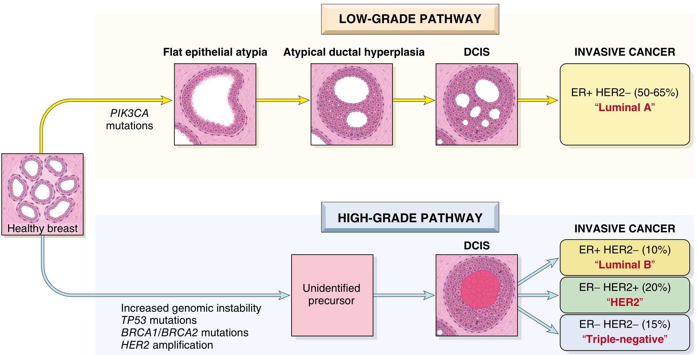
II. PATHOGENESIS
A. General Concept
B. Low-Grade Pathway (ER-Positive / Luminal)
- Proceeds through recognizable ER-positive precursor lesions: Normal breast → Flat epithelial atypia (columnar cell lesions) → Atypical ductal hyperplasia (ADH) → Low-grade DCIS → Invasive ER-positive carcinoma (Luminal A)
- Key genetic changes: loss of 16q, gain of 1q, PIK3CA mutations
- Result: Well-differentiated, slow-growing, ER-positive tumors
- Associated with germline BRCA2 mutations in familial cases
C. High-Grade Pathway (TNBC / HER2)
- Precursor lesion is high-grade DCIS; earliest steps are unidentified
- Key genetic changes: TP53 mutations, BRCA1 inactivation, increased genomic instability, HER2 amplification (17q12), loss of 13q, gain of 11q13
- Results in: HER2+ tumors, Luminal B, or Triple-negative tumors
- Associated with germline BRCA1 mutations (→ TNBC) and TP53 mutations (→ HER2+)
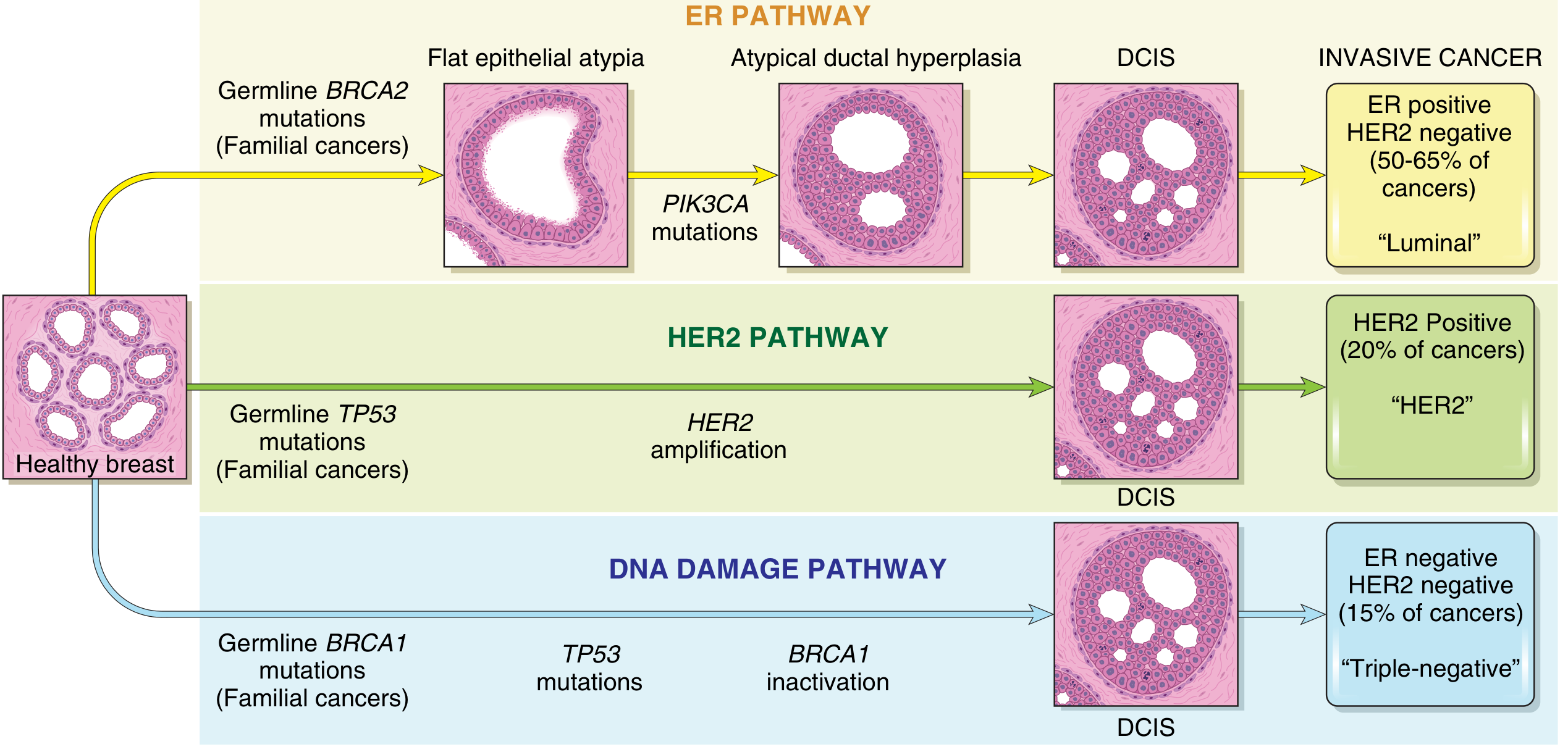
D. TNBC/Basal-like Specific Pathogenesis
- Loss of BRCA1 function impairs differentiation of ER-negative progenitor cells to ER-positive luminal cells → accumulation of genomically unstable ER-negative progenitors
- TP53 mutation: found in ~70-80% of TNBC somatic cases
- Cancers are ER-negative, HER2-negative with high genomic instability (chromosomal rearrangements, copy number changes)
- These tumors have homologous recombination deficiency (HRD) → susceptibility to PARP inhibitors (synthetic lethality)
E. Role of Estrogen in Pathogenesis
- Estrogen promotes proliferation of luminal breast epithelial cells → increases the number of cell divisions → higher probability of acquiring somatic mutations
- Estrogen may facilitate survival of BRCA1-deficient cells
- This explains why increased estrogen exposure (early menarche, late menopause, nulliparity, HRT, obesity) increases breast cancer risk - especially Luminal cancers
F. Tumor Microenvironment
- Cancer-associated fibroblasts: facilitate cell adhesion, ECM remodeling, immunosuppression
- Myoepithelial cells: their reduction creates gaps in the basement membrane, setting the stage for stromal invasion
- PD-L1 upregulation: tumor cells suppress host immune response via immune checkpoint proteins
- Tumor-infiltrating lymphocytes (TILs): paradoxically, high TILs in TNBC/HER2+ cancers indicate better prognosis and predict response to immunotherapy
G. In Situ → Invasive Transition
- Most genomic driver mutations present in invasive cancer are already present in in situ (DCIS) precursors
- The transition from in situ to invasive likely depends on tumor microenvironment changes (not new tumor cell mutations)
III. CLINICAL FEATURES
A. Local Breast Findings
| Sign/Symptom | Mechanism | Notes |
|---|---|---|
| Palpable breast mass | Desmoplastic stromal reaction | Most common presenting feature; hard, irregular, non-tender; UOQ most common (~50%) |
| Skin dimpling/retraction | Cooper's ligament invasion by tumor | Classic sign of malignancy |
| Peau d'orange ("orange peel") | Dermal lymphatic obstruction → skin edema tethered by Cooper's ligaments | Pathognomonic of inflammatory or advanced breast cancer |
| Nipple retraction/inversion | Central breast involvement | New-onset inversion is suspicious |
| Paget disease of nipple | DCIS extending into lactiferous ducts → nipple skin | Unilateral crusting/eczematous nipple discharge |
| Skin ulceration | Advanced/locally advanced disease | Indicates T4 stage |
| Skin fixation | Chest wall invasion | Indicates T4 stage |
| Bloody nipple discharge | Intraductal tumor / Paget disease |
B. Nodal/Systemic Features
- Axillary lymphadenopathy: first/most common metastatic site; hard, matted nodes
- Supraclavicular nodes (N3 disease)
- Lymph node spread: In unscreened populations, >50% already have node involvement at presentation; in screened populations, only ~15%
C. Inflammatory Breast Carcinoma (Special Clinical Syndrome)
- Stage IIIB; <3% of all breast cancers
- Brawny erythema with raised edges + skin edema (peau d'orange) over >33% of breast skin
- No discrete palpable mass - diffusely infiltrative
- Caused by plugging of dermal lymphovascular spaces by tumor cells (not true inflammation)
-
75% have palpable axillary lymphadenopathy; 25% have distant metastases at diagnosis
- Frequently misdiagnosed as bacterial mastitis
D. Screening vs Symptomatic Presentation
| Screened (older women) | Unscreened / young women | |
|---|---|---|
| Detection | Mammographic density or calcification | Palpable mass |
| Typical size at detection | Small (<2 cm) | ≥2-3 cm |
| Node involvement | ~15% | >50% |
| Common subtype | Luminal A | TNBC and HER2+ more common |
| In situ proportion | ~20% are DCIS | Rare in situ at presentation |
E. Metastatic Disease - Subtype-Specific Patterns
| Subtype | Sites | Timing |
|---|---|---|
| Luminal (ER+) | Bone (70-80%), viscera, brain | Low rate over many years; late recurrence >10 years possible; long survival with bone mets |
| HER2+ | Bone (70%), viscera (45%), brain (30%) | Bimodal - early and late peaks |
| TNBC | Bone (40%), viscera (35%), brain (25%) | Early peak <8 years; late recurrence rare; survival with metastases poor |
| Lobular | CSF, GI tract, serosal surfaces, ovary, uterus, bone marrow | Unique pattern - rarely bone predominant |
IV. MOLECULAR CLASSIFICATION
Three Major Clinical Groups (IHC-Based)
| Group | Definition | Frequency |
|---|---|---|
| Luminal (ER+/HER2-) | ER+, HER2- | 50-65% |
| HER2 | HER2+, ER± | ~20% |
| Triple Negative (TNBC) | ER-, PR-, HER2- | 10-20% |
Six Intrinsic Subtypes (Gene Expression Profiling)
| Subtype | IHC Equivalent | Ki-67 | Prognosis |
|---|---|---|---|
| Luminal A | ER+, PR+, HER2- | Low (<14%) | Best |
| Luminal B (HER2-) | ER+, PR low/-, HER2- | High (>20%) | Intermediate |
| Luminal B (HER2+) | ER+, HER2+ | Any | Intermediate |
| HER2-enriched | ER-, PR-, HER2+ | High | Poor (improved with targeted Rx) |
| Basal-like | ER-, PR-, HER2-; CK5/6+, EGFR+ | High | Worst |
| Normal-like / Claudin-low | Variable | Low-moderate | Poor |
TNBC Deep Classification (Lehmann Subtypes)
| Subtype | Key Molecular Features | Implication |
|---|---|---|
| BL1 (Basal-like 1) | High DNA damage response | Highest pCR to platinum-based chemo |
| BL2 (Basal-like 2) | Growth factor receptor signaling | Lower pCR |
| M (Mesenchymal) | EMT, stem cell features | PI3K/mTOR inhibitors |
| LAR (Luminal Androgen Receptor) | Androgen receptor expression | Enzalutamide |
V. PROGNOSTIC AND PREDICTIVE MARKERS
A. Anatomic/Histologic
| Factor | Significance |
|---|---|
| Tumor size (T) | Independent prognostic factor; correlates with nodal risk |
| Lymph node status (N) | Single most important prognostic factor |
| Histologic Grade (Nottingham) | Tubule formation + Nuclear pleomorphism + Mitotic rate = Grade 1-3 |
| Lymphovascular invasion | Adverse; predicts nodal and distant spread |
| Surgical margins | Positive margins = higher local recurrence |
B. Biological Markers
| Marker | Significance |
|---|---|
| ER/PR | Positive = better prognosis; predicts endocrine therapy response |
| HER2 | Positive = aggressive; predicts trastuzumab/pertuzumab response |
| Ki-67 | <14% = Luminal A; >20% = Luminal B; higher = worse prognosis |
| pCR to neoadjuvant chemo | pCR in TNBC/HER2+ = better outcome |
| TILs | High TILs in TNBC/HER2+ = better prognosis + response to immunotherapy |
| PD-L1 (CPS ≥10) | Predicts pembrolizumab benefit in TNBC |
C. Multi-Gene Genomic Assays (ER+ only)
| Assay | Genes | Use |
|---|---|---|
| Oncotype DX | 21 genes (RS 0-100) | Chemo vs. endocrine in ER+/HER2-/N0-1 |
| MammaPrint | 70-gene signature | Binary low/high risk |
| Prosigna (PAM50) | 50 genes | Intrinsic subtype + RoR score |
| IHC4 score | ER + PR + HER2 + Ki-67 | Equivalent prognostic value to Oncotype DX |
VI. IHC AND LABORATORY TESTS
Standard Mandatory IHC Panel (all invasive breast cancers)
| Test | Method | Interpretation |
|---|---|---|
| ER | IHC (nuclear) | ≥1% staining = positive |
| PR | IHC (nuclear) | ≥1% staining = positive |
| HER2 | IHC (0-3+) | 3+ = positive; 2+ → FISH/ISH |
| HER2 FISH/ISH | Gene amplification | HER2:CEP17 ratio ≥2.0 = amplified |
| Ki-67 | IHC (% nuclear, MIB-1 clone) | <14% = low; >20% = high |
Additional Diagnostic/Subtype IHC
| Marker | Purpose |
|---|---|
| E-cadherin | Lost in lobular carcinoma (ILC) |
| CK5/6, CK14 | Confirm basal-like/TNBC phenotype |
| EGFR | Overexpressed in basal-like TNBC |
| AR (Androgen Receptor) | LAR subtype of TNBC; enzalutamide target |
| p63 / SMA | Myoepithelial markers - confirm in situ vs invasive |
Immunotherapy Biomarkers
| Marker | Test | Significance |
|---|---|---|
| PD-L1 (CPS ≥10) | IHC (22C3 assay) | Pembrolizumab benefit in TNBC |
| MSI/MMR | IHC / PCR | Rare in breast; checkpoint inhibitor indication |
| TMB | NGS panel | Emerging predictive marker |
Genetic/Molecular Tests
| Test | Indication | Therapeutic Relevance |
|---|---|---|
| BRCA1/2 germline sequencing | ALL TNBC patients, strong family history | PARP inhibitors (olaparib, talazoparib) |
| PIK3CA mutation | ER+/HER2- metastatic at progression | Alpelisib eligibility |
| ESR1 mutation (ctDNA) | ER+ metastatic progressing on endocrine Rx | Elacestrant eligibility |
| HRD score | TNBC neoadjuvant setting | Platinum/PARP inhibitor benefit |
| ctDNA | TNBC with residual disease post-neoadjuvant | Predicts recurrence |
| NTRK gene fusion | Secretory carcinoma | Larotrectinib/entrectinib |
VII. STAGING (AJCC 8th Edition)
- Anatomic Stage: T (tumor) + N (nodes) + M (metastasis)
- Prognostic Stage: Anatomic stage adjusted by Grade + ER + PR + HER2 + multigene assay
- TNBC is "up-staged" to reflect its aggressive biology
- Luminal A is "down-staged" to reflect its favorable biology
- Inflammatory carcinoma = Stage III regardless of T/N
EXAM QUICK-RECALL SUMMARY
ETIOLOGY KEY POINTS:
  • Most important risk factors: Sex (female), Age, Estrogen exposure, Genetics
  • BRCA1 mutation → TNBC; BRCA2 mutation → ER+ Luminal
  • Protective: Early pregnancy (<20 yrs), prolonged breastfeeding

PATHOGENESIS PATHWAYS:
  Low-grade: PIK3CA mutation → Flat atypia → ADH → low-grade DCIS → LUMINAL A
  High-grade: TP53 + HER2 amplification → DCIS → HER2+
  DNA damage: BRCA1 inactivation + TP53 mutation → DCIS → TRIPLE NEGATIVE

CLINICAL FEATURES (remember "SNIP-LAMP"):
  Skin dimpling, Nipple retraction/Paget disease, Inflammatory signs (peau d'orange)
  Palpable lump (UOQ most common), Axillary lymphadenopathy, Metastases, Peau d'orange

MOLECULAR SUBTYPE PROGNOSIS (best → worst):
  Luminal A → Luminal B → HER2 → TNBC

TNBC TREATMENT:
  Chemo (anthracycline + taxane ± carboplatin)
  + Pembrolizumab (early TNBC, CPS-unselected - KEYNOTE-522)
  + PARP inhibitors (BRCA1/2 mutated)
  + Sacituzumab govitecan (metastatic)

IHC PANEL: ER + PR + HER2 (± FISH) + Ki-67 + Grade
GENETIC: BRCA1/2 testing for ALL TNBC
- Robbins & Kumar Basic Pathology, 10th ed. (Chapter 17 - Breast, pp. 709-713, Tables 17.7-17.9, Figs 17.28-17.30)
- Robbins, Cotran & Kumar Pathologic Basis of Disease, 10th ed. (Chapter 23 - Breast, pp. 954-966, Figs 23.15-23.17)
- Fischer's Mastery of Surgery, 8th ed. (pp. 1779-1781, Table 66.2)
- Schwartz's Principles of Surgery, 11th ed. (Chapter 17)
- Chen et al., 2025 - Classifications of TNBC (PMID: 39893480)