I'll look up hypersensitivity in the medical library for a thorough, textbook-quality answer.**Searching sources...**Excellent results. Let me now pull the richest sources - Roitt's Essential Immunology (dedicated immunology textbook), Goldman-Cecil Medicine, and the Barash Clinical Anesthesia block for the Types I-IV breakdown.Good - now let me get the detailed content on each type from Roitt's and Goldman-Cecil, plus the hypersensitivity table from Scott-Brown's and Jawetz.Excellent content. Now let me get the detailed per-type descriptions from Roitt's (the best immunology text) and fetch the key diagram image.I now have an excellent diagram and rich content. Let me get the detailed descriptions of Type II, III, and IV from Roitt's to complete the picture.I have rich, comprehensive content across all four types. I now have enough to write a thorough, well-structured answer. Let me compile everything.
Hypersensitivity
Hypersensitivity refers to immune responses that are excessive, misdirected, or occur at inappropriate locations - causing tissue damage (immunopathology) rather than protection. The underlying effector mechanisms are the same ones used in normal host defense; the problem is that they are overactivated against antigens that pose no real threat, such as innocuous environmental antigens, self-antigens, or antigens from another species.
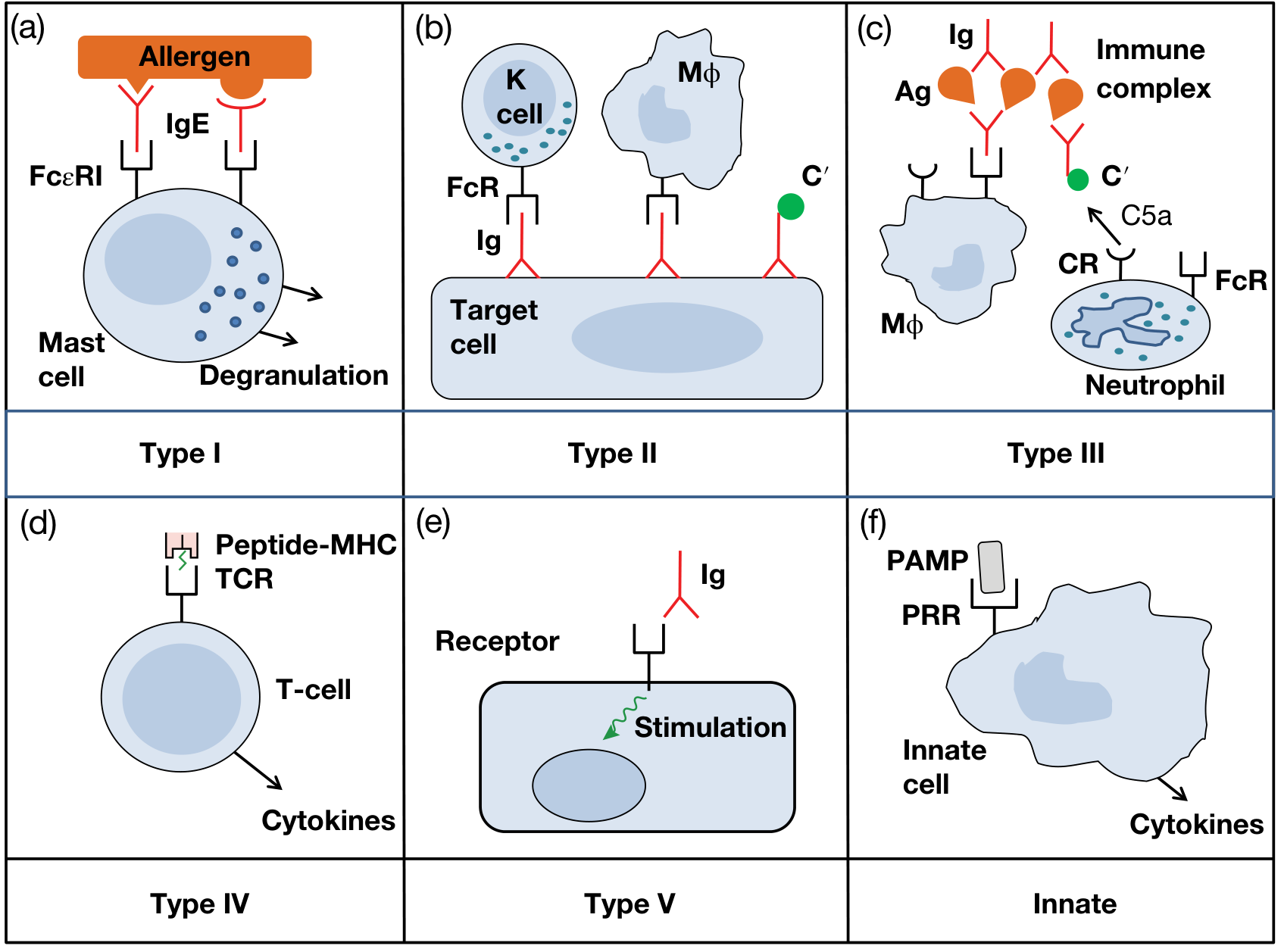
The classification system introduced by Gell and Coombs (1963) divides hypersensitivity into four types (I-IV). Subsequent work has added a Type V and recognition of innate hypersensitivity reactions.
Figure 14.1 - Six categories of hypersensitivity (Roitt's Essential Immunology, 13th Ed.)
Summary Table (Gell and Coombs Classification)
| Feature | Type I | Type II | Type III | Type IV |
|---|
| Name | Immediate / Anaphylactic | Cytotoxic | Immune Complex | Cell-Mediated / Delayed |
| Immune reactant | IgE | IgG | IgG | T cells |
| Effector cells | Mast cells, basophils, eosinophils | Complement, phagocytes | Complement, phagocytes, neutrophils | Macrophages, cytotoxic T cells |
| Onset | Minutes | Minutes-hours | Hours | 48-72 hours |
| Disease examples | Allergic rhinitis, anaphylaxis, asthma | Haemolytic anaemia, Goodpasture's | Serum sickness, SLE, post-streptococcal GN | Contact dermatitis, tuberculin reaction, graft rejection |
- Scott-Brown's Otorhinolaryngology Head & Neck Surgery, Table 14.1
Type I - Immediate (IgE-Mediated) Hypersensitivity
Mechanism: Allergen exposure in a sensitized individual cross-links IgE molecules already bound to the high-affinity FcεRI receptors on mast cells and basophils. This triggers explosive degranulation, releasing preformed mediators (histamine, tryptase, heparin) and prompting synthesis of lipid mediators (prostaglandins, leukotrienes) and cytokines.
Two phases:
- Early phase (within minutes): Histamine-mediated vasodilation, bronchoconstriction, increased vascular permeability, and mucus secretion.
- Late phase (4-12 hours): Eosinophil-dominated inflammation driven by Th2 cytokines (IL-4, IL-5, IL-13), causing prolonged tissue damage.
Sensitization: Initial antigen exposure drives Th2 differentiation, IL-4/IL-13 production, and B-cell class switching to IgE production. IgE then coats mast cells throughout the body, "priming" for future exposure.
Clinical examples:
- Anaphylaxis (penicillin, bee sting, peanuts)
- Allergic rhinitis (hayfever)
- Atopic asthma
- Urticaria and atopic eczema
- Food allergy (cow's milk, eggs, nuts, shellfish)
Treatment principles: Epinephrine (reverses bronchospasm and vasodilation - the cornerstone of anaphylaxis treatment), antihistamines (H1 blockers), corticosteroids, mast cell stabilizers (sodium cromoglycate), leukotriene antagonists (montelukast), and anti-IgE biologics (omalizumab).
- Roitt's Essential Immunology, 13th Ed., Chapter 14
Type II - Cytotoxic (Antibody-Dependent) Hypersensitivity
Mechanism: IgG (or IgM) antibodies bind directly to antigens on cell surfaces or extracellular matrix. This triggers cell destruction via three pathways:
- Complement activation - classical pathway generates MAC (C5b-C9), causing direct cell lysis
- Opsonization and phagocytosis - C3b and Fc receptors on phagocytes mediate cell engulfment
- Antibody-Dependent Cellular Cytotoxicity (ADCC) - NK cells, monocytes, and eosinophils bind antibody-coated targets via FcγR and kill them without phagocytosis
Clinical examples:
- ABO transfusion reactions - pre-existing antibodies against ABO antigens lyse incompatible red cells
- Haemolytic disease of the newborn (Rh incompatibility) - maternal anti-D IgG crosses placenta
- Goodpasture's syndrome - anti-GBM antibodies destroy glomerular and alveolar basement membranes
- Autoimmune haemolytic anaemia and immune thrombocytopenia (ITP)
- Bullous pemphigoid - IgG against hemidesmosomal proteins (BP180/BP230), causing subepidermal blisters with eosinophilic infiltrate
- Roitt's Essential Immunology, 13th Ed.; Goldman-Cecil Medicine
Type III - Immune Complex-Mediated Hypersensitivity
Mechanism: Soluble antigen-antibody (IgG) complexes form in the circulation or in tissues. Normally these are cleared by the mononuclear phagocyte system (MPS). When complexes are produced in excess or not cleared efficiently, they deposit in vessel walls, glomeruli, and synovium. Deposited complexes activate complement, generating C3a and C5a (anaphylatoxins), which attract neutrophils. Neutrophil degranulation releases proteases and reactive oxygen species, causing local tissue injury.
Types I, II, and III are all antibody-mediated. Type III is distinguished by the fact that the antigen is soluble (not on a cell surface).
Clinical examples:
- Serum sickness - systemic reaction 1-2 weeks after injection of foreign protein (horse antitoxin); fever, arthralgia, urticaria, lymphadenopathy
- Systemic lupus erythematosus (SLE) - anti-dsDNA immune complexes deposit in kidneys, skin, and joints
- Post-streptococcal glomerulonephritis - streptococcal antigen-antibody complexes deposit in glomeruli
- Hypersensitivity pneumonitis (Farmer's lung) - inhaled fungal spores form immune complexes in lung parenchyma
- Arthus reaction - local tissue necrosis from intradermal antigen injection in a pre-immunized individual
- Roitt's Essential Immunology, 13th Ed.; Goldman-Cecil Medicine
Type IV - Delayed-Type (Cell-Mediated) Hypersensitivity
Mechanism: Unlike Types I-III, Type IV does not involve antibodies. It is entirely T cell-mediated. Sensitized CD4+ Th1 cells recognize antigen (presented on MHC II by antigen-presenting cells) and release cytokines - particularly IFN-γ, which activates macrophages, causing inflammation and tissue destruction. CD8+ cytotoxic T cells are also involved in some forms (e.g., contact dermatitis).
Onset is delayed (48-72 hours) because it takes time to recruit and activate sufficient effector T cells and macrophages at the site of antigen deposition.
Subtypes (Cher classification):
- IVa - Th1-mediated, macrophage activation (tuberculin test, Crohn's disease)
- IVb - Th2-mediated, eosinophilic inflammation (contact dermatitis to nickel)
- IVc - Cytotoxic T cell-mediated (Stevens-Johnson syndrome)
- IVd - Neutrophil-mediated, T cell-dependent (AGEP)
Clinical examples:
- Tuberculin (Mantoux) test - induration at 48-72 h is classic delayed hypersensitivity
- Contact dermatitis - nickel, poison ivy, latex
- Granulomatous diseases - tuberculosis, sarcoidosis (macrophages fail to clear persistent antigen, forming granulomas)
- Graft rejection - T cell-mediated destruction of allogeneic tissue
- Type 1 diabetes mellitus (CD8+ T cell destruction of pancreatic beta cells)
- Roitt's Essential Immunology, 13th Ed.; Goldman-Cecil Medicine
Type V - Stimulatory Hypersensitivity (Addition to Classic Classification)
Mechanism: Antibodies bind to cell surface receptors and act as agonists, mimicking the normal ligand and stimulating the cell rather than destroying it. This is distinct from Type II because the outcome is cell overstimulation, not cytotoxicity.
Clinical example:
- Graves' disease - TSI (thyroid-stimulating immunoglobulin) antibodies bind TSH receptor on thyroid follicular cells, mimicking TSH and causing unregulated hyperthyroidism
- Roitt's Essential Immunology, 13th Ed.
Innate Hypersensitivity
Excessive or inappropriately activated pattern recognition receptors (PRRs) - such as Toll-like receptors (TLRs) - can trigger cytokine storms and inflammatory tissue damage without involving adaptive immunity. This is increasingly recognized in conditions like sepsis and cytokine release syndrome.
Key Points to Remember
| Point | Detail |
|---|
| Types I, II, III = antibody-mediated | Type IV = cell-mediated |
| Type I antigen = soluble; extracellular | Type II antigen = cell surface or matrix-bound |
| Type III antigen = soluble (circulating) | Type IV antigen = intracellular or cell-surface (presented by MHC) |
| Most allergies (hayfever, asthma, eczema, food) | Type I (IgE); some eczema is Type IV |
| Serum sickness prototype | Type III |
| Tuberculin test prototype | Type IV |
| A single disease can involve multiple types | e.g., SLE involves Types II, III, and IV |
Sources:
- Roitt's Essential Immunology, 13th Edition, Chapter 14 (Allergy and Other Hypersensitivities)
- Goldman-Cecil Medicine, International Edition, Chapter on Hypersensitivity
- Scott-Brown's Otorhinolaryngology Head & Neck Surgery, Vol. 1, Table 14.1
- Barash, Cullen & Stoelting's Clinical Anesthesia, 9th Edition, Chapter on Immune FunctionLet me search the textbooks for dedicated diagrams for each hypersensitivity type.Good - Barash's Clinical Anesthesia has individual figures for each type. Let me pull all of them at once.I have all four image URLs. Let me fetch all four diagrams in parallel now.I have the URLs for Type III and Type IV figures. Let me fetch them both now.All four diagrams retrieved. Here they are, one per type.
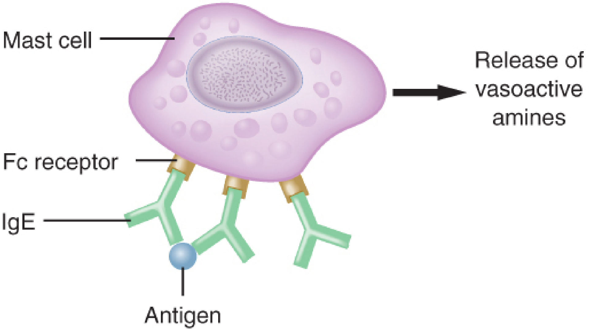
Type I - Immediate (IgE-Mediated) Hypersensitivity
Mechanism: Antigen binds to IgE antibodies already attached to Fc receptors on mast cells/basophils. Cross-linking of IgE triggers degranulation and release of vasoactive amines (histamine, etc.). Complement is not involved. Examples: anaphylaxis, asthma, allergic rhinitis.
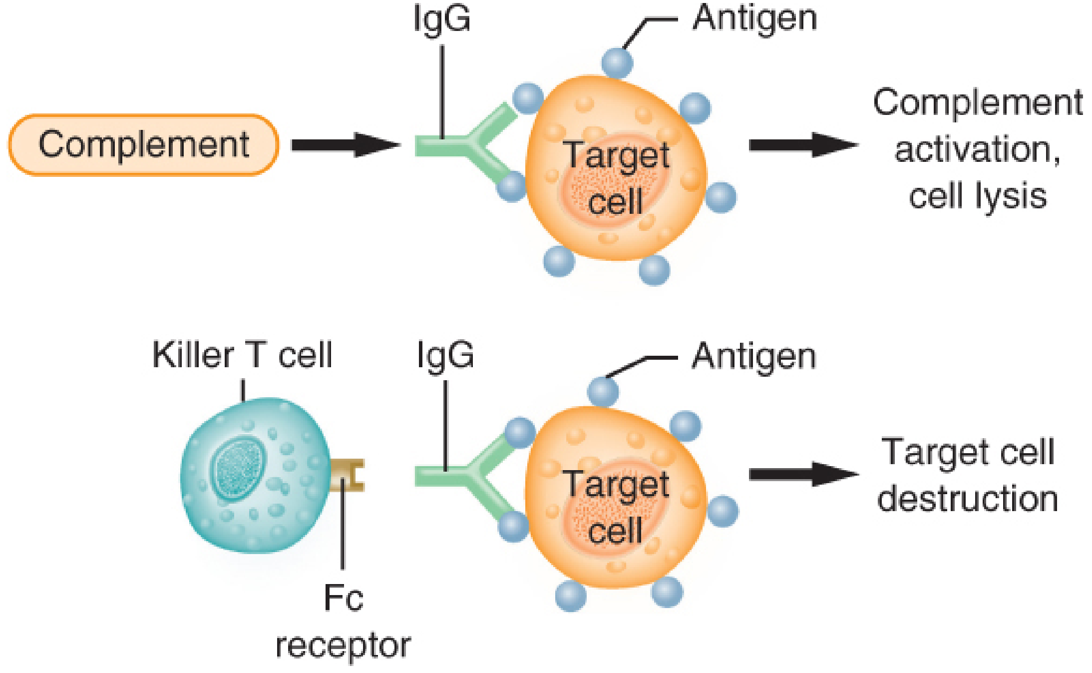
Type II - Cytotoxic (Antibody-Dependent) Hypersensitivity
Mechanism: IgG (or IgM) targets antigens on the surface of cells. Destruction occurs via two routes shown: (1) complement activation leading to cell lysis; (2) killer T cells binding via Fc receptors (ADCC) causing target cell destruction. Examples: ABO transfusion reactions, autoimmune haemolytic anaemia, Goodpasture's syndrome.
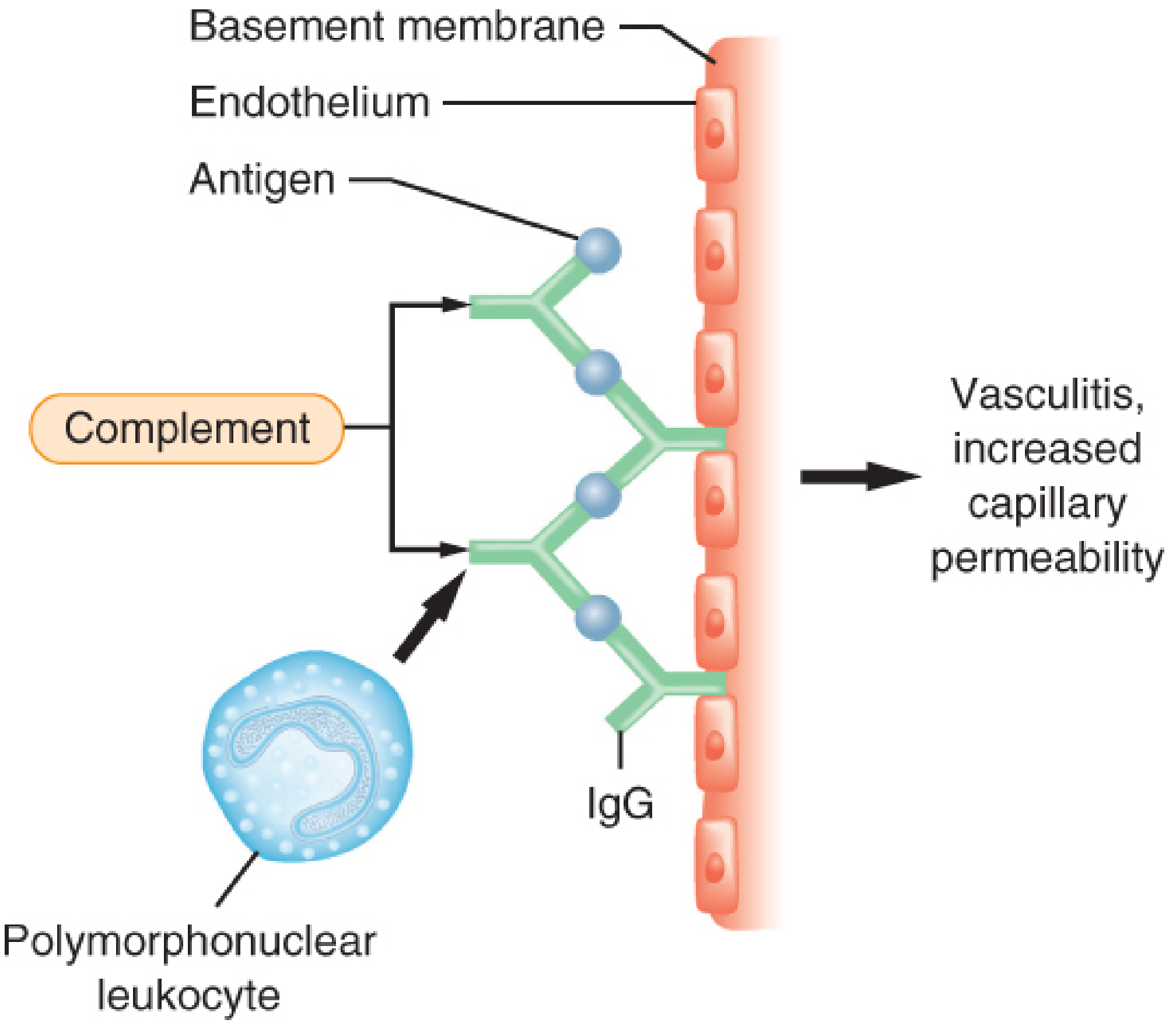
Type III - Immune Complex-Mediated Hypersensitivity
Mechanism: Soluble antigen-IgG complexes deposit in the microvasculature basement membrane. Complement activation generates C5a, attracting polymorphonuclear leukocytes (neutrophils), which degranulate and cause vasculitis and increased capillary permeability. Examples: serum sickness, SLE, post-streptococcal GN.
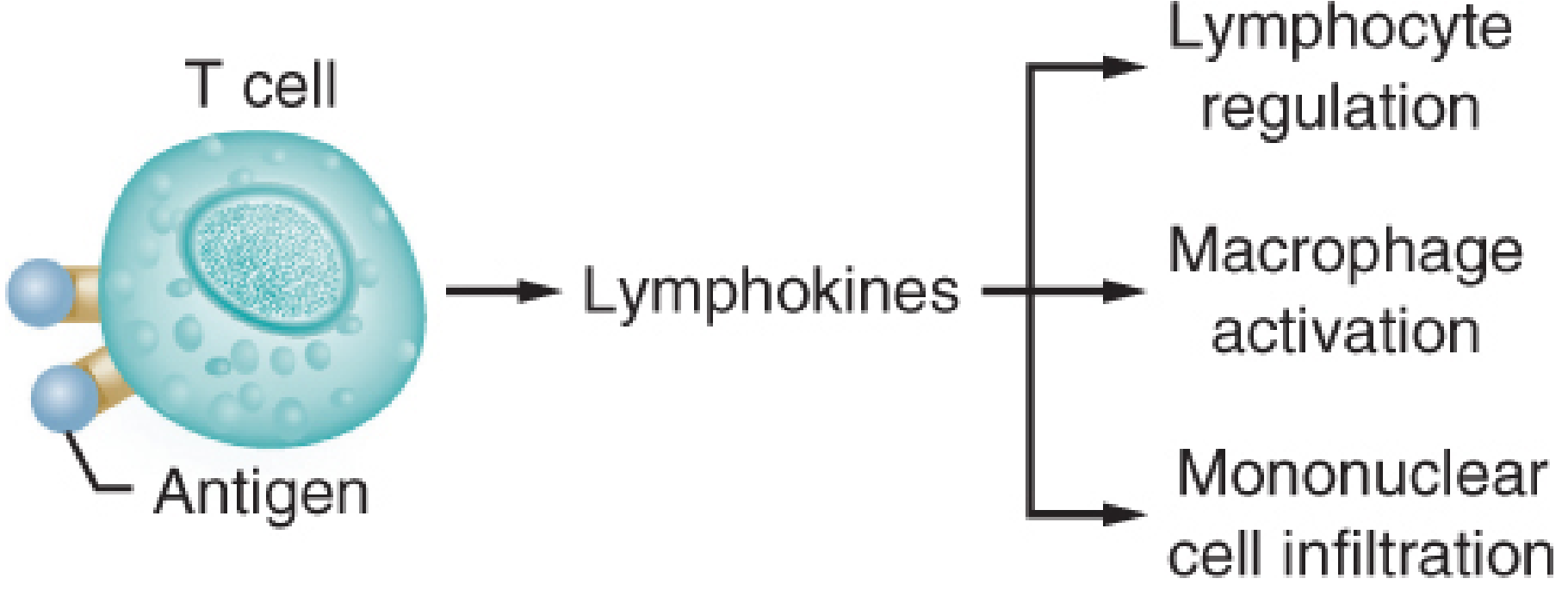
Type IV - Delayed (Cell-Mediated) Hypersensitivity
Mechanism: Antigen binds to sensitized T cells, which release lymphokines (cytokines) causing three downstream effects: lymphocyte regulation, macrophage activation, and mononuclear cell infiltration. No antibody or complement involved. Onset is 18-96 hours after antigen exposure. Examples: tuberculin test, contact dermatitis, graft rejection, granulomatous disease.
All diagrams from Barash, Cullen & Stoelting's Clinical Anesthesia, 9th Edition (Figures 9-3 to 9-6)
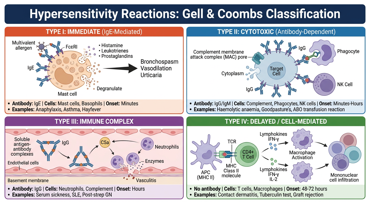
Here's your side-by-side comparison poster covering all four Gell & Coombs types. Each panel shows:
- Type I (orange) - IgE/mast cell axis, immediate onset, classic allergies
- Type II (blue) - IgG targeting cell-surface antigens, complement lysis + ADCC
- Type III (purple) - Soluble immune complex deposition, neutrophil-driven vasculitis
- Type IV (green) - T cell/macrophage mediated, no antibody, delayed 48-72 h onset**Searching sources...**Excellent. Let me pull the richest sources in parallel - Henry's Clinical Diagnosis (lab medicine bible), Quick Compendium of Clinical Pathology, Schwartz's Surgery, and Bailey & Love's table.Excellent content. Now let me pull Henry's diagnostic pattern table, the Quick Compendium section on bilirubin/aminotransferases, and search for LFT images.Excellent content. Let me now get the Henry's Table 9.5 (diagnostic patterns), the bilirubin metabolism table, and search for LFT images in the textbooks.Let me fetch the bilirubin metabolism diagrams and the Henry's diagnostic patterns table simultaneously.I have all the content I need. Now let me fetch all key images in parallel - bilirubin metabolism diagram and Henry's bilirubin transport diagram.Outstanding images retrieved. I now have comprehensive content and diagrams. Let me compile the full LFT explanation.
Liver Function Tests (LFT) - Full Explanation
Liver function tests (LFTs) are a panel of blood tests used to assess hepatocellular integrity, biliary function, and the liver's synthetic capacity. The liver performs three broad categories of function, each tested by different analytes:
- Hepatocellular integrity - tested by aminotransferases (AST, ALT)
- Biliary/cholestatic function - tested by ALP, GGT, bilirubin
- Synthetic capacity - tested by albumin, total protein, prothrombin time (INR)
"The liver is the site of synthesis of over 90% of the proteins in the body, including all of the albumin. Unlike with the two aminotransferases, liver pathology is recognized by decreases in serum levels of total protein and albumin. These decreases do not occur unless 80% or more of liver tissue is destroyed." - Henry's Clinical Diagnosis and Management by Laboratory Methods
1. Bilirubin
Physiology
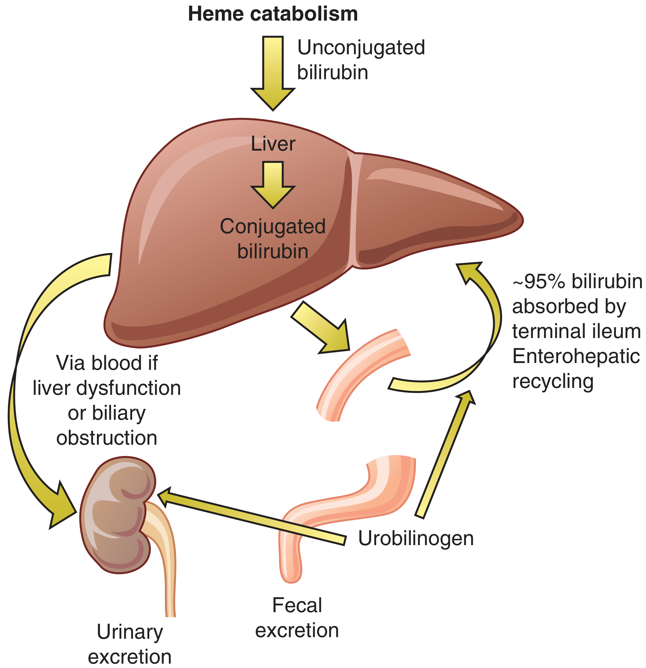
Bilirubin is the end product of haem catabolism. Understanding its metabolism is essential for interpreting LFTs.
Figure 26-1: Bilirubin metabolism and excretion - Symptom to Diagnosis, 4th Ed.
Steps:
- Haem catabolism in the spleen (Kupffer cells) → unconjugated (indirect) bilirubin - water insoluble, bound to albumin in blood
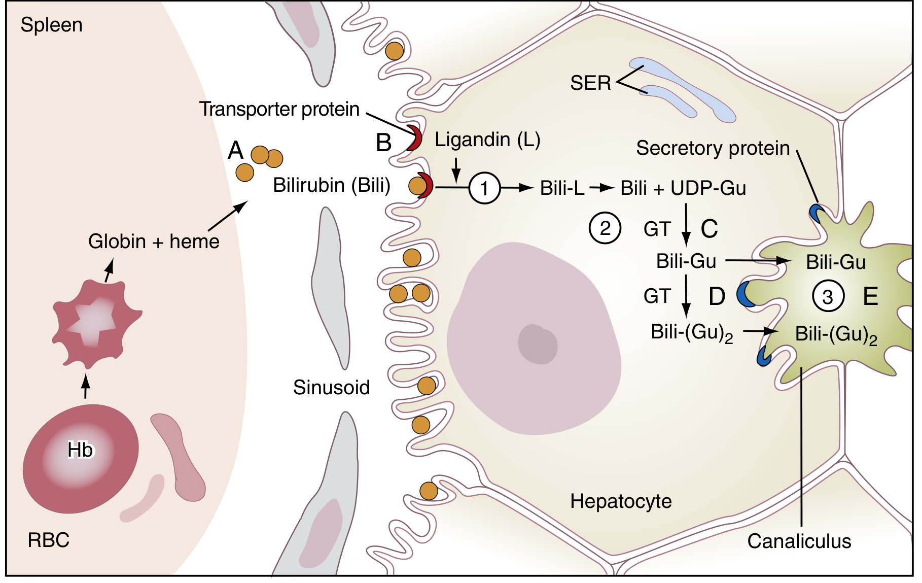
- Enters hepatocyte via transporter proteins → binds ligandin → transported to smooth endoplasmic reticulum (SER)
- Conjugation by UDP-glucuronosyltransferase (UGT1A1) with glucuronic acid → conjugated (direct) bilirubin - water soluble
- Secreted into bile canaliculi via MRP2 transporter → flows into intestines
- Gut bacteria convert to urobilinogen → 95% reabsorbed (enterohepatic recycling) → remainder excreted in faeces (stercobilin - gives stool brown colour) and a small amount in urine (urobilinogen)
Figure 22.2: Bilirubin transport and metabolism at hepatocyte level - Henry's Clinical Diagnosis, Laboratory Methods
Normal Values
| Fraction | Normal Range |
|---|
| Total bilirubin | 5-17 µmol/L (0.3-1.2 mg/dL) |
| Direct (conjugated) | 0-5 µmol/L (<0.3 mg/dL) |
| Indirect (unconjugated) | 3-12 µmol/L |
- Jaundice becomes clinically visible when total bilirubin >34-51 µmol/L (>2-3 mg/dL)
- Conjugated hyperbilirubinaemia: >30% of total bilirubin is conjugated → indicates excretory defect
Causes of Elevated Bilirubin by Type
| Type | Cause | Examples |
|---|
| Unconjugated (pre-hepatic) | Excess haemolysis OR impaired conjugation | Haemolytic anaemia, Gilbert syndrome, Crigler-Najjar, neonatal jaundice |
| Conjugated (hepatic/post-hepatic) | Impaired excretion or biliary obstruction | Viral hepatitis, cirrhosis, cholestasis, bile duct obstruction, Dubin-Johnson syndrome |
| Mixed | Combined hepatocellular damage | Liver failure, sepsis, advanced cirrhosis |
2. Alanine Aminotransferase (ALT)
ALT is the most liver-specific of the aminotransferases. It is found predominantly in hepatocyte cytoplasm.
| Value |
|---|
| Normal range | 5-40 IU/L (7-56 U/L by some labs) |
| Mild elevation | <5× upper limit of normal (ULN) = <175-200 U/L |
| Moderate elevation | 5-10× ULN |
| Marked elevation | >10× ULN (>1000 U/L) |
Causes by degree:
- >1000 U/L (massive): Acute viral hepatitis, ischaemic hepatitis ("shock liver"), drug/toxin hepatitis (paracetamol), autoimmune hepatitis, acute bile duct obstruction, Budd-Chiari
- Moderate (5-10× ULN): Chronic viral hepatitis B/C, drug reactions, Wilson's disease
- Mild (<5× ULN): NAFLD/NASH, alcoholic liver disease, coeliac disease, thyroid disease, strenuous exercise
3. Aspartate Aminotransferase (AST)
AST is less liver-specific than ALT - also found in heart, skeletal muscle, kidney, and brain. It is elevated in both hepatic and non-hepatic conditions.
| Value |
|---|
| Normal range | 5-40 IU/L |
AST:ALT Ratio - Key Diagnostic Clue
| Ratio | Interpretation |
|---|
| ALT > AST (ratio <1) | Viral hepatitis, NAFLD, cholestasis |
| AST:ALT >2:1 | Alcoholic liver disease (hallmark sign) |
| AST:ALT >3:1 | Strongly suggests alcoholic hepatitis |
| Both very high (>1000) | Acute viral/ischaemic/drug hepatitis |
"In alcoholic hepatitis, AST elevation exceeds that of ALT." - Goldman-Cecil Medicine, 22nd Ed.
4. Alkaline Phosphatase (ALP)
ALP is located on the canalicular surface of hepatocytes and biliary epithelium. It is most sensitive for cholestatic disease and biliary obstruction.
| Value |
|---|
| Normal range (adults) | 30-140 IU/L |
| Children | Higher (due to bone growth - separate reference intervals required) |
| Pregnancy | Elevated (placental isoform) |
Important: ALP is not liver-specific - it also comes from bone, placenta, intestine. To confirm hepatic origin, check GGT (if GGT is elevated with ALP, the source is hepatic; if GGT is normal, ALP is likely from bone).
Causes of elevated ALP:
| Liver origin | Bone origin | Other |
|---|
| Biliary obstruction (choledocholithiasis, cholangiocarcinoma) | Paget's disease | Pregnancy |
| Primary biliary cholangitis (PBC) | Bone metastases | Childhood growth |
| Primary sclerosing cholangitis (PSC) | Osteomalacia | Intestinal (postprandial) |
| Hepatic metastases (most sensitive marker) | Hyperparathyroidism | - |
| Cholestatic drug reactions | Healing fractures | - |
Low ALP: Hypophosphatasia, malnutrition, Wilson's disease, theophylline/oestrogen therapy
5. Gamma-Glutamyltransferase (GGT)
GGT is derived primarily from biliary epithelial cells and hepatocytes. It is a highly sensitive but non-specific marker of hepatobiliary injury.
| Value |
|---|
| Normal range | 10-48 IU/L (slightly higher in men) |
Key uses:
- Confirms hepatic origin of elevated ALP - if both ALP and GGT are elevated, the source is hepatic
- Alcohol marker - elevated in up to 70% of chronic alcoholics; correlates with alcohol consumption; elevated to 2-3× ULN in heavy drinkers
- Elevated by enzyme-inducing drugs: warfarin, barbiturates, phenytoin, valproate, methotrexate
6. Albumin
Albumin is synthesised exclusively by the liver. Because its half-life is ~20 days, it is a marker of chronic (not acute) synthetic function.
| Value |
|---|
| Normal range | 35-50 g/L (3.5-5 g/dL) |
| Hypoalbuminaemia | <35 g/L |
Causes of low albumin:
- Chronic liver disease/cirrhosis (>80% hepatocyte destruction needed)
- Malnutrition, malabsorption
- Nephrotic syndrome (urinary loss)
- Protein-losing enteropathy
- Acute phase response (albumin is a negative acute-phase reactant)
"Albumin levels below normal do not occur unless 80% or more of liver tissue is destroyed, as in cirrhosis and fulminant hepatic failure." - Henry's Clinical Diagnosis
7. Total Protein
| Value |
|---|
| Normal range | 60-85 g/L (6-8.5 g/dL) |
Total protein = albumin + globulins. In cirrhosis, albumin falls but immunoglobulins rise (polyclonal gammopathy), so total protein may appear normal or even elevated despite severe liver disease - always interpret albumin and globulins separately.
8. Prothrombin Time (PT) / INR
The liver synthesises the majority of clotting factors (I, II, V, VII, IX, X). Factor VII has the shortest half-life (~6 hours), making PT/INR the most sensitive and rapidly changing test of hepatic synthetic function.
| Value |
|---|
| Normal PT | 12-16 seconds |
| Normal INR | 0.8-1.2 |
- INR becomes prolonged within hours of a major hepatic insult - much faster than albumin (days-weeks)
- Used to assess severity in acute liver failure (Kings College Criteria uses INR)
- INR >1.5 with liver disease = significant synthetic dysfunction
9. Ammonia
| Value |
|---|
| Normal range | 15-45 µmol/L (11-35 µg/dL) |
The liver is the sole site of ammonia detoxification via the urea cycle. Elevated ammonia occurs when >80% of liver is non-functional. Note: ammonia levels do not correlate with the degree of hepatic encephalopathy.
Pre-examination errors are common - specimen must be chilled on ice, processed immediately, haemolysis avoided, patient should not smoke for several hours beforehand.
Diagnostic Patterns - How to Read an LFT Panel
Based on Henry's Clinical Diagnosis 6 classic patterns:
| Pattern | AST/ALT | ALP/GGT | Bilirubin | Albumin | PT/INR | Diagnosis |
|---|
| Acute hepatitis | ↑↑↑ (>10× ULN) | ↑ | ↑ (both types) | Normal | Normal | Viral/drug/ischaemic hepatitis |
| Cirrhosis | Normal or mildly ↑ | ↑ | ↑ (both) | ↓↓ | ↑ (prolonged) | End-stage chronic liver disease |
| Biliary obstruction | Normal or mildly ↑ | ↑↑↑ | ↑ (conjugated) | Normal | Normal | Stone, stricture, cholangiocarcinoma, pancreatic head mass |
| Space-occupying lesion | Normal | ↑↑ (ALP) | Normal | Normal | Normal | Hepatic metastases, HCC |
| Passive congestion | Mildly ↑ | Normal/↑ | ↑ (if severe) | Normal | Normal | Right heart failure, Budd-Chiari |
| Fulminant hepatic failure | ↑↑↑↑ (>10,000 U/L possible) | ↑ | ↑↑ | ↓↓ | ↑↑ prolonged | Paracetamol overdose, fulminant viral hepatitis |
Pattern Recognition at a Glance
- Transaminases >> ALP: Hepatocellular pattern (hepatitis, NAFLD, ischaemia)
- ALP >> Transaminases: Cholestatic pattern (obstruction, PBC, PSC)
- Low albumin + prolonged INR: Impaired synthetic function - chronic or severe acute disease
- AST:ALT >2: Think alcohol
- Isolated ALP ↑ + normal GGT: Think bone, not liver
- Progressive bilirubin rise with known liver disease: Deteriorating function - urgent review
Quick Reference: Normal Values Summary
| Test | Normal Range | What It Measures |
|---|
| Bilirubin (total) | 5-17 µmol/L | Excretory function |
| Bilirubin (direct) | <5 µmol/L | Conjugated fraction |
| ALT | 5-40 IU/L | Hepatocellular damage (liver-specific) |
| AST | 5-40 IU/L | Hepatocellular damage (less specific) |
| ALP | 30-140 IU/L | Cholestasis / biliary obstruction |
| GGT | 10-48 IU/L | Cholestasis / alcohol marker |
| Albumin | 35-50 g/L | Chronic synthetic function |
| Total protein | 60-85 g/L | Synthetic function (global) |
| PT | 12-16 s | Acute synthetic function |
| INR | 0.8-1.2 | Acute synthetic function |
| Ammonia | 15-45 µmol/L | Urea cycle / encephalopathy |
Sources:
- Henry's Clinical Diagnosis and Management by Laboratory Methods - Chapter 9 & 22
- Goldman-Cecil Medicine, International Edition - Laboratory Studies in Liver Disease
- Bailey & Love's Short Practice of Surgery, 28th Ed. - Table 69.1
- Quick Compendium of Clinical Pathology, 5th Ed. - Liver Function Tests
- Symptom to Diagnosis: An Evidence-Based Guide, 4th Ed. - Figure 26-1
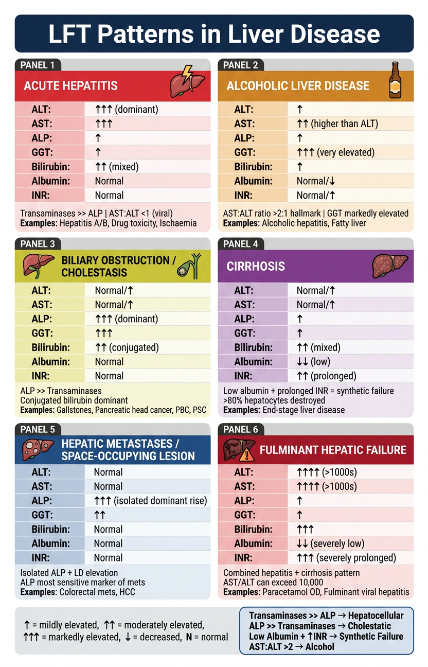
Here is your LFT pattern comparison poster covering all 6 major diagnostic patterns:
| Panel | Key Pattern | Hallmark Clue |
|---|
| Acute Hepatitis | ALT/AST ↑↑↑ dominant | Transaminases >> ALP |
| Alcoholic Liver Disease | GGT ↑↑↑, AST:ALT >2 | AST higher than ALT |
| Biliary Obstruction | ALP/GGT ↑↑↑ dominant | Conjugated bilirubin ↑ |
| Cirrhosis | Low albumin + ↑ INR | Synthetic failure pattern |
| Hepatic Metastases | Isolated ALP ↑↑↑ | Transaminases normal |
| Fulminant Failure | Everything severely deranged | AST/ALT can hit >10,000 |
The bottom legend summarises the two master pattern rules:
- Transaminases >> ALP = hepatocellular injury
- ALP >> Transaminases = cholestatic/obstructiveGood. Let me pull Goodman & Gilman (the definitive pharmacology textbook), Ganong's Physiology, and Katzung simultaneously.Excellent! Let me pull the Lippincott Pharmacology table (perfect for this topic) and Goodman & Gilman's drug table simultaneously.Excellent data. Let me get more from Goodman & Gilman's table (anticholinesterases section) and fetch key images.Now let me get one more image - the cholinergic neurotransmission/receptor diagram from Lippincott, and the Ganong's effector organ table.I now have all the content and images I need. Let me compile the full answer.# ANS Pharmacology - Cholinergic Drugs
Step 1: The Foundation - Cholinergic Neurotransmission
Before classifying the drugs, understand what they are acting on.
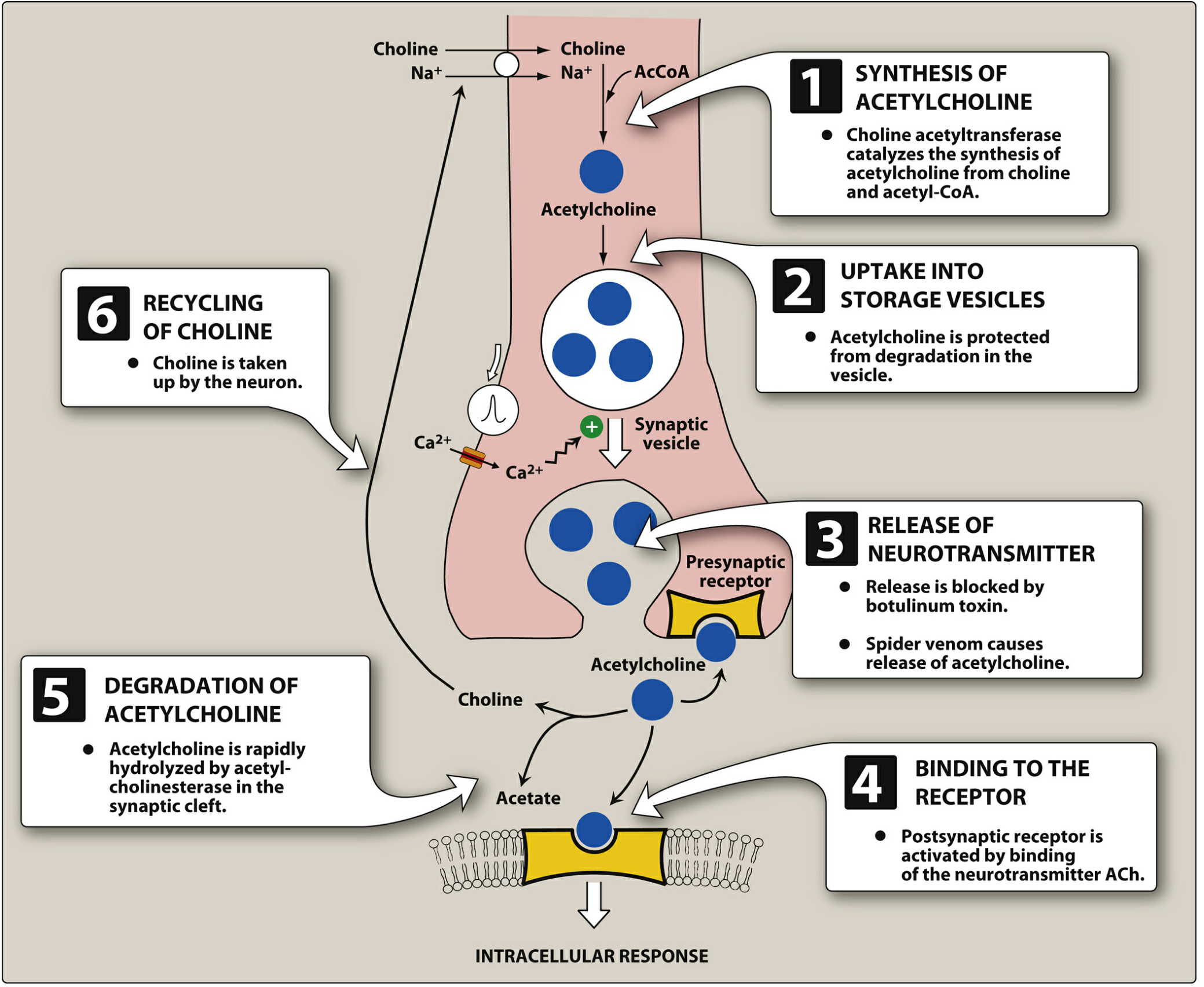
Figure 4.3 - Synthesis and release of ACh from the cholinergic neuron. Lippincott Illustrated Reviews: Pharmacology
Key steps drugs can target:
- Synthesis (choline transport - rate-limiting step)
- Storage in vesicles
- Release (Ca²⁺-triggered exocytosis)
- Receptor binding - where direct agonists act
- Degradation by AChE - where indirect agonists (anticholinesterases) act
- Choline recycling
Step 2: Cholinoceptor Types
| Receptor | Type | Location | Signal Mechanism | Blocked by |
|---|
| Muscarinic (M1-M5) | G protein-coupled (metabotropic) | Postganglionic parasympathetic targets (heart, smooth muscle, glands), CNS | M1/M3: ↑PLC → IP3/DAG → ↑Ca²⁺; M2: ↓adenylyl cyclase, ↑K⁺ channels | Atropine |
| Nicotinic-N (NN) | Ligand-gated ion channel (ionotropic) | Autonomic ganglia (sympathetic + parasympathetic), adrenal medulla, CNS | Opens Na⁺/K⁺ channels → depolarisation | Hexamethonium |
| Nicotinic-M (NM) | Ligand-gated ion channel (ionotropic) | Neuromuscular junction (skeletal muscle) | Opens Na⁺/K⁺ channels → end-plate potential | Tubocurarine (curare) |
"Nicotinic receptors at ganglia are called NN receptors, blocked by hexamethonium. At the neuromuscular junction they are NM receptors, blocked by d-tubocurarine. Muscarinic receptors at postganglionic targets are blocked by atropine." - Ganong's Review of Medical Physiology, 26th Ed.
Muscarinic Receptor Subtypes and Locations
| Subtype | Location | Effect of activation |
|---|
| M1 | Gastric parietal cells, CNS, ganglia | ↑ Gastric acid, CNS excitation, slow EPSP in ganglia |
| M2 | Heart (SA node, AV node, atria) | ↓ Heart rate (bradycardia), ↓ conduction velocity, ↓ atrial contractility |
| M3 | Smooth muscle (gut, bronchi, bladder, eye), glands | Contraction of smooth muscle, ↑ secretions, miosis, accommodation |
| M4 | CNS | Modulation of dopamine release |
| M5 | CNS, iris | Pupil dilation, CNS effects |
Part A: DIRECT-ACTING CHOLINERGIC AGONISTS
These drugs bind directly to muscarinic or nicotinic receptors and mimic ACh.
Classification by Receptor Selectivity
Direct-Acting Cholinergic Agonists
├── Muscarinic-Selective (act mainly on M receptors)
│ ├── Bethanechol
│ ├── Pilocarpine
│ ├── Methacholine
│ └── Cevimeline
└── Non-Selective (act on both M and N receptors)
├── Acetylcholine (ACh)
└── Carbachol
1. Acetylcholine (ACh)
| Property | Detail |
|---|
| Selectivity | Non-selective (M + N receptors) |
| Route | Intraocular injection only (not systemic - rapidly hydrolysed by AChE and plasma cholinesterase) |
| Therapeutic use | Produce miosis during ophthalmic surgery (e.g., cataract surgery) |
| Side effects | Bradycardia, hypotension, bronchospasm (if systemic absorption) |
| Note | No oral bioavailability; not used systemically |
2. Bethanechol
| Property | Detail |
|---|
| Selectivity | Muscarinic-selective (preferentially M3) |
| Resistant to | AChE hydrolysis (carbamic ester - not hydrolysed by AChE) |
| Routes | Oral, subcutaneous |
Therapeutic uses:
- Urinary retention (neurogenic bladder, postoperative/postpartum) - stimulates detrusor muscle (M3)
- Postoperative ileus / neurogenic ileus - stimulates GI motility
- Gastro-oesophageal reflux (increases lower oesophageal sphincter tone)
Side effects (predictable muscarinic stimulation - "DUMBELS/SLUDGE"):
- Diarrhoea, abdominal cramps
- Urination (urgency)
- Miosis, lacrimation
- Bradycardia, hypotension
- Excessive secretions (salivation, sweating)
- Bronchospasm
Contraindications: Asthma/COPD, GI or urinary obstruction, peptic ulcer, recent GI surgery, hyperthyroidism
3. Pilocarpine
| Property | Detail |
|---|
| Selectivity | Muscarinic-selective |
| Key property | Tertiary amine - uncharged, lipid-soluble → penetrates CNS and eye |
| Routes | Topical (eye drops), oral |
Therapeutic uses:
- Open-angle glaucoma - causes miosis (pupil constriction) → pulls iris away from trabecular meshwork → increases aqueous humour outflow → ↓ intraocular pressure
- Angle-closure (narrow-angle) glaucoma emergency - same mechanism
- Xerostomia (dry mouth) - due to Sjögren syndrome, or after head/neck irradiation - take on empty stomach to minimise nausea
- Cholinergic crisis diagnosis - differentiates from myasthenic crisis
Side effects:
- Topical (eye): Miosis, blurred near vision, brow ache, retinal detachment risk (myopic patients)
- Systemic: Sweating, salivation, nausea, vomiting, diarrhoea, bradycardia, bronchospasm
4. Carbachol (Carbamylcholine)
| Property | Detail |
|---|
| Selectivity | Non-selective - binds both M and N receptors |
| Resistant to | AChE hydrolysis |
| Routes | Topical (ophthalmic), intraocular |
Therapeutic uses:
- Glaucoma (topical) - especially when patients develop tolerance to pilocarpine
- Miosis during ocular surgery (intraocular injection)
Side effects:
- Strong muscarinic AND nicotinic effects if absorbed systemically - more side effects than pilocarpine
- Local: ciliary spasm, blurred vision, headache
5. Methacholine
| Property | Detail |
|---|
| Selectivity | Muscarinic-selective |
| Resistant to | AChE (partially) |
Therapeutic use:
- Bronchial provocation (methacholine challenge test) - inhaled to diagnose bronchial airway hyperreactivity (asthma diagnosis). A positive test = ≥20% fall in FEV1 at ≤16 mg/mL methacholine.
Side effects: Bronchospasm, bradycardia, hypotension, GI cramps - should be performed only with resuscitation available
6. Cevimeline
| Property | Detail |
|---|
| Selectivity | Muscarinic-selective (M1, M3) |
Therapeutic use:
- Dry mouth in Sjögren syndrome (M3 stimulation of salivary glands)
Side effects: Sweating, nausea, rhinitis
Part B: INDIRECT-ACTING CHOLINERGIC AGONISTS (Anticholinesterases / AChE Inhibitors)
These drugs do not bind receptors directly. They inhibit acetylcholinesterase (AChE), the enzyme that breaks down ACh in the synapse → ACh accumulates → amplifies effects at both muscarinic AND nicotinic receptors.
Classification by Duration/Mechanism
Indirect-Acting Cholinergic Agonists (AChE Inhibitors)
├── Reversible
│ ├── Short-acting
│ │ └── Edrophonium (electrostatic bond only - minutes)
│ ├── Medium-acting (carbamic acid esters)
│ │ ├── Neostigmine (quaternary - does NOT cross CNS)
│ │ ├── Pyridostigmine (quaternary - does NOT cross CNS)
│ │ └── Physostigmine (tertiary - CROSSES CNS)
│ └── Long-acting (reversible)
│ ├── Rivastigmine
│ ├── Donepezil
│ └── Galantamine
└── Irreversible (organophosphates - phosphorylate AChE)
├── Therapeutic: Echothiophate (now discontinued)
└── Toxic: Sarin, Tabun, VX (nerve agents); Parathion, Malathion (pesticides)
How Reversible vs. Irreversible Inhibition Works
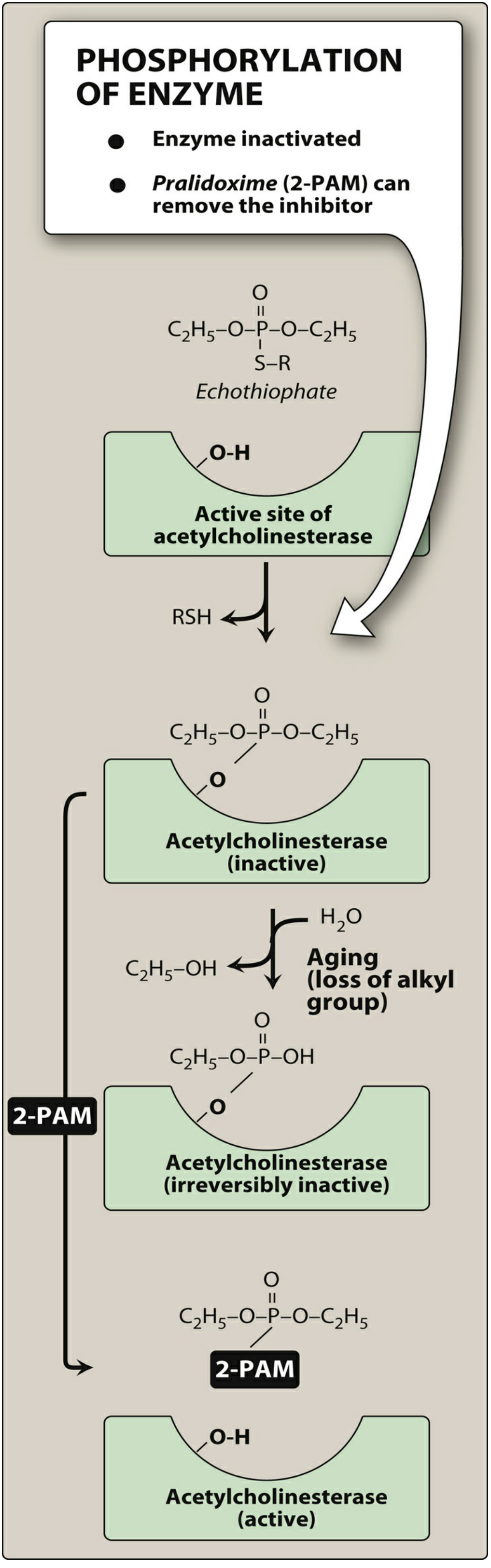
Figure 4.10 - Covalent modification of AChE by organophosphate (echothiophate) and reactivation with pralidoxime (2-PAM). Lippincott Illustrated Reviews: Pharmacology
Key concept - "Aging": After organophosphate binding, the enzyme-phosphate complex undergoes progressive loss of an alkyl group ("aging"), making it permanently irreversible. Pralidoxime (2-PAM) must be given before aging to reactivate the enzyme.
Detailed Drug Profiles
Edrophonium
| Property | Detail |
|---|
| Mechanism | Electrostatic attachment only (no covalent bond) |
| Duration | Very short: 5-15 minutes |
| CNS penetration | None (quaternary) |
| Therapeutic use | Tensilon test - IV injection to diagnose myasthenia gravis (transient improvement in muscle strength = positive) |
| Side effects | Bradycardia, excessive secretions (brief) |
Neostigmine
| Property | Detail |
|---|
| Mechanism | Carbamylates AChE (covalent but hydrolysable) |
| Duration | Medium: 0.5-2 hours |
| CNS penetration | NO (quaternary nitrogen = charged = cannot cross BBB) |
| Therapeutic uses | 1. Myasthenia gravis (long-term treatment) 2. Reversal of non-depolarising NMJ blockers (e.g., vecuronium, atracurium) post-operatively - given with atropine to block muscarinic side effects 3. Postoperative ileus and urinary retention |
| Side effects | Bradycardia, salivation, lacrimation, bronchospasm, GI cramps, increased urination (all muscarinic); excessive muscular weakness at high doses (nicotinic) |
Pyridostigmine
| Property | Detail |
|---|
| Mechanism | Carbamylates AChE |
| Duration | Longer than neostigmine (3-6 h), oral preparation available |
| CNS penetration | NO (quaternary) |
| Therapeutic uses | First-line for chronic myasthenia gravis (preferred over neostigmine for long-term); military prophylaxis against nerve agent (soman) |
| Side effects | Similar to neostigmine but slightly milder and better tolerated orally |
Physostigmine
| Property | Detail |
|---|
| Mechanism | Carbamylates AChE |
| Duration | 0.5-2 hours |
| CNS penetration | YES (tertiary amine = uncharged = crosses BBB) |
| Therapeutic uses | 1. Antidote for anticholinergic (atropine) poisoning - reverses CNS + peripheral effects 2. Antidote for tricyclic antidepressant overdose (cardiac + CNS effects) 3. Glaucoma (historically; now replaced by pilocarpine) 4. Increases intestinal/bladder motility |
| Side effects | ALL muscarinic + nicotinic effects; convulsions possible (CNS penetration); avoid in asthma, cardiovascular disease |
Donepezil, Rivastigmine, Galantamine (Centrally-Acting AChE Inhibitors)
| Drug | Selectivity | Duration | Notes |
|---|
| Donepezil | Central AChE | 24 h (once daily) | Reversible; no hepatotoxicity; preferred in moderate-severe AD |
| Rivastigmine | Central AChE + BuChE | 12 h / patch available | Also inhibits butyrylcholinesterase; useful in Lewy body dementia and Parkinson's dementia |
| Galantamine | Central AChE + allosteric nicotinic modulator | 12 h | Also has nicotinic receptor modulation |
Therapeutic use: Alzheimer's disease (first-line treatment) - modest improvement in cognition and daily function; can be combined with memantine (NMDA antagonist) in moderate-severe disease
"Used as first-line treatments for Alzheimer disease, though conferring modest benefit. Have not been shown to reduce healthcare costs or delay institutionalization." - Lippincott Illustrated Reviews: Pharmacology
Side effects (all three): Nausea, vomiting, diarrhoea, insomnia, muscle cramps, bradycardia, anorexia, weight loss - all predictable cholinergic effects
Organophosphate Toxicity - Cholinergic Crisis
Irreversible AChE inhibitors cause cholinergic crisis = overstimulation of ALL cholinergic receptors.
Symptoms - remembered as SLUDGE + Killer B's:
| System | Muscarinic (SLUDGE) | Nicotinic | CNS |
|---|
| Features | Salivation, Lacrimation, Urination, Defecation, GI cramps, Emesis | Muscle fasciculations → weakness → paralysis (respiratory failure) | Anxiety → seizures → coma |
| Additional | Bradycardia, bronchospasm, miosis, sweating | Tachycardia (early, from ganglionic stimulation) | |
Treatment of organophosphate poisoning:
- Atropine (high doses, IV) - blocks muscarinic effects (secretions, bronchospasm, bradycardia); does NOT reverse NMJ paralysis
- Pralidoxime (2-PAM) - reactivates AChE if given before aging occurs; reverses both muscarinic and nicotinic effects peripherally (not CNS - quaternary)
- Diazepam - controls seizures
- Supportive care - airway management, O2, ventilation
Master Summary Table
| Category | Drug | Receptor Target | CNS Penetration | Key Use | Key Side Effects |
|---|
| Direct - Non-selective | Acetylcholine | M + N | No | Intraoperative miosis | Bradycardia, bronchospasm |
| Direct - Non-selective | Carbachol | M + N | No | Glaucoma, ocular surgery | Strong M+N effects |
| Direct - M-selective | Bethanechol | M (M3) | No | Urinary retention, ileus | SLUDGE symptoms |
| Direct - M-selective | Pilocarpine | M (M3) | Yes | Glaucoma, xerostomia | Sweating, miosis, nausea |
| Direct - M-selective | Methacholine | M | No | Bronchial provocation test | Bronchospasm |
| Direct - M-selective | Cevimeline | M1, M3 | Partial | Sjögren dry mouth | Sweating, nausea |
| Indirect - Reversible | Edrophonium | AChE | No | Myasthenia diagnosis | Bradycardia (brief) |
| Indirect - Reversible | Neostigmine | AChE | No | MG treatment, NMJ reversal | SLUDGE + muscle effects |
| Indirect - Reversible | Pyridostigmine | AChE | No | MG (first-line chronic) | SLUDGE (mild) |
| Indirect - Reversible | Physostigmine | AChE | Yes | Atropine/TCA antidote | Seizures, bradycardia |
| Indirect - Central | Donepezil | Central AChE | Yes (CNS-selective) | Alzheimer's disease | Nausea, vomiting, bradycardia |
| Indirect - Central | Rivastigmine | AChE + BuChE | Yes | Alzheimer's, Lewy body, Parkinson's dementia | GI upset |
| Indirect - Central | Galantamine | AChE + Nicotinic modulator | Yes | Alzheimer's disease | GI upset, insomnia |
| Indirect - Irreversible | Organophosphates | AChE (permanent) | Yes | Toxic (insecticides, nerve agents) | Cholinergic crisis → death |
Memory Aid: Direct vs. Indirect
DIRECT agonists → "Act directly on receptor"
Muscarinic-selective: Bethanechol, Pilocarpine, Methacholine, Cevimeline
Non-selective (M+N): Acetylcholine, Carbachol
INDIRECT agonists → "Block the destroyer (AChE), so ACh builds up"
Reversible:
No CNS: Neostigmine, Pyridostigmine, Edrophonium (Quaternary = Charged = Can't cross BBB)
Yes CNS: Physostigmine (Tertiary = Uncharged = Crosses BBB)
CNS-selective: Donepezil, Rivastigmine, Galantamine (Alzheimer's)
Irreversible: Organophosphates (Nerve agents, pesticides)
Sources:
- Lippincott Illustrated Reviews: Pharmacology - Chapter 4 (Cholinergic Pharmacology)
- Goodman & Gilman's Pharmacological Basis of Therapeutics - Chapter 11 (Muscarinic Receptor Agonists/Antagonists)
- Ganong's Review of Medical Physiology, 26th Ed. - Chapter 13 (Autonomic Neurotransmission)
- Rosen's Emergency Medicine - Cholinergic Toxidrome
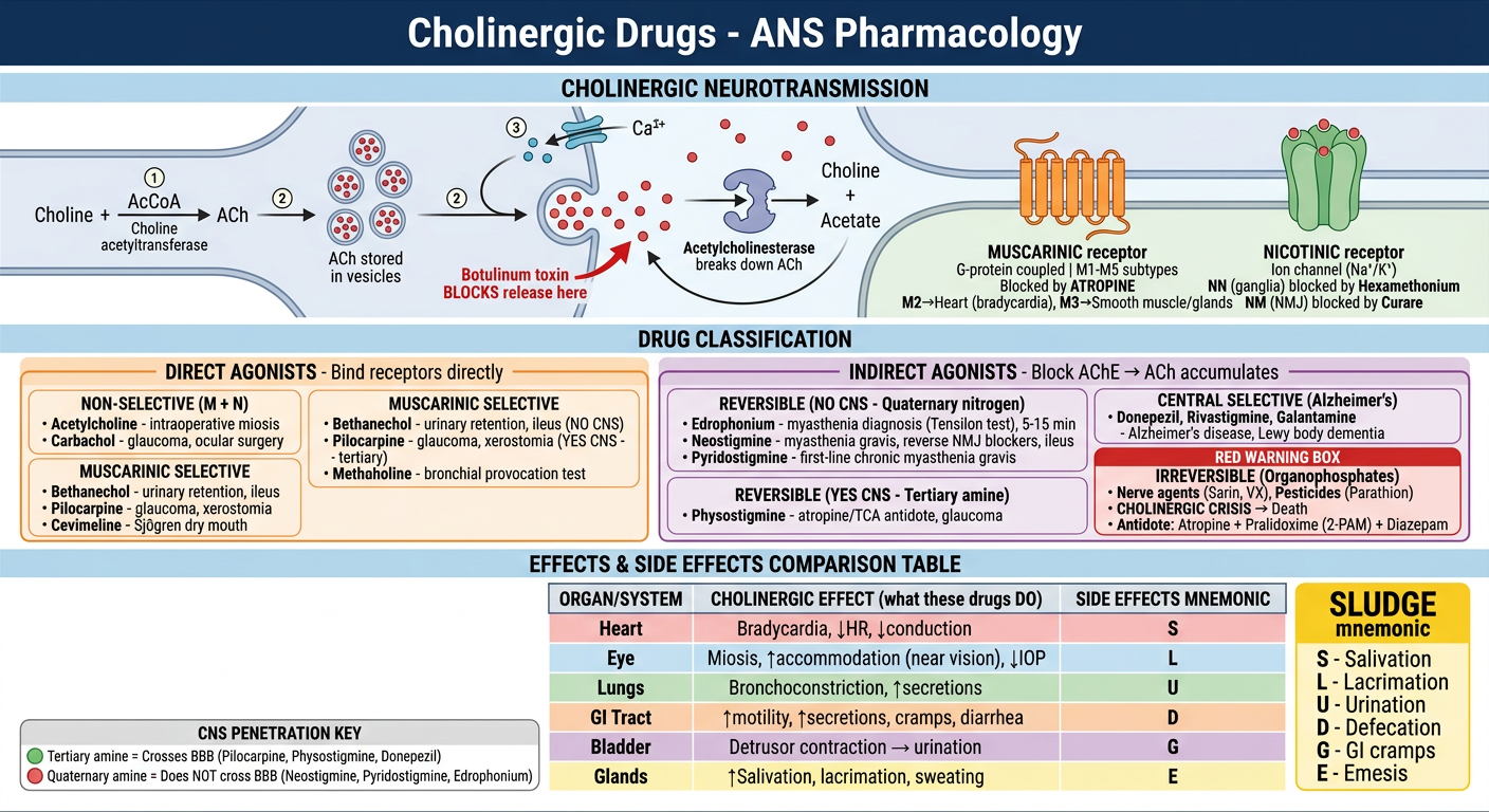
Here's the full cholinergic pharmacology diagram covering all 3 layers:
Section 1 (Top) - The cholinergic synapse: ACh synthesis → vesicle storage → Ca²⁺-triggered release → receptor binding → AChE degradation → choline recycling. Shows where botulinum toxin blocks and where drugs intervene.
Section 2 (Middle) - Drug classification split into:
- Direct agonists (orange): Non-selective (ACh, Carbachol) vs. Muscarinic-selective (Bethanechol, Pilocarpine, Methacholine, Cevimeline)
- Indirect agonists/AChE inhibitors (purple): Reversible no-CNS (Neostigmine, Pyridostigmine, Edrophonium) → Reversible+CNS (Physostigmine) → Central-selective (Donepezil, Rivastigmine, Galantamine) → Irreversible organophosphates (red warning)
Section 3 (Bottom) - Organ-by-organ effects table + SLUDGE mnemonic + the critical quaternary/tertiary amine CNS-penetration rule.I have the atropine/anticholinergic toxidrome content from Tintinalli's. Let me now read the specific management sections from that block.Excellent - I got the full anticholinergic management section. Let me now read the next section for the complete treatment details including physostigmine use, and the TCA/organophosphate content.The read went past the anticholinergic section. Let me search for the TCA and organophosphate management content in the Rosen's emergency medicine and Goodman & Gilman blocks I already identified.I now have comprehensive content on all three poisonings. Let me compile the complete answer with the textbook content already gathered, plus the Goodman & Gilman and Lippincott content from the previous session.
Poisoning Management: Atropine, TCA, and Organophosphate
FRAMEWORK: Understanding the Three Toxidromes
Before treatment, recognise which syndrome you are dealing with - they are mechanistically opposite to each other in many ways:
| Toxidrome | Core Mechanism | Receptor Effect | Key Mnemonic |
|---|
| Atropine / Anticholinergic | Blocks muscarinic receptors | ↓ Parasympathetic | "Hot, Dry, Red, Blind, Mad" |
| Organophosphate / Cholinergic | Inhibits AChE → ACh excess | ↑ Parasympathetic + Nicotinic | SLUDGE / DUMBELS |
| TCA overdose | Blocks Na⁺ channels + anticholinergic + α-blockade | Mixed (Na⁺ channel + anticholinergic + CNS) | "Cardiac + CNS + Anticholinergic" |
1. ATROPINE (ANTICHOLINERGIC) POISONING
Sources
- Atropine, scopolamine (medications)
- Antihistamines (diphenhydramine, hydroxyzine)
- Antipsychotics (olanzapine, chlorpromazine - partial anticholinergic)
- Plants: Jimsonweed (Datura), belladonna, deadly nightshade, henbane
- Cyclopentolate, tropicamide eye drops (especially in children via nasolacrimal absorption)
Clinical Features - "The Classic Anticholinergic Toxidrome"
"The classic features of the anticholinergic toxidrome can be stated as: Dry as a bone, Red as a beet, Hot as a hare, Blind as a bat, Mad as a hatter, Stuffed as a pipe." - Tintinalli's Emergency Medicine
| Feature | Organ | Mechanism |
|---|
| Dry skin / dry mouth | Skin, salivary glands | ↓ Sweat and salivary secretion |
| Red, flushed skin | Peripheral vasculature | Cutaneous vasodilation |
| Hyperthermia | Thermoregulation | ↓ Sweating + agitation-induced heat production |
| Blind (mydriasis + blurred vision) | Eye | Dilated pupils, loss of accommodation (cycloplegia) - often delayed 12-24 h |
| Tachycardia | Heart | ↓ Vagal tone (M2 block) |
| Urinary retention | Bladder | ↓ Detrusor contraction (M3 block) |
| Absent bowel sounds / ileus | GI | ↓ Peristalsis |
| Delirium, agitation, hallucinations | CNS | Central muscarinic blockade |
| Coma | CNS | High doses |
Life-threatening complications:
- Hyperthermia → rhabdomyolysis → multiorgan failure
- Wide-complex tachyarrhythmias (from sodium channel blockade - especially diphenhydramine)
- Status epilepticus
- Cardiovascular collapse
Diagnosis
- Clinical (history + toxidrome)
- Routine labs: electrolytes, glucose, CK (usually normal in isolated anticholinergic toxicity)
- ECG: sinus tachycardia; QRS widening if Na⁺ channel blocking agent (diphenhydramine)
- Urine drug screen: often does not detect plant alkaloids or atropine
Key differentiator from sympathomimetic toxicity:
- Anticholinergic: Red DRY skin, absent bowel sounds
- Sympathomimetic (cocaine): Wet, diaphoretic skin, hyperactive bowel sounds
Management
| Step | Intervention | Details |
|---|
| 1. Decontamination | Activated charcoal | If within 1 hour of ingestion, patient awake and can protect airway |
| 2. Supportive care | Temperature monitoring and cooling | Hyperthermia is the biggest killer - ice packs, cooling blankets |
| 3. Agitation / seizures | Benzodiazepines (lorazepam, diazepam) | First line - cool the patient AND sedate safely. Avoid phenothiazines (add anticholinergic burden) |
| 4. Specific antidote | Physostigmine | Indicated for severe CNS toxicity (delirium, hallucinations) not responding to benzodiazepines |
| 5. Dysrhythmias | Sodium bicarbonate (if QRS widening from Na⁺ channel blockade) | Especially for diphenhydramine-associated wide-complex tachyarrhythmia |
| 6. Urinary retention | Urinary catheterisation | If bladder distension |
Physostigmine - The Specific Antidote for Atropine Poisoning
| Property | Detail |
|---|
| Why it works | Tertiary amine AChE inhibitor → crosses BBB → reverses central AND peripheral muscarinic blockade |
| Dose | 1-2 mg IV slowly (over 5 min) in adults; repeat every 20-30 min if needed |
| Indication | Severe delirium, agitation, seizures, haemodynamic instability FROM anticholinergic toxicity |
| Contraindications | TCA overdose (risk of asystole, seizures), bradycardia, AV block, reactive airway disease, bowel/bladder obstruction |
| Monitoring | Atropine must be at bedside; watch for cholinergic excess (bradycardia, bronchospasm, seizure) |
| Duration | Short (1-2 hours) - patient may re-lapse into delirium and repeat dosing may be needed |
"Slow intravenous injection of physostigmine will rapidly abolish the delirium and coma caused by large doses of atropine but carries some risk of overdose in mild atropine intoxication. Because physostigmine is metabolized rapidly, the patient may again lapse into coma within 1 to 2 h, and repeated doses may be needed." - Goodman & Gilman's
"If physostigmine does not elicit the expected salivation, sweating, bradycardia, and intestinal hyperactivity, intoxication with atropine or a related agent is almost certain." - Goodman & Gilman's
2. ORGANOPHOSPHATE (CHOLINERGIC) POISONING
Sources
- Agricultural pesticides: parathion, malathion, chlorpyrifos
- Nerve agents (weapons): sarin, soman, tabun, VX, Novichok agents
- Ophthalmic agents (historical): echothiophate (now discontinued)
Mechanism
Organophosphates irreversibly phosphorylate AChE → ACh cannot be broken down → accumulates at ALL cholinergic synapses → overstimulation of muscarinic AND nicotinic receptors.
"Aging": After phosphorylation, the enzyme-drug complex undergoes progressive dealkylation ("aging") - once aged, the enzyme is permanently inactive and 2-PAM cannot reactivate it. Aging time varies by agent (soman = seconds; parathion = hours-days).
Clinical Features - DUMBELS + Nicotinic Signs
Muscarinic (DUMBELS):
| Letter | Feature |
|---|
| D | Diarrhoea, Diaphoresis |
| U | Urination (incontinence) |
| M | Miosis (pinpoint pupils - hallmark) |
| B | Bradycardia, Bronchorrhea, Bronchospasm |
| E | Emesis |
| L | Lacrimation, Lethargy |
| S | Salivation |
Nicotinic (Days of Week mnemonic - MTWThF):
- Mydriasis (can override miosis in severe cases)
- Tachycardia (early - ganglionic stimulation)
- Weakness → paralysis
- Tremor, Fasciculations
- Flaccid paralysis (respiratory muscles → respiratory failure = primary cause of death)
CNS effects: Anxiety → agitation → seizures → coma
Key life-threatening: Bronchorrhea + bronchospasm + respiratory muscle paralysis = "drowning in own secretions"
Management of Organophosphate Poisoning
Priority: Airway, Atropine, 2-PAM, Diazepam
Step 1 - Decontamination (BEFORE touching patient)
- Remove clothing - skin absorption ongoing; rescuers must wear PPE
- Copious water/soap washing of skin
- Activated charcoal if oral ingestion within 1-2 hours
Step 2 - ATROPINE (cornerstone - titrated to secretions)
| Property | Detail |
|---|
| Mechanism | Competitive muscarinic antagonist → blocks muscarinic effects only |
| Initial dose | 2-4 mg IV in adults; 0.05 mg/kg in children |
| Titration | Double every 5-10 min until secretions dry up (not until heart rate normalises - that's the wrong endpoint!) |
| Doses needed | Can be massive (10s to 100s of mg in severe poisoning) |
| Endpoint | Dry secretions, clear chest, cessation of bronchospasm |
| Does NOT reverse | Nicotinic effects (NMJ paralysis, tachycardia, fasciculations) |
"Atropine is administered to prevent muscarinic side effects... including increased bronchial and salivary secretion, bronchoconstriction, and bradycardia." - Lippincott Pharmacology
Step 3 - PRALIDOXIME / 2-PAM (oxime - reactivates AChE)
| Property | Detail |
|---|
| Mechanism | Quaternary pyridinium oxime - displaces phosphate group from inhibited AChE → regenerates enzyme |
| Effect | Reverses BOTH muscarinic and nicotinic peripheral effects; does NOT cross BBB |
| Critical timing | Must be given before aging of the enzyme complex |
| Dose | 1-2 g IV over 15-30 min, then 200-400 mg/hr infusion |
| Ineffective against | Soman (ages in seconds) and other rapidly-aging agents; carbamate insecticides (not needed as they are reversible) |
| Note | Gives the nicotinic reversal that atropine cannot provide |
Step 4 - DIAZEPAM (for seizures)
| Property | Detail |
|---|
| Mechanism | GABA-A receptor agonist → CNS sedation + anticonvulsant |
| Indication | Seizures from organophosphate CNS toxicity |
| Dose | 5-10 mg IV; repeat as needed |
| Note | Phenytoin is NOT effective for toxin-induced seizures |
Step 5 - Supportive Care
- Airway management / intubation if respiratory failure
- Suction secretions aggressively
- Ventilatory support
- Avoid succinylcholine (prolonged paralysis - AChE is inhibited, so succinylcholine is not metabolised)
Organophosphate Antidote Summary
ATROPINE → Blocks muscarinic effects (secretions, bradycardia, bronchospasm)
2-PAM → Reactivates AChE (reverses both M and N effects peripherally)
DIAZEPAM → Stops seizures
3. TRICYCLIC ANTIDEPRESSANT (TCA) POISONING
Drugs
Amitriptyline, imipramine, clomipramine, doxepin, nortriptyline, amoxapine
Mechanism of Toxicity - Triple Threat
| Mechanism | Effect | Clinical Consequence |
|---|
| Fast Na⁺ channel blockade | Slows phase 0 depolarisation in cardiac conduction | QRS widening → ventricular tachycardia/fibrillation → cardiac arrest |
| Anticholinergic (M blockade) | Blocks muscarinic receptors | Tachycardia, dry mouth, urinary retention, ileus, delirium |
| Alpha-1 adrenergic blockade | Peripheral vasodilation | Hypotension |
| GABA-A antagonism | CNS excitability | Seizures |
| Histamine H1 blockade | CNS depression | Sedation, coma |
Clinical Features
Rapid progression is the hallmark - patient can deteriorate from mildly altered to cardiac arrest in minutes.
- Cardiac: Sinus tachycardia → QRS widening (>100ms is dangerous; >160ms = high risk of VT/VF) → right axis deviation → S wave in lead I, R wave in aVR → ventricular arrhythmias
- CNS: Agitation → seizures → coma
- Anticholinergic: Tachycardia, dry mouth, urinary retention, mydriasis
- Haemodynamic: Hypotension (vasodilation + cardiac depression)
ECG - Key findings:
- Prolonged QRS (>100-120 ms) = most predictive of arrhythmia
- Prolonged QTc
- Tall R wave in aVR (>3 mm)
- S wave in lead I and aVL
Management
"Physostigmine should never be prophylactically administered and its use in TCA overdose is generally considered contraindicated, particularly in patients with bradycardia, AV block, and seizures." - Rosen's Emergency Medicine
Step 1 - Decontamination
- Activated charcoal (within 1 hour of ingestion if airway is protected) - first-line decontamination
- No gastric lavage (not indicated)
Step 2 - SODIUM BICARBONATE (cornerstone of TCA management)
| Property | Detail |
|---|
| Mechanism 1 | Increases extracellular Na⁺ → overcomes Na⁺ channel blockade (mass effect) |
| Mechanism 2 | Alkalinisation (pH 7.45-7.55) → decreases TCA binding to Na⁺ channels (TCAs bind less at higher pH) |
| Indication | QRS >100 ms, ventricular arrhythmia, hypotension refractory to fluids |
| Dose | 1-2 mEq/kg IV bolus; repeat until QRS narrows or pH reaches 7.50-7.55 |
| Target | Serum pH 7.45-7.55; QRS narrowing |
| NOT prophylactic | Give only when QRS widens - not preemptively |
Step 3 - Seizures: BENZODIAZEPINES
| Drug | Dose | Notes |
|---|
| Lorazepam | 2-4 mg IV | First-line |
| Phenobarbital | Second-line if benzos fail | |
| Propofol | Third-line with intubation | |
| AVOID Phenytoin | - | Ineffective for toxin-induced seizures + worsens Na⁺ channel blockade → more ventricular tachycardia |
| AVOID Physostigmine | - | Contraindicated in TCA - can cause asystole and worsen seizures |
Step 4 - Hypotension
- IV fluid bolus (0.9% NaCl) first
- Norepinephrine (alpha + beta) preferred vasopressor if fluids fail
- Avoid dopamine (indirect mechanism less reliable)
Step 5 - Airway
- Low threshold for early intubation in deteriorating patients
- Avoid succinylcholine for RSI (if seizures have caused hyperkalaemia)
- Maintain alkalosis (pH 7.45-7.55) post-intubation via hyperventilation + bicarbonate
Step 6 - LIPID EMULSION THERAPY (rescue)
- Intravenous lipid emulsion (Intralipid 20%) - "lipid sink" mechanism
- For life-threatening cardiac toxicity refractory to all other measures
- Dose: 1.5 mL/kg IV bolus, then infusion
MASTER COMPARISON TABLE
| Feature | Atropine Poisoning | Organophosphate Poisoning | TCA Overdose |
|---|
| Mechanism | Muscarinic blockade | AChE inhibition → ACh excess | Na⁺ channel block + anticholinergic + α-block |
| Pupils | Mydriasis (dilated) | Miosis (pinpoint) | Mydriasis (anticholinergic) |
| Heart rate | Tachycardia | Bradycardia | Tachycardia |
| Secretions | Dry (no secretions) | Profuse (bronchorrhoea, sweating) | Dry (anticholinergic) |
| Skin | Hot, dry, flushed | Wet, diaphoretic | Dry (anticholinergic) |
| Bowel sounds | Absent | Hyperactive | Absent |
| Seizures | Yes (high dose) | Yes (CNS AChE inhibition) | Yes (GABA antagonism) |
| ECG | Sinus tachycardia | Bradycardia, QTc prolongation | QRS widening, QTc, R in aVR |
| Specific antidote | Physostigmine | Atropine + 2-PAM | Sodium bicarbonate |
| Seizure Rx | Benzodiazepines | Diazepam | Benzodiazepines (NOT phenytoin) |
| Avoid | Phenothiazines | Succinylcholine, morphine | Physostigmine, phenytoin |
DRUG CAUTIONS - What NOT to Use and Why
| Drug | Poisoning | Why Avoid |
|---|
| Physostigmine | TCA overdose | Causes bradycardia, AV block, asystole, worsens seizures |
| Phenytoin | TCA or any toxin-induced seizure | Na⁺ channel blocker - worsens cardiac toxicity; ineffective for toxin seizures |
| Phenothiazines | Atropine poisoning | Add anticholinergic burden, lower seizure threshold |
| Succinylcholine | Organophosphate poisoning | AChE is inhibited → succinylcholine not metabolised → prolonged paralysis (hours) |
| Morphine | Organophosphate | Releases histamine, worsens bronchospasm |
| Sodium bicarbonate (prophylactic) | TCA without QRS widening | Not indicated prophylactically |
| Dopamine | TCA hypotension | Less reliable than norepinephrine in severe toxicity |
Sources:
- Tintinalli's Emergency Medicine, 9th Ed. - Anticholinergic Toxidrome (Chapter 202)
- Rosen's Emergency Medicine - Cholinergic/Anticholinergic Toxidromes (Chapter 135, 141)
- Goodman & Gilman's Pharmacological Basis of Therapeutics - Muscarinic Agonists/Antagonists
- Lippincott Illustrated Reviews: Pharmacology - Chapter 4 (Anticholinesterases, 2-PAM)Excellent content. Let me get the treatment section from Bradley's Neurology and the Nephrology Primer, and search for any images.Excellent - I have the Bartter-Schwartz criteria in full. Let me get the rest of those criteria and the treatment algorithm, and search for a hyponatremia algorithm image.I now have comprehensive, authoritative content from multiple top textbooks. Let me compile the full SIADH answer.
SIADH - Syndrome of Inappropriate Antidiuretic Hormone Secretion
Definition
SIADH is the most common cause of euvolemic hyponatremia in clinical practice. It is characterised by:
- Hypotonic hyponatraemia (low serum Na⁺ and low serum osmolality)
- Inappropriately concentrated urine (urine not maximally dilute despite low plasma osmolality)
- Euvolaemia (no oedema, no dehydration)
- Elevated urinary sodium (kidneys continue to excrete Na⁺)
The fundamental problem: ADH (vasopressin / AVP) is secreted in the absence of the normal osmotic or haemodynamic stimuli - leading to free water retention, dilutional hyponatraemia, and volume expansion.
"Euvolemic hyponatremia is caused by an excess in body water. The most common cause of euvolemic hyponatremia is the syndrome of inappropriate ADH secretion (SIADH), which occurs when the stimulus for ADH secretion is not osmotic." - Textbook of Family Medicine, 9th Ed.
Pathophysiology
Inappropriate ADH secretion (no osmotic/haemodynamic stimulus)
↓
ADH binds V2 receptors on renal collecting duct
↓
Aquaporin-2 channels inserted → collecting duct becomes permeable to water
↓
Free water reabsorption ↑ → urine concentrated → plasma diluted
↓
Plasma osmolality ↓ | Serum Na⁺ ↓ | Total body water ↑
↓
Volume expansion → suppresses renin-angiotensin-aldosterone → natriuresis
(urinary Na⁺ remains HIGH despite hyponatraemia)
Causes
Mnemonic: "CNS-PAID" (CNS, Neoplasm, Pulmonary, ADH analogues, Inflammatory, Drugs)
1. Malignancy / Neoplastic (Ectopic ADH production)
- Small cell carcinoma of the lung - most common tumour cause
- Bronchial adenoma, mesothelioma, thymoma
- Non-thoracic: pancreatic, duodenal, ureteral, prostate, uterine carcinoma
- Lymphoma, leukaemia, carcinoid
2. CNS Disorders
- Head injury, neurosurgery, pituitary stalk section
- Brain abscess, tumours, subdural haematoma, hydrocephalus
- Meningitis, encephalitis
- Subarachnoid haemorrhage
- Stroke (ischaemic or haemorrhagic)
- Guillain-Barré syndrome
- Acute intermittent porphyria
- Acute psychosis, delirium tremens, alcohol withdrawal
3. Pulmonary / Chest Disorders
- Pneumonia (bacterial, viral, tuberculosis)
- Aspergillosis, empyema
- Positive-pressure mechanical ventilation (increases ADH secretion by reducing venous return)
- COPD, pneumothorax, cystic fibrosis
4. Drugs (most common cause in hospital inpatients)
| Mechanism | Drugs |
|---|
| Stimulate ADH release | Nicotine, phenothiazines, tricyclic antidepressants, cyclophosphamide |
| Potentiate ADH action | Desmopressin (DDAVP), oxytocin, NSAIDs |
| Mixed/Uncertain | SSRIs, carbamazepine, chlorpropamide, clofibrate, vincristine, clozapine, omeprazole, tacrolimus, MDMA ("Ecstasy"), ACE inhibitors, thiazide diuretics, opiates |
Ecstasy (MDMA) - causes massive ADH release + causes hyponatraemia by drinking large amounts of water at clubs; a well-known cause of acute fatal hyponatraemia in young people.
5. Other / Miscellaneous
- Postoperative state (surgery itself triggers ADH release)
- HIV/AIDS
- Prolonged strenuous exercise (marathon, triathlon)
- Idiopathic
- Acute/chronic pain, nausea (physiological ADH stimuli)
- Beer potomania (very low solute intake limits free-water excretion)
Diagnostic Criteria (Bartter & Schwartz, 1967 - Modified by Verbalis)
Essential Criteria (ALL must be present)
| Criterion | Value |
|---|
| 1. Decreased effective plasma osmolality | Posm < 275 mOsm/kg H₂O |
| 2. Inappropriate urinary concentration | Uosm > 100 mOsm/kg H₂O (urine not maximally dilute) |
| 3. Clinical euvolaemia | No signs of dehydration (orthostasis, tachycardia, dry skin) AND no oedema/ascites |
| 4. Elevated urinary sodium | Urine Na⁺ ≥ 30 mEq/L on normal salt intake |
| 5. Normal thyroid, adrenal, and renal function | Exclude hypothyroidism, adrenal insufficiency, CKD |
"SIADH remains a diagnosis of exclusion. No patient with chronic hyponatraemia should be diagnosed with SIADH without a thorough evaluation of adrenal function." - NKF Primer on Kidney Diseases, 8th Ed.
Supplemental Criteria
- Fractional uric acid excretion (FEuric acid) >12% (hyponatraemia + hypouricaemia suggests SIADH)
- Failure to raise serum [Na⁺] with 0.9% saline infusion (isotonic saline can worsen SIADH)
- Improvement (↑ Na⁺) after fluid restriction
- Inappropriately elevated plasma AVP level for serum osmolality
What Must Be Excluded Before Diagnosing SIADH
| Condition | Why it mimics SIADH |
|---|
| Dehydration (hypovolaemia) | Activates ADH via haemodynamic stimulus |
| Heart failure, cirrhosis, nephrotic syndrome | Effective arterial hypovolaemia → ADH release |
| Primary renal disease | ↓ Free water excretion |
| Hypothyroidism | ↓ GFR + ADH effect → dilutional hyponatraemia |
| Adrenal insufficiency (Addison's) | ↑ ADH via reduced cortisol + ACTH → clinically indistinguishable |
| Diuretic use | Stimulates ADH; sodium loss |
| Pseudohyponatraemia | Hyperlipidaemia, hyperproteinaemia → artefactually low Na⁺ |
| Hyperglycaemia (translocation hyponatraemia) | Glucose draws water from ICF → dilutes Na⁺ |
SIADH vs Cerebral Salt Wasting (CSW) - Critical Distinction
This is a high-yield differentiation, especially in neurosurgical/SAH patients:
| Feature | SIADH | Cerebral Salt Wasting (CSW) |
|---|
| Mechanism | ↑ ADH → water retention | ↑ Natriuretic peptide → renal Na⁺ wasting |
| Volume status | Euvolaemic | Hypovolaemic |
| Body sodium | Normal (diluted) | Depleted |
| Urine Na⁺ | High (>30 mEq/L) | High (>30 mEq/L) |
| Serum uric acid | Low (uricosuric) | Low |
| CVP/BP | Normal | Low |
| Treatment | Fluid restriction | IV fluid + salt replacement ← opposite! |
| Risk of wrong Rx | Fluid expansion → worsens hyponatraemia + cerebral oedema | Fluid restriction → worsens hypovolaemia + ischaemia |
Clinical Features
Features are neurological and related to the degree and rate of hyponatraemia:
| Serum Na⁺ | Symptoms |
|---|
| 130-135 mmol/L | Asymptomatic or mild: fatigue, malaise, loss of appetite, nausea |
| 125-130 mmol/L | Headache, confusion, cognitive impairment |
| 115-125 mmol/L | Disorientation, obtundation, abnormal gait |
| <115 mmol/L | Seizures, coma, respiratory arrest, death |
"The more rapidly this condition develops, the more symptomatic the patient. Serum sodium less than 115 mmol/L is almost always associated with confusion or obtundation, and seizures can occur." - Bradley & Daroff's Neurology in Clinical Practice
Chronic hyponatraemia is often "asymptomatic" but carries real risk:
- Increased falls and fractures
- Reduced bone density
- Subtle cognitive impairment
Investigation
| Test | SIADH Result |
|---|
| Serum Na⁺ | Low (<135 mmol/L, often <125 in significant SIADH) |
| Serum osmolality | Low (<275 mOsm/kg) |
| Urine osmolality | >100 mOsm/kg (inappropriately concentrated) |
| Urine Na⁺ | ≥30 mEq/L |
| Serum uric acid | Low (hypouricaemia) |
| BUN / Creatinine | Normal or low (volume-replete) |
| Serum K⁺ | Normal |
| Cortisol, TFTs | Normal (to exclude adrenal insufficiency, hypothyroidism) |
| CXR / CT chest | Screen for lung malignancy |
| CT/MRI brain | Screen for CNS cause |
Management
Step 1 - Identify and Treat Underlying Cause
Always address the root cause - remove offending drug, treat pneumonia, resect tumour where possible.
Step 2 - Assess Severity and Duration
| Category | Criteria | Approach |
|---|
| Acute (<48 h) + symptomatic | Seizures, coma | Rapid partial correction with 3% NaCl |
| Chronic (>48 h) + mild symptoms | Confusion, gait instability | Controlled slow correction |
| Chronic + asymptomatic | Mild hyponatraemia, no symptoms | Fluid restriction ± pharmacological therapy |
Step 3 - Correction Rate Rules (CRITICAL)
"An expert consensus panel suggests that the serum sodium level be raised by no more than 10-12 mmol/L during the first 24 hours and by less than 18 mmol/L over 48 hours." - Bradley & Daroff's Neurology
| Rule | Value |
|---|
| Maximum correction rate | 8-10 mmol/L per 24 hours |
| Absolute limit over 48 hours | <18 mmol/L |
| Target in acute symptomatic | Raise by 1-2 mmol/L/hour until seizures stop, then slow down |
| Overshoot correction | Extremely dangerous → Osmotic Demyelination Syndrome (ODS) |
Step 4 - Specific Treatments
A. Fluid Restriction (First-line for mild-moderate SIADH)
| Detail | Value |
|---|
| Target intake | <800 mL/day (insensible losses only) |
| Indication | Asymptomatic or mildly symptomatic SIADH |
| Limitation | Poor patient compliance; slow acting; often not tolerated long-term |
B. Hypertonic Saline 3% NaCl (for severe/symptomatic)
| Detail | Value |
|---|
| Indication | Seizures, coma, acute severe hyponatraemia (<120 mmol/L with symptoms) |
| Rate | 0.1-2 mL/kg/hour via central line |
| Goal | Raise Na⁺ by 1-2 mmol/L/hour initially, then limit to <10 mmol/L/24h |
| Often combined with | Furosemide (1 mg/kg IV) to prevent volume overload and promote free-water loss |
C. Vaptans - V2 Receptor Antagonists (Aquaretics)
Vaptans block ADH at the V2 receptor on the collecting duct → produce aquaresis (excretion of free water without sodium loss).
| Drug | Route | Notes |
|---|
| Tolvaptan | Oral | FDA-approved for SIADH, heart failure, ADPKD-related hyponatraemia; most used |
| Conivaptan | IV only | FDA-approved for hospitalised hyponatraemia from SIADH; non-selective (V1a + V2) |
| Lixivaptan | Oral | Selective V2 antagonist; may be more effective than demeclocycline |
"Antagonists of the vasopressin receptor in the kidney (e.g., lixivaptan and tolvaptan) may be more effective than demeclocycline at managing SIADH. Conivaptan is approved by the FDA for the treatment of hyponatraemia caused by SIADH." - Bradley & Daroff's Neurology
Cautions with vaptans:
- Initiate only in hospital - risk of overcorrection
- Contraindicated with strong CYP3A4 inhibitors
- Tolvaptan - risk of hepatotoxicity with long-term use (>30 days) in SIADH (different from its use in ADPKD)
- Do NOT use in hypovolaemic hyponatraemia
D. Demeclocycline (Historical - now rarely used)
| Detail | Value |
|---|
| Mechanism | Induces nephrogenic diabetes insipidus - blocks ADH action in collecting duct |
| Dose | 300-600 mg twice daily |
| Indication | Chronic SIADH where fluid restriction fails and vaptans unavailable |
| Onset | Slow (2-5 days) |
| Side effects | Nephrotoxicity (especially in cirrhosis), photosensitivity, GI upset, antianabolic effects |
| Avoid in | Liver disease, renal impairment |
E. Oral Sodium + Urea (supplemental)
- Oral NaCl tablets (3-6 g/day) - increase solute load → allow more free water excretion
- Oral urea (15-30 g/day) - osmotic diuresis → free water loss; used in neurological SIADH (e.g., SAH)
Step 5 - Osmotic Demyelination Syndrome (ODS) Prevention
The most feared complication of treatment - too rapid correction of hyponatraemia.
| Feature | Detail |
|---|
| Mechanism | Brain cells lost osmolytes (taurine, myoinositol) during adaptation to hyponatraemia; rapid correction causes hypertonic stress → oligodendrocyte death → demyelination |
| Classic lesion | Central pontine myelinolysis (CPM) |
| Also affects | Cerebellum, thalamus, putamen, basal ganglia (extrapontine myelinolysis) |
| Symptoms | Appear 1-3 days after overcorrection: paraparesis, quadriparesis, dysarthria, dysphagia, diplopia, "locked-in syndrome", ataxia, mutism, coma |
| Risk factors | Alcoholism, malnutrition, hypokalaemia, liver transplantation, serum Na⁺ <120 mmol/L |
If overcorrection occurs:
- Re-lower plasma Na⁺ using dextrose water (D5W) infusion ± DDAVP to slow correction and potentially prevent/attenuate ODS
Treatment Summary Table
| Situation | Treatment |
|---|
| Mild, asymptomatic, chronic SIADH | Fluid restriction (<800-1000 mL/day) + treat cause |
| Moderate, symptomatic (confusion) | Fluid restriction ± oral NaCl/urea ± tolvaptan |
| Severe, symptomatic (seizures, coma) | 3% NaCl + furosemide IV (hospital setting) |
| Chronic SIADH, fluid restriction failing | Tolvaptan (oral) or demeclocycline |
| Hospitalised, IV therapy needed | Conivaptan IV |
| ODS prevention | Never exceed 8-10 mmol/L Na⁺ rise per 24 h |
| Overcorrection occurred | D5W infusion + DDAVP to re-lower Na⁺ |
Key Mnemonic Summary
SIADH = "You keep water, lose sodium in urine, but volume is normal"
Diagnose with:
↓ Serum Na⁺ | ↓ Serum Osm | ↑ Urine Osm | ↑ Urine Na⁺ | Euvolaemic
Treat with:
1. Fix the cause
2. Fluid restrict (800 mL/day)
3. 3% NaCl if seizures/coma
4. Tolvaptan if chronic, refractory
5. NEVER correct >8-10 mmol/L per 24 hours → ODS risk!
Sources:
- NKF Primer on Kidney Diseases, 8th Ed. - Chapter 7 (Hyponatraemia, Diagnostic Criteria Box 7.2)
- Harrison's Principles of Internal Medicine, 22nd Ed. - Clinical Features & ODS
- Bradley & Daroff's Neurology in Clinical Practice - SIADH Etiology, Management
- Barash, Cullen & Stoelting's Clinical Anesthesia, 9th Ed. - Table 16-15, 16-16 (SIADH Associations & Criteria)
- Textbook of Family Medicine, 9th Ed. - Euvolaemic HyponatraemiaYour credits are exhausted. You can purchase a top-up or subscribe to a plan to continue using Orris. If you're already subscribed, your daily credits will reset tomorrow.Your credits are exhausted. You can purchase a top-up or subscribe to a plan to continue using Orris. If you're already subscribed, your daily credits will reset tomorrow.For an Internal Medicine-focused digestive-system examination, I can cover these sections:
- Preparation and general survey: consent, position, exposure, vitals, nutritional status, anaemia, jaundice, dehydration, lymph nodes, oedema, and stigmata of chronic liver disease.
- Abdominal examination: inspection, auscultation, percussion, light/deep palpation, liver, spleen, kidneys, bladder, aorta, ascites, and hernias.
- Hepatobiliary examination: jaundice, chronic liver disease signs, hepatomegaly, splenomegaly, ascites, hepatic encephalopathy, and portal hypertension.
- Gastrointestinal examination beyond the abdomen: oral cavity, perianal inspection, digital rectal examination, and stool assessment where indicated.
- Special bedside tests and interpretation: shifting dullness, fluid thrill, Murphy sign, scratch test, succussion splash, Courvoisier sign, and relevant systemic examination.
- Clinical synthesis: how to present normal findings, identify major syndromes, and formulate likely diagnoses and differentials.
This is a large exam-note topic. Which section would you like in full detail first?Please select one section for a detailed Internal Medicine examination note:
- Complete abdominal examination
- Liver disease and ascites examination
- Per rectal examination
- Special bedside tests
- How to present findings and formulate diagnoses