
Group of nucleus in hypothalamus
Reading File
The hypothalamus contains several well-defined nuclear groups arranged in anterior, middle (tuberal), and posterior regions. Let me pull the full textbook coverage now.
Finding Sources
Finding Sources
Good. I have a clear lead from Adams & Victor's - let me read the full section plus the Color Atlas image and the Ganong's physiology section in parallel.
Reading File
Reading File
Finding Sources
Excellent detail retrieved. Let me now read the Neuroanatomy textbook section for more detail and fetch the diagram image.
Reading File
Reading File
Loading Image
I now have comprehensive information from multiple authoritative textbooks plus an excellent diagram. Here is the full answer:
Nuclei of the Hypothalamus
The hypothalamus contains discrete nuclear groups organized by two classification systems: anterior-to-posterior (rostrocaudal) and medial-to-lateral (zonal).
Classification 1: Rostrocaudal (A-P) Regions
The hypothalamus is divided into four rostrocaudal regions, each containing specific nuclei:
1. Preoptic Region (most anterior)
| Zone | Nuclei |
|---|---|
| Periventricular | Periventricular nuclei |
| Medial | Medial preoptic nucleus |
| Lateral | Lateral preoptic nucleus |
- Also contains the median preoptic nucleus and suprachiasmatic nucleus (SCN)
- SCN controls circadian rhythms
- Preoptic area regulates thermoregulation and reproductive behavior
2. Anterior (Supraoptic) Region
| Zone | Nuclei |
|---|---|
| Periventricular | Suprachiasmatic nucleus, Periventricular nuclei |
| Medial | Anterior hypothalamic nucleus, Paraventricular nucleus (PVN), Supraoptic nucleus (SON) |
| Lateral | Lateral hypothalamic nucleus |
Key nuclei:
- Supraoptic nucleus - synthesizes ADH (vasopressin) and oxytocin
- Paraventricular nucleus - synthesizes ADH and oxytocin; also key autonomic control center
- Both SON and PVN send axons via the hypothalamicohypophysial tract to the posterior pituitary
3. Middle (Tuberal/Infundibular) Region
| Zone | Nuclei |
|---|---|
| Periventricular | Arcuate nucleus (= infundibular nucleus) |
| Medial | Dorsomedial nucleus, Ventromedial nucleus |
| Lateral | Lateral tuberal nuclei, Lateral hypothalamic nucleus |
Key nuclei:
- Arcuate nucleus - releases GnRH, GHRH, dopamine (inhibits prolactin); key in appetite regulation (NPY/AgRP, POMC neurons)
- Ventromedial nucleus - "satiety center"; lesion causes hyperphagia and obesity; also involved in reproductive behavior
- Dorsomedial nucleus - feeding behavior, emotional responses, autonomic regulation
4. Posterior (Mammillary) Region
| Zone | Nuclei |
|---|---|
| Medial | Mammillary nuclei (medial + lateral), Posterior hypothalamic nucleus |
| Lateral | Lateral hypothalamic nucleus |
Key nuclei:
- Mammillary bodies - receive hippocampal input via the fornix; project to anterior thalamus via the mammillothalamic tract (Vicq d'Azyr tract) - critical for memory (Papez circuit)
- Posterior hypothalamic nucleus - involved in arousal; reciprocal connections with the amygdala
Classification 2: Mediolateral Zones
| Zone | Description |
|---|---|
| Periventricular zone | Narrow band lining 3rd ventricle; neurosecretory cells controlling anterior pituitary |
| Medial zone | Rich in nuclei; contains most named hypothalamic nuclei; neurosecretory and visceral control |
| Lateral zone | Sparsely cellular; contains medial forebrain bundle; lateral hypothalamic "feeding center" |
Summary by 3-Group Classification (Adams & Victor)
| Group | Nuclei |
|---|---|
| Anterior | Preoptic, Supraoptic, Paraventricular |
| Middle (Tuberal) | Tuberal, Arcuate, Ventromedial, Dorsomedial |
| Posterior | Mammillary, Posterior hypothalamic |
Diagram of Hypothalamic Nuclei
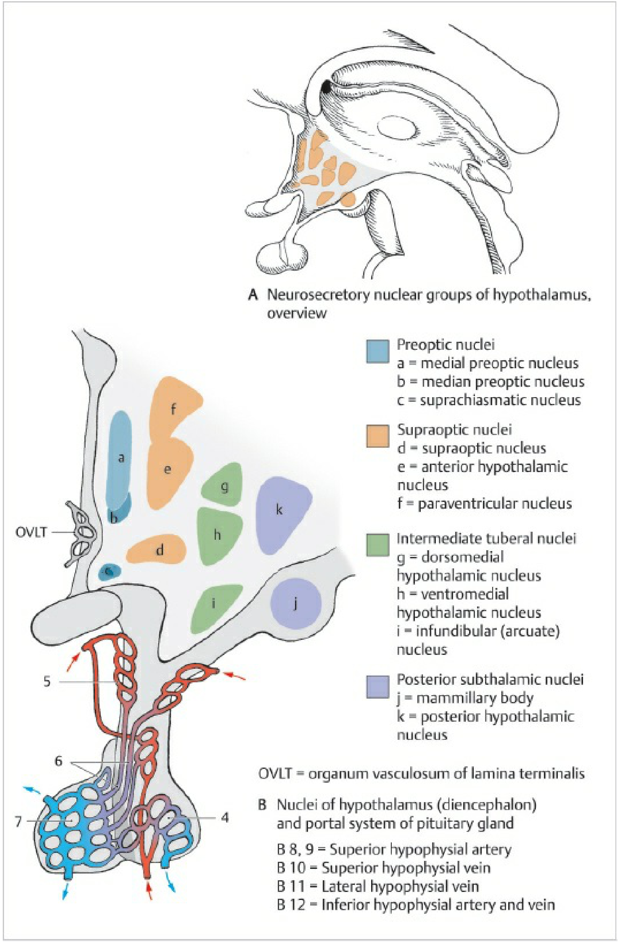
Color Atlas of Human Anatomy Vol. 2 - Nuclei of the hypothalamus and portal system of the pituitary gland. Color coding: Blue = Preoptic nuclei (medial, median, suprachiasmatic). Orange = Supraoptic nuclei (supraoptic, anterior hypothalamic, paraventricular). Green = Intermediate tuberal nuclei (dorsomedial, ventromedial, arcuate/infundibular). Purple = Posterior subthalamic nuclei (mammillary body, posterior hypothalamic nucleus).
Key Functional Mnemonics
| Nucleus | Key Function |
|---|---|
| Supraoptic + Paraventricular | ADH, Oxytocin → posterior pituitary |
| Suprachiasmatic | Circadian rhythm (master clock) |
| Arcuate | GnRH, GHRH, dopamine; appetite |
| Ventromedial | Satiety center (lesion → obesity) |
| Lateral hypothalamus | Feeding/hunger center (lesion → anorexia) |
| Mammillary bodies | Memory (Papez circuit) |
| Preoptic area | Thermoregulation, reproductive |
| Posterior nucleus | Arousal, sympathetic activation |
Sources: Adams and Victor's Principles of Neurology 12e, p. 577 | Kaplan & Sadock's Comprehensive Textbook of Psychiatry, Table 1.2-4 | Color Atlas of Human Anatomy Vol. 2, Fig. 9.6 | Neuroanatomy through Clinical Cases 3e, Table 17.1
This is a shared conversation. Sign in to Orris to start your own chat.