
Vertoplasty
Reading File
Finding Sources
Finding Sources
Searching PubMed
vertebroplasty
Searching the Web
vertebroplasty procedure indications complications
Reading File
Reading File
Vertebroplasty
Vertebroplasty is a minimally invasive percutaneous procedure in which bone cement (polymethylmethacrylate, PMMA) is injected into a fractured or weakened vertebral body to relieve pain and stabilize the bone - Gray's Anatomy for Students, p. 91.
How it's done
Under fluoroscopic (and sometimes CT) guidance, a needle/cannula is passed through the vertebral pedicle into the vertebral body, and liquid cement is injected. As the cement hardens it:
- Restores mechanical strength to the vertebral body, preventing further height loss
- Generates heat during polymerization, which is thought to disrupt local pain nerve endings
A related technique, kyphoplasty, first inflates a balloon in the vertebral body to create a cavity and partially restore lost height before injecting a more viscous cement - Grainger & Allison's Diagnostic Radiology, p. 1087.
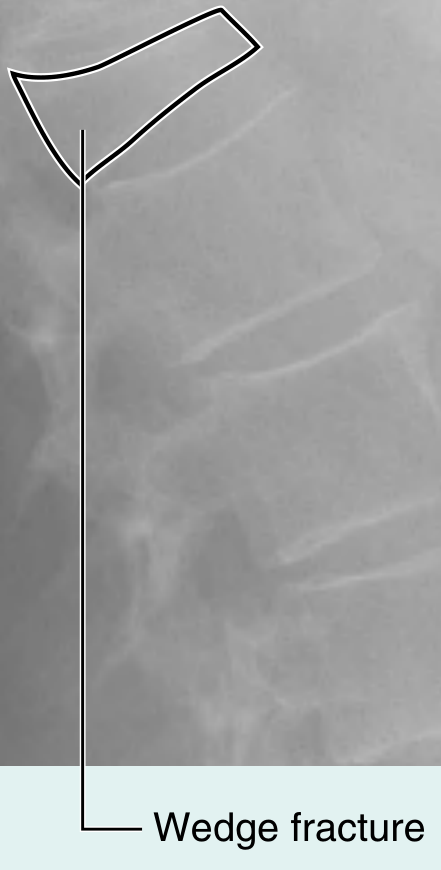
Indications
- Painful osteoporotic vertebral compression fractures (the most common indication), ideally treated within 4-6 weeks of symptom onset in patients with severe pain requiring opioids or hospitalization
- Painful vertebral metastases or multiple myeloma
- Kümmell's disease
- Painful vertebral hemangiomas
Patient selection matters: pain should correlate temporally with the fracture, and MRI (fluid-sensitive/fat-suppressed sequences showing marrow edema) helps confirm an acute, "actionable" fracture rather than an old, healed one - Grainger & Allison's Diagnostic Radiology, p. 1087.
Effectiveness
Reported pain relief rates range from 70-95% for osteoporotic fractures. Benefits cited include earlier hospital discharge, lower readmission, and in some studies reduced mortality when performed within 6 weeks of fracture - Grainger & Allison's Diagnostic Radiology, p. 1087.
Important caveat: Not all evidence is favorable. Two well-known blinded, sham-controlled randomized trials found no benefit of vertebroplasty over a placebo (sham) procedure for painful osteoporotic compression fractures, which sparked ongoing controversy about how much of the benefit is a placebo/regression-to-mean effect versus a true treatment effect - Firestein & Kelley's Textbook of Rheumatology, p. 2112-2127. A 2023 systematic review specifically examined how the choice of control group (sham vs. usual care) shapes these divergent results (Eneling et al., PMID 36566694).
Complications (~1% overall serious complication rate, no reported procedural mortality)
- Needle-related: pedicle fracture, needle breakage, pneumothorax, hemorrhage, spinal infection
- Cement-related: nerve root or cord compression, pulmonary cement embolism, cement leakage into epidural/foraminal/paravertebral veins
- Procedure-related: fat embolism, adjacent rib or new vertebral fracture (about 10% of patients develop a new compression fracture, often at an adjacent level)
- Sedation/anesthesia-related: respiratory or cardiac arrest, airway injury
- Drug-related: allergic reaction
Complication rates are notably higher in malignant/tumor cases (up to 10%) and vertebral hemangiomas (2-5%) than in osteoporotic fractures (1-3%).
Vertebroplasty vs. kyphoplasty
Meta-analyses comparing the two find similar pain and disability outcomes, no difference in symptomatic cement leakage, but kyphoplasty tends to have lower overall (asymptomatic) leakage rates and better restoration of vertebral height/kyphosis correction - Grainger & Allison's Diagnostic Radiology, p. 1087. This is consistent with more recent meta-analyses (e.g., Daher et al. 2023, PMID 36455843).
Recent evidence (last 2-3 years)
- A 2025 systematic review/meta-analysis compared newer "curved" (unipedicular, bilateral cement distribution) vertebroplasty techniques against standard unipedicular approaches, generally favoring the curved technique for better cement distribution and pain outcomes (PMID 40342107, 37839572).
- A 2024 meta-analysis of RCTs comparing percutaneous vertebroplasty to nonoperative treatment continues to show mixed results depending on control group design, reinforcing the sham-controlled-trial controversy noted above (PMID 39053849).
If you want, I can go deeper into any specific angle - e.g., the sham-trial controversy, technique/anesthesia details, or oncologic (metastatic) use.
Steps
Reading File
Reading File
Reading File
Steps of the Vertebroplasty Procedure
Based on Gray's Anatomy for Students, Grainger & Allison's Diagnostic Radiology, and Barash's Clinical Anesthesia:
1. Patient preparation and workup
- Confirm indication with imaging: plain radiograph shows osteopenia/wedge or crush deformity; MRI (fluid-sensitive, fat-suppressed sequences) confirms an acute fracture by detecting bone marrow edema - this is the key criterion for selecting patients likely to benefit.
- Ensure the patient is on or started on bone-protective/anti-osteoporotic therapy if the fracture is osteoporotic.
- Position the patient prone on the fluoroscopy table.
2. Anesthesia/sedation
- Most cases are done under conscious (moderate) sedation with local anesthesia and analgesia at the needle entry site.
- General anesthesia is used in select patients (e.g., those who cannot lie still, significant pain, or multi-level procedures).
3. Imaging setup
- High-quality fluoroscopy is set up in biplane or C-arm configuration (sometimes combined with CT guidance) to allow accurate, real-time visualization of the vertebral body and pedicles.
- A sterile field is established.
4. Needle/cannula placement
- Under fluoroscopic guidance, the pedicle of the target vertebra is identified on the anteroposterior view.
- A trocar/cannula is advanced percutaneously through the pedicle (transpedicular approach) into the anterior third of the vertebral body. This can be done unipedicularly (one side) or bipedicularly (both sides) depending on how well cement can be expected to fill the body from one approach.
5. (Kyphoplasty only - optional additional step)
- If kyphoplasty is being performed instead of/in addition to vertebroplasty, a balloon (bone tamp) is introduced through the cannula and inflated within the vertebral body to create a cavity and partially restore lost vertebral height before cement injection. This allows use of a thicker, more viscous cement.
6. Cement injection
- Liquid PMMA (polymethylmethacrylate) bone cement, mixed to a specific viscosity, is injected slowly through the cannula under continuous fluoroscopic monitoring.
- Injection is stopped as soon as cement approaches the posterior vertebral cortex, epidural space, or an intervertebral disc space, or if venous filling is seen, to avoid leakage/embolization.
- Filling the entire vertebral body is not necessary for pain relief - studies show no correlation between cement volume and clinical outcome.
7. Cement setting
- The cement is allowed to harden in place. As it polymerizes it releases heat, which is thought to help ablate pain nerve endings in addition to mechanically stabilizing the fracture.
- The needle/cannula is withdrawn once the cement has sufficiently hardened (to avoid a cement "tail" tracking back along the needle path).
8. Post-procedure monitoring
- The patient is monitored for immediate complications (neurologic changes, hypotension, respiratory issues) and typically observed for a short period before discharge, since the procedure is usually done as an outpatient or short-stay case.
- Follow-up imaging (radiograph or CT) may be done to confirm cement placement and rule out extravasation.
9. Ongoing management
- Patients treated for osteoporotic fractures should remain under the care of a clinician managing their underlying osteoporosis to reduce future fracture risk, since vertebroplasty treats the current fracture's pain but does not address the systemic bone disease.
Key technical notes
- Access route: transpedicular (most common) or parapedicular/posterolateral for levels where the pedicle is too narrow (e.g., mid-thoracic spine).
- Cement is injected slowly and incrementally, with imaging checked between small aliquots - this stepwise approach is the main technique for minimizing leakage, the most common complication.
Would you like the steps specifically for kyphoplasty (balloon technique) broken out in more detail, or the anesthetic/sedation protocol in more depth?
This is a shared conversation. Sign in to Orris to start your own chat.