I now have all the content needed to write a comprehensive answer. Let me compile the full explanation with flowcharts (as ASCII/Markdown diagrams), tables, and integrated images from Robbins & Cotran.
Atherosclerosis - Robbins & Cotran Pathologic Basis of Disease
Overview
Atherosclerosis (from Greek: "gruel" + "hardening") is the most frequent and clinically important vasculopathy, underlying the pathogenesis of coronary, cerebral, and peripheral vascular disease. It causes roughly half of all deaths in the Western world - more morbidity and mortality than any other disorder. Myocardial infarction alone accounts for nearly one-quarter of all deaths in the United States.
Types of Arteriosclerosis
ARTERIOSCLEROSIS
(Hardening of the arteries)
|
┌─────────────────┼─────────────────┐
▼ ▼ ▼ ▼
Arteriolosclerosis Mönckeberg Fibromuscular ATHEROSCLEROSIS
(small arteries/ Medial Sclerosis Intimal (Most clinically
arterioles) (medial Hyperplasia important)
Hyaline or calcification, (due to
Hyperplastic; >50 yrs, NOT inflammation
related to HTN) clinically or mechanical
significant) injury)
Epidemiology
| Region | Relative Risk vs. USA |
|---|
| United States | Baseline |
| Africa, India, SE Asia | Now EXCEEDS the USA |
| Eastern Europe | 3-5x higher |
| Japan | 7-12x LOWER than Eastern Europe |
- Risk reduction + improved therapies have sharply decreased morbidity/mortality in high-resource nations.
- Adoption of Western diet/lifestyle has increased prevalence in lower-resource nations.
Risk Factors
| Category | Risk Factor | Mechanism/Notes |
|---|
| Non-modifiable | Genetic variation / Family history | Most important independent risk factor; familial hypercholesterolemia (LDL receptor mutations); polygenic basis |
| Increasing age | Incidence of MI increases 5-fold between ages 40-60; CHIP (clonal hematopoiesis) worsens inflammation with age |
| Male sex | Premenopausal women are relatively protected; post-menopause, risk equalizes and can exceed males |
| Modifiable | Hyperlipidemia (elevated LDL) | Most important modifiable risk factor; LDL deposited and oxidized in intima; HDL is protective |
| Hypertension | Increases shear stress, promotes endothelial dysfunction; single factor doubles risk |
| Cigarette smoking | Promotes endothelial injury, oxidizes LDL, promotes thrombosis |
| Diabetes mellitus | Promotes hypercholesterolemia; increases risk 2-fold even after controlling for other factors |
| Inflammation (elevated CRP) | Even in patients with low traditional risk, elevated CRP adds independent prognostic risk |
Note: Two risk factors = ~4-fold increased risk; Three factors (hyperlipidemia + HTN + smoking) = ~7-fold increased risk.
Pathogenesis - "Response to Injury" Hypothesis
The Response to Injury Hypothesis is the cornerstone of Robbins' explanation of atherogenesis. Atherosclerosis is viewed as a chronic inflammatory and healing response of the arterial wall to endothelial injury.
Step-by-Step Pathogenesis Flowchart
┌─────────────────────────────────────────────────────────────────┐
│ STEP 1: ENDOTHELIAL INJURY / DYSFUNCTION │
│ Causes: Shear stress (HTN), oxidized LDL, cigarette toxins, │
│ homocysteine, infections, immune injury │
└─────────────────────────┬───────────────────────────────────────┘
▼
┌─────────────────────────────────────────────────────────────────┐
│ STEP 2: INCREASED VASCULAR PERMEABILITY │
│ LDL (and oxidized LDL) accumulates in the intima │
│ Upregulation of VCAM-1, ICAM-1 on endothelium │
└─────────────────────────┬───────────────────────────────────────┘
▼
┌─────────────────────────────────────────────────────────────────┐
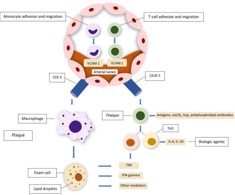
│ STEP 3: MONOCYTE RECRUITMENT & ADHESION │
│ Monocytes adhere to endothelium (via VCAM-1) │
│ → Migrate into intima → differentiate into MACROPHAGES │
│ T lymphocytes also recruited (adaptive immunity) │
└─────────────────────────┬───────────────────────────────────────┘
▼
┌─────────────────────────────────────────────────────────────────┐
│ STEP 4: FOAM CELL FORMATION (FATTY STREAK) │
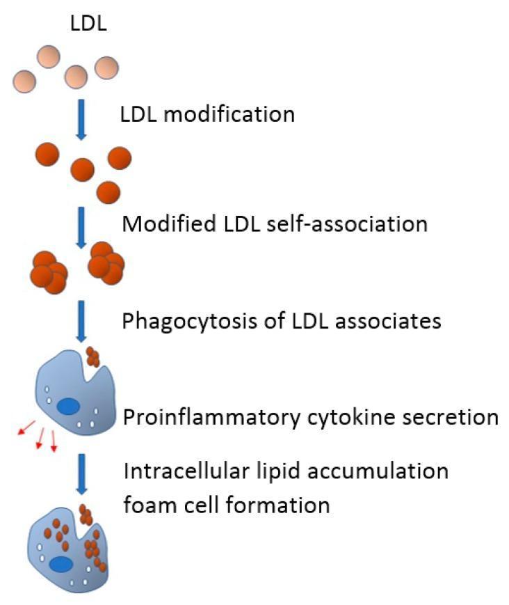
│ Macrophages engulf oxidized LDL via scavenger receptors │
│ (CD36, SR-A, Lox-1) → Lipid-laden FOAM CELLS │
│ = First visible lesion: FATTY STREAK │
└─────────────────────────┬───────────────────────────────────────┘
▼
┌─────────────────────────────────────────────────────────────────┐
│ STEP 5: PLATELET ADHESION & FACTOR RELEASE │
│ Activated macrophages + foam cells + platelets release: │
│ → PDGF, FGF (recruit SMCs from media) │
│ → TGF-β, TNF, IL-1 (pro-inflammatory amplification) │
│ → MCP-1 (more monocyte recruitment) │
└─────────────────────────┬───────────────────────────────────────┘
▼
┌─────────────────────────────────────────────────────────────────┐
│ STEP 6: SMOOTH MUSCLE CELL (SMC) MIGRATION & │
│ PROLIFERATION │
│ SMCs migrate from media → intima │
│ Proliferate + synthesize ECM (collagen, elastin, proteo- │
│ glycans) → forms FIBROUS CAP │
└─────────────────────────┬───────────────────────────────────────┘
▼
┌─────────────────────────────────────────────────────────────────┐
│ STEP 7: ADVANCED PLAQUE (ATHEROMATOUS PLAQUE) │
│ Fibrous cap + necrotic lipid core + calcification │
│ Neovascularization at periphery │
│ Chronic inflammation sustains lesion growth │
└─────────────────────────────────────────────────────────────────┘
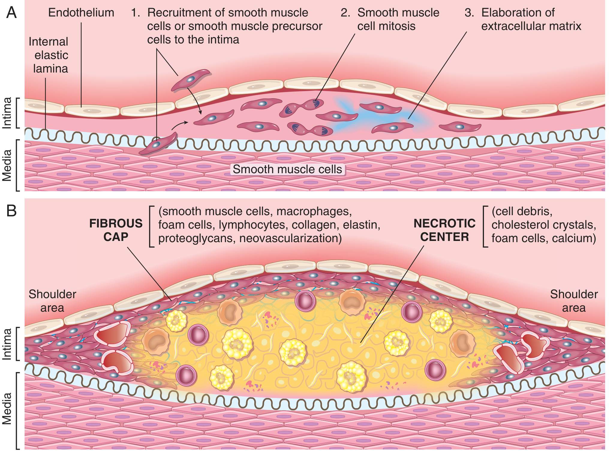
Vascular Response to Endothelial Injury (Fig. 8.5 - Robbins Basic Pathology):
(A) Shows SMC recruitment to the intima, proliferation, and ECM elaboration.
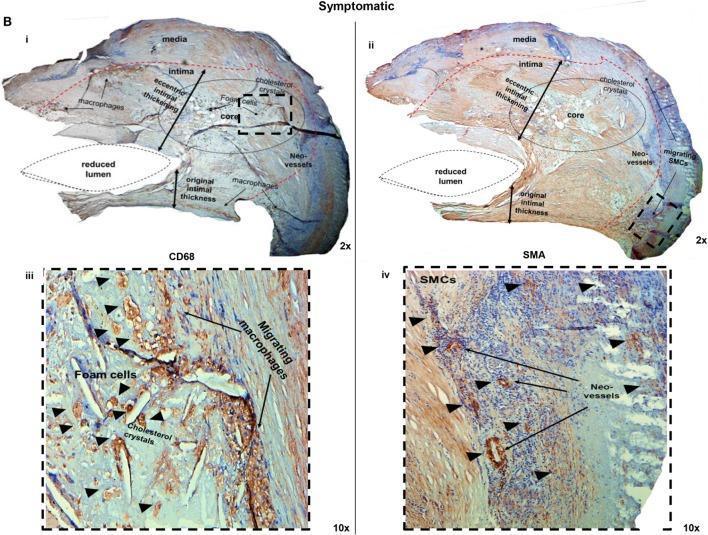
(B) Shows the mature atheromatous plaque: fibrous cap (SMCs, macrophages, foam cells, lymphocytes, collagen, elastin, proteoglycans) and necrotic center (cell debris, cholesterol crystals, foam cells, calcium).
Key Cellular Players
| Cell Type | Role in Atherogenesis |
|---|
| Endothelial Cells (ECs) | First injured; become dysfunctional; upregulate VCAM-1, ICAM-1; increase permeability |
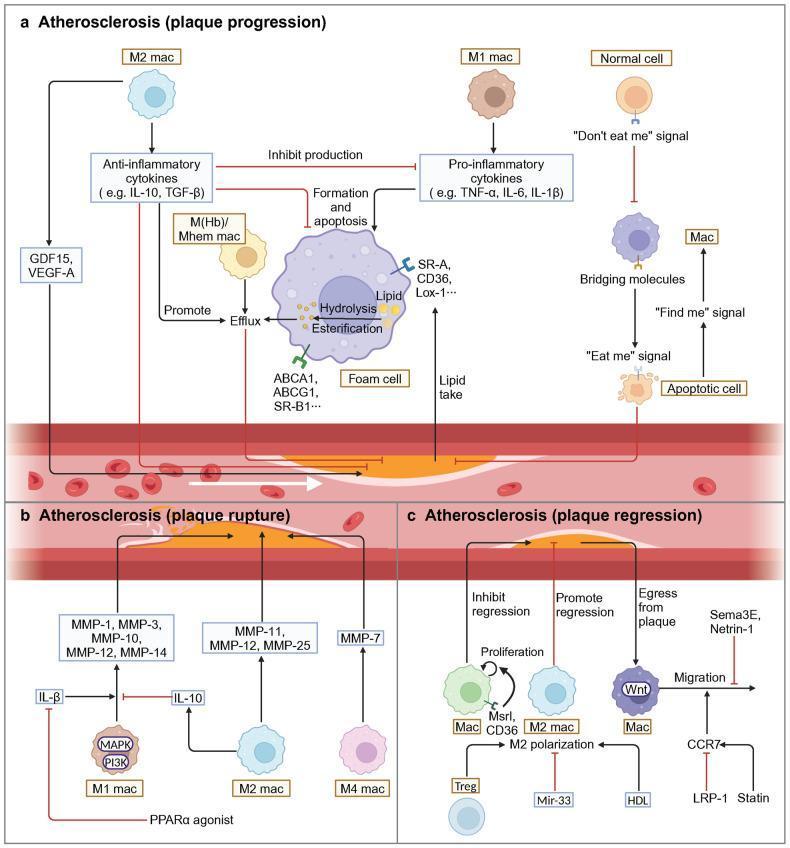
| Monocytes/Macrophages | Recruited from blood; engulf oxidized LDL; become foam cells; release cytokines (TNF, IL-1, IL-6) and MMPs |
| Foam Cells | Lipid-laden macrophages and SMCs; core of fatty streak; release further pro-inflammatory mediators |
| T Lymphocytes | Recruited to plaque; TH1 cells release IFN-γ (inhibits SMC collagen synthesis, weakens cap) |
| Smooth Muscle Cells (SMCs) | Recruited from media; proliferate in intima; synthesize collagen for fibrous cap; can also become foam cells |
| Platelets | Adhere at sites of injury; release PDGF, TGF-β → promote SMC migration and ECM production |
Foam Cell Formation (Macrophage Pathway)
Circulating LDL
│
▼
Enters arterial intima
(facilitated by endothelial dysfunction)
│
▼
Oxidation of LDL → oxLDL
(oxidative stress in intima)
│
▼
Monocyte → Macrophage in intima
│
▼
Macrophage engulfs oxLDL via
Scavenger Receptors (CD36, SR-A, Lox-1)
[NOT regulated by negative feedback - unlike LDL receptor]
│
▼
Intracellular lipid droplet accumulation
│
▼
══════════════════════════
FOAM CELL FORMED
══════════════════════════
│
┌────┴────┐
▼ ▼
Release Apoptosis/Necrosis
cytokines → contributes to
(TNF, IL-1, necrotic core
MMP) of plaque
Morphology of Atherosclerotic Lesions
Lesion Progression (Sequential)
NORMAL INTIMA
│
▼
FATTY STREAK
• Earliest visible lesion
• Flat, yellow intimal streaks
• Composed of lipid-laden foam cells (macrophages)
• Start near ostia of branch vessels
• Found even in children and young adults
• NOT clinically significant alone
│
▼
INTERMEDIATE LESION / FIBRO-FATTY PLAQUE
• Foam cells + extracellular lipid + SMC proliferation begins
• ECM deposition starts
│
▼
ADVANCED ATHEROMATOUS PLAQUE (ATHEROMA)
• Fibrous cap (SMCs + collagen + proteoglycans)
• Shoulder region: macrophages, T cells, SMCs
• Necrotic lipid core: cholesterol crystals, debris, foam cells
• Peripheral neovascularization
• Calcification of ECM and necrotic debris
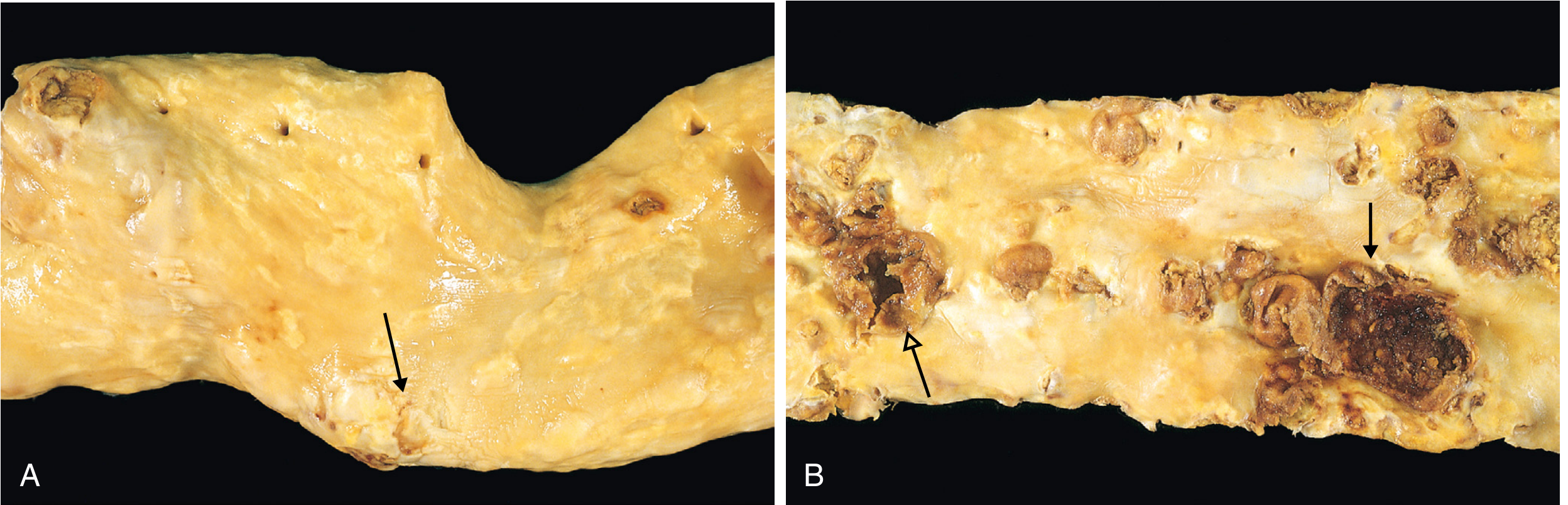
Gross Pathology of Atherosclerotic Aorta:
(A) Early/intermediate plaque (arrow). (B) Advanced disease: ulceration (open arrow) and calcified, ruptured plaques with dark hemorrhagic debris (closed arrow).
Composition of the Atherosclerotic Plaque
| Component | Location in Plaque | Details |
|---|
| Fibrous cap | Superficial (luminal side) | SMCs, dense collagen, proteoglycans |
| Shoulder region | Cap edges | Most cellular area: macrophages, T lymphocytes, SMCs; highest vulnerability to rupture |
| Necrotic core | Deep to fibrous cap | Cholesterol, cholesterol esters, cell debris, foam cells, fibrin, thrombus, plasma proteins |
| Cholesterol crystals | Within core | Empty "clefts" seen histologically (washed out in processing) |
| Neovascularization | Periphery of plaque | Thin-walled vessels; source of intraplaque hemorrhage |
| Calcification | Throughout (late) | Dystrophic calcification of phospholipids and necrotic debris |
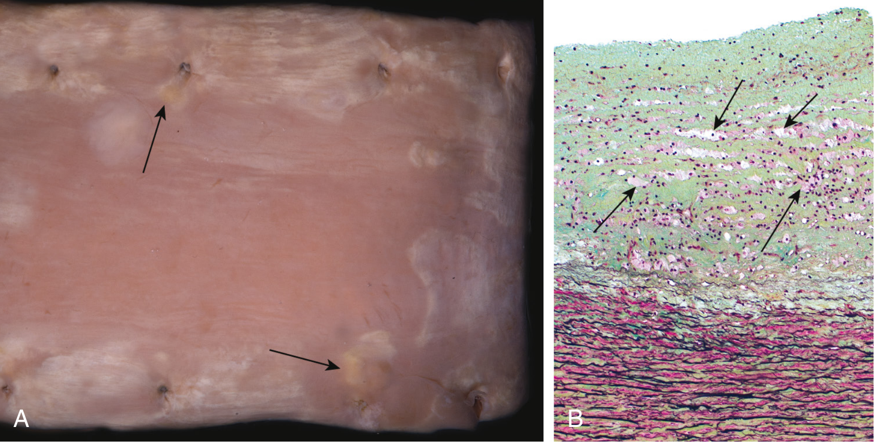
Fatty Streak (Earliest Lesion)
(A) Aorta with fatty streaks (arrows), near ostia of branch vessels. (B) Histology demonstrating foam cells (arrows) within the intima.
Complications of Advanced Plaques
ADVANCED ATHEROSCLEROTIC PLAQUE
│
┌─────────────┼──────────────┬──────────────┐
▼ ▼ ▼ ▼
RUPTURE/ INTRAPLAQUE ATHEROEMBOLISM ANEURYSM
EROSION/ HEMORRHAGE FORMATION
ULCERATION
│ │ │ │
▼ ▼ ▼ ▼
Thrombogenic Rupture of Plaque debris Medial
material fibrous cap discharged ischemia +
exposed → or thin → microemboli loss of
THROMBUS neovessels elastin →
FORMATION → hematoma weakening →
→ plaque RUPTURE
expansion/
rupture
│
▼
Partial occlusion → Unstable angina
Complete occlusion → ACUTE MI / STROKE
Organization of thrombus → further plaque growth
Clinical Consequences by Vessel
| Vessel Affected | Clinical Consequence |
|---|
| Coronary arteries | Stable angina (>70% stenosis), unstable angina, acute MI, sudden cardiac death |
| Cerebral / carotid arteries | TIA, ischemic stroke, carotid bruits |
| Aorta | Abdominal aortic aneurysm (AAA), aortic dissection |
| Renal arteries | Renovascular hypertension, renal ischemia |
| Iliac / femoral / popliteal | Peripheral vascular disease, intermittent claudication, gangrene |
| Mesenteric arteries | Bowel ischemia |
Stable vs. Vulnerable Plaque
| Feature | Stable Plaque | Vulnerable Plaque |
|---|
| Fibrous cap | Thick, rich in collagen | Thin, collagen-poor |
| Lipid core | Small | Large (>40% plaque volume) |
| Inflammation | Minimal | Heavy macrophage infiltration |
| SMC content | High (protective) | Low |
| IFN-γ | Low | High (inhibits collagen synthesis) |
| MMP activity | Low | High (degrades ECM of cap) |
| Risk | Causes stable angina at high stenosis | Ruptures even at <50% stenosis |
Stenosis and Flow Limitation
Luminal Narrowing
│
< 70% stenosis
(no resting symptoms)
│
≥ 70% stenosis
(critical stenosis)
│
┌┴──────────────┐
▼ ▼
At REST On EXERTION
Adequate flow Demand > Supply
(OK) │
▼
STABLE ANGINA
(reversible ischemia)
Summary Flowchart - Overall Atherosclerosis
Risk Factors (LDL↑, HTN, Smoking, DM, Age, Genetics)
│
▼
ENDOTHELIAL INJURY / DYSFUNCTION
│
┌─────────┴──────────┐
▼ ▼
Permeability ↑ Leukocyte adhesion
(LDL enters intima) (VCAM-1, ICAM-1 ↑)
│ │
▼ ▼
LDL oxidized Monocytes → Macrophages
(oxLDL) T cells recruited
│ │
└──────────┬─────────┘
▼
FOAM CELL FORMATION
(Fatty streak)
│
▼
Cytokine release (TNF, IL-1, PDGF)
SMC recruitment from media
SMC proliferation + ECM synthesis
│
▼
FIBROUS PLAQUE / ATHEROMA
(Fibrous cap + Necrotic lipid core)
│
┌────────┴────────┐
▼ ▼
STABLE PLAQUE VULNERABLE PLAQUE
(thick cap) (thin cap, large core)
│ │
▼ ▼
Gradual stenosis PLAQUE RUPTURE
→ Stable angina → Thrombosis
→ ACUTE MI / STROKE
→ Sudden death
Key Mediators Summary Table
| Mediator | Source | Effect |
|---|
| oxLDL | Oxidized LDL in intima | Endothelial dysfunction, foam cell formation, pro-inflammatory |
| VCAM-1, ICAM-1 | Dysfunctional endothelium | Monocyte/T cell adhesion |
| MCP-1 | Endothelium, SMCs, macrophages | Monocyte recruitment into intima |
| PDGF | Platelets, macrophages, ECs | SMC migration and proliferation |
| TGF-β | Platelets, macrophages | SMC ECM production (fibrous cap) |
| IFN-γ | T lymphocytes (TH1) | Inhibits SMC collagen synthesis → destabilizes cap |
| MMPs (MMP-1,3,9) | Macrophages | Degrade fibrous cap ECM → plaque rupture |
| TNF, IL-1, IL-6 | Macrophages | Systemic inflammation (↑CRP), local vascular injury |
| CRP | Liver (acute phase) | Marker of inflammation; independent CV risk predictor |
Sources
- Robbins, Cotran & Kumar Pathologic Basis of Disease, Chapter 11 (Arteries, pp. 465-475)
- Robbins & Kumar Basic Pathology (Robbins Pathology), Chapter 8 (pp. 313-325)