
Cerebral Angiogram simplify
Reading File
A cerebral angiogram is an imaging procedure that uses X-rays and contrast dye injected into brain blood vessels to visualize the cerebral circulation and detect abnormalities like aneurysms, AVMs, or stenosis.
Searching sources...
Finding Sources
Searching the Web
cerebral angiogram procedure explained simply
Reading File
Reading File
Loading Image
Here is a simplified breakdown of the cerebral angiogram:
Cerebral Angiogram - Simplified
What Is It?
A cerebral angiogram (also called cerebral angiography) is the gold standard X-ray-based test for looking at blood vessels in and around the brain. Think of it as a detailed "road map" of your brain's plumbing system.
How It's Done (Step by Step)
- Access - A thin plastic tube (catheter) is inserted into an artery, usually at the wrist or groin
- Navigation - The catheter is guided up through the blood vessels toward the neck and brain
- Dye injection - A contrast dye is injected through the catheter into the brain's blood vessels
- X-ray images - X-rays are taken as the dye flows through, making the vessels clearly visible
Here is what the result looks like - a real cerebral angiogram showing brain blood vessels:
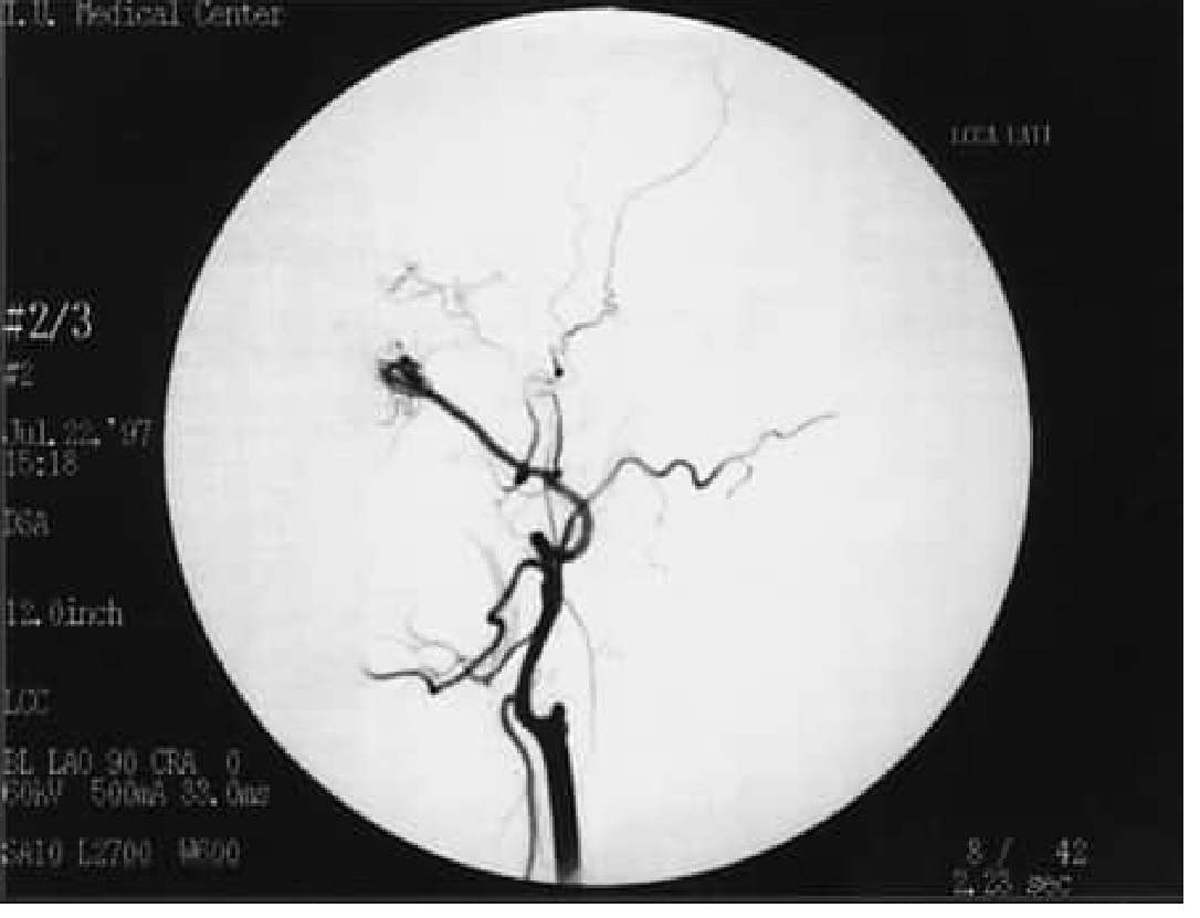
The dark branching lines are the arteries of the brain lit up by contrast dye.
Why Is It Done? (Indications)
| Condition | Why Angiogram Helps |
|---|---|
| Brain aneurysm | Shows exact size and location |
| AVM (arteriovenous malformation) | Maps the abnormal tangle of vessels |
| Stroke / vessel blockage | Confirms the blocked artery; guides thrombectomy |
| Arterial dissection | Detects a tear in the vessel wall |
| Vasculitis / Moyamoya disease | Identifies abnormal vessel narrowing |
| CNS vasculitis | Confirms diagnosis when other tests are unclear |
Angiogram vs. Other Tests
| Test | Pros | Cons |
|---|---|---|
| Conventional angiogram | Most accurate; guides intervention | Invasive; small risk of stroke |
| CTA (CT with contrast) | Fast, widely available | Radiation; less detail for small vessels |
| MRA (MRI of vessels) | No radiation; no catheter | Tends to overestimate stenosis; motion artifact |
The conventional catheter angiogram remains superior when exact diagnosis matters or when a procedure (like thrombectomy or stenting) will be performed right after.
Risks (Rare but Real)
- Overall complication rate: ~1-5%
- Most common: minor groin bruising/hematoma
- Permanent neurological damage: ~0.2%
- Death: ~0.05%
Before and After
- Before: You may be told to fast 4-8 hours prior; blood thinners may be adjusted
- During: You lie flat; local anesthesia at the entry site; sedation may be given
- After: Rest and pressure at the catheter site; monitored for a few hours
Bottom Line
A cerebral angiogram is a minimally invasive but direct way to see the brain's blood vessels with the highest possible detail. It is most useful when non-invasive tests (CTA/MRA) give uncertain results, or when a same-session treatment is planned.
- Bradley and Daroff's Neurology in Clinical Practice, p. 1389
This is a shared conversation. Sign in to Orris to start your own chat.