
Neet imp pyqs .
Which subject do you want NEET important PYQs for? · Any specific topic/chapter? (optional - select if applicable)
Pg
What do you mean by 'PG'?
Which subject or specialty do you want important NEET PG PYQs from?
Any specific topic within Medicine, or all high-yield topics?
NEET PG - Medicine: High-Yield Important PYQs
🫀 CARDIOLOGY
- A) LAD B) RCA C) LCX D) Diagonal
- ✅ Answer: B) RCA - Inferior MI (II, III, aVF) = RCA territory
- A) Digoxin B) Verapamil C) Procainamide D) Adenosine
- ✅ Answer: C) Procainamide (Digoxin & Verapamil are contraindicated in WPW)
- OS is best heard at apex; short S2-OS interval = severe MS (high LA pressure)
- ✅ Key: Short S2-OS interval = Severe MS
- A) Hydralazine B) Amiodarone C) Captopril D) Metoprolol
- ✅ Answer: A) Hydralazine (also Procainamide, Isoniazid)
- ✅ HOCM (most murmurs decrease; HOCM & MVP increase with Valsalva)
🫁 RESPIRATORY / PULMONOLOGY
- A) D-dimer B) CTPA C) V/Q scan D) Pulmonary angiography
- ✅ Answer: B) CTPA (Gold standard in clinical practice; pulmonary angiography is historical gold standard)
- ✅ PaO2 ≤ 55 mmHg OR SpO2 ≤ 88% (or PaO2 ≤ 59 with cor pulmonale/polycythemia)
- Involves apex of lung, causes Horner's syndrome, C8-T1-T2 nerve involvement, rib erosion
- ✅ Key: Does NOT cause phrenic nerve palsy (that's a central/mediastinal tumor feature)
- ✅ Silicosis (also Sarcoidosis rarely)
- ✅ Co-trimoxazole (TMP-SMX) - both treatment and prophylaxis
🧠 NEUROLOGY
- ✅ Brown-Sequard syndrome - ipsilateral motor loss + contralateral pain/temp loss
- A) Cholangitis B) Multiple Sclerosis C) Wernicke's D) Wilson's
- ✅ A) Cholangitis (fever, jaundice, RUQ pain); MS has Charcot's triad of Nystagmus + Intention tremor + Scanning speech
- ✅ Vitamin B12 deficiency - affects dorsal columns + lateral corticospinal tracts
- ✅ Streptococcus pneumoniae
- ✅ Neurosyphilis (also DM rarely)
🩸 HAEMATOLOGY
- ✅ Megaloblastic anemia (B12 / Folate deficiency)
- ✅ Flow cytometry (CD55/CD59 deficiency) - Ham's test is classical but outdated
- ✅ Myelofibrosis
- ✅ Hemophilia A (Factor VIII deficiency); X-linked recessive
- ✅ Imatinib (Gleevec) - BCR-ABL tyrosine kinase inhibitor; Philadelphia chromosome t(9;22)
🫘 NEPHROLOGY
- ✅ Membranous nephropathy (in adults); MCNS in children
- ✅ Malignant hypertension
- ✅ Cannot acidify urine below pH 5.5 - H+ secretion defect; causes hypokalemia + nephrocalcinosis
- ✅ Standard ATT (HRZE regimen) for 6-9 months; same as pulmonary TB
- ✅ Rapid osmotic shifts causing cerebral edema during first dialysis session
🦠 INFECTIOUS DISEASES
- ✅ Leptospira (severe form with jaundice + renal failure + hemorrhage)
- ✅ Typhoid fever (Salmonella typhi)
- ✅ Babesiosis
- ✅ Artemisinin-based Combination Therapy (ACT) - e.g., Artesunate + Lumefantrine
- ✅ Urea Breath Test (UBT) (post-treatment); Biopsy/CLO test for active diagnosis
🔬 ENDOCRINOLOGY
- ✅ Hypocalcemia (tetany)
- ✅ Exogenous steroid use (Iatrogenic); Cushing's disease = pituitary ACTH-secreting adenoma
- ✅ Graves' disease (hyperthyroidism, not hypothyroidism)
- ✅ Medullary thyroid carcinoma + Pheochromocytoma + Hyperparathyroidism (RET mutation)
- ✅ Insulinoma (symptoms with hypoglycemia, low blood glucose, relief with glucose)
🦴 RHEUMATOLOGY
- ✅ Rheumatoid Arthritis (most specific; RF is sensitive but less specific)
- ✅ SLE (anti-dsDNA + anti-Sm are most specific for SLE)
- ✅ Low-dose Aspirin (high-dose aspirin is uricosuric)
- ✅ Ankylosing Spondylitis (HLA-B27 associated)
- ✅ NSAIDs (Indomethacin) OR Colchicine
🫃 GASTROENTEROLOGY
- ✅ Ulcerative Colitis (loss of haustrations)
- ✅ Crohn's Disease
- ✅ Appendix (overall); Ileum (most common to cause carcinoid syndrome)
- Parameters: Bilirubin, Albumin, PT/INR, Ascites, Encephalopathy
- ✅ NOT AFP or ALT - those are not part of Child-Pugh
- ✅ Lactulose + Rifaximin (reduces ammonia production)
💊 HIGH-YIELD PHARMACOLOGY IN MEDICINE
| Drug | Key Point |
|---|---|
| Metformin | DOC in Type 2 DM; contraindicated in renal failure (GFR <30) |
| Digoxin toxicity | Yellow vision, bradycardia; treat with Digibind |
| Warfarin reversal | Vitamin K + Fresh Frozen Plasma (FFP) |
| Heparin overdose | Protamine sulfate |
| Lithium toxicity | Coarse tremor, polyuria, nephrogenic DI; narrow TI |
| Amiodarone side effects | Thyroid, pulmonary, hepatic, corneal deposits, photosensitivity |
📊 QUICK HIGH-YIELD MNEMONICS
- MUDPILES = Causes of high anion gap metabolic acidosis (Methanol, Uremia, DKA, Propylene glycol, INH/Iron, Lactic acidosis, Ethylene glycol, Salicylates)
- AEIOU TIPS = Causes of coma
- HACEK organisms = Infective endocarditis in culture-negative cases
Explain cardio 1
inferior MI ST elevation leads II III aVF ECG RCA occlusion
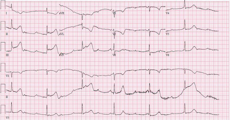
A standard 12-lead electrocardiogram (ECG) with rhythm strips (V1, II, V5) showing an acute inferior ST-elevation myocardial infarction (STEMI) complicated by high-grade atrioventricular (AV) block. Significant ST-segment elevation is present in the inferior leads (II, III, and aVF), with the magnitude of elevation in lead III exceeding that in lead II, suggesting a right coronary artery (RCA) occlusion. Reciprocal ST-segment depression and T-wave inversion are visible in the high lateral leads (I, aVL) and anterior lead V2. The rhythm analysis reveals complete heart block (third-degree AV block) characterized by atrioventricular dissociation, with an atrial rate significantly faster than the ventricular escape rate (approximately 37 bpm). The QRS complexes are wide, and P waves do not maintain a fixed relationship with the ventricular activity. This visual findings are pathognomonic for an inferior wall MI with ischemia to the AV node, commonly associated with a proximal RCA lesion.
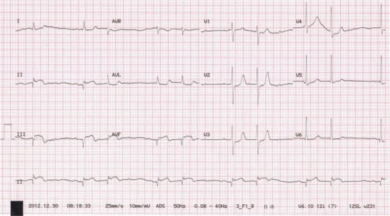
A 12-lead diagnostic electrocardiogram (ECG) tracing displaying classic signs of an inferior wall myocardial infarction (MI). The tracing exhibits significant convex-upwards ST-segment elevation (Pardee waves) and T-wave inversions in the inferior leads (II, III, and aVF). Notably, deep and wide pathological Q waves are present in these same leads, suggesting myocardial necrosis or a completed infarction sequence. Additionally, the rhythm strip demonstrates evidence of a second-degree atrioventricular (AV) block, characterized by a non-conducted P wave following a consistent PR interval pattern. Reciprocal ST-segment depression is visible in the lateral leads (I, aVL), a common finding in acute inferior ST-elevation myocardial infarction (STEMI). This visual provides critical educational evidence for diagnosing coronary artery occlusion, specifically involving the right coronary artery (RCA) or left circumflex artery, and illustrates the potential conduction system complications associated with inferior wall damage.
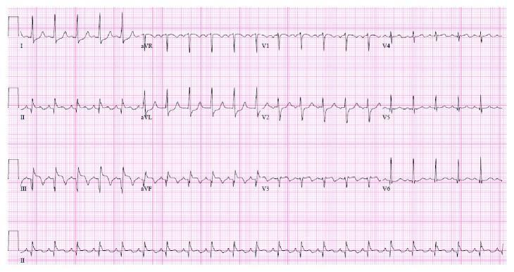
A 12-lead electrocardiogram (ECG) demonstrating signs of an acute inferior wall ST-elevation myocardial infarction (STEMI). The visual shows prominent, convex ST-segment elevation in the inferior leads: II, III, and aVF. Concurrent reciprocal ST-segment depression is visible in the lateral leads, specifically I and aVL, a hallmark finding that supports the diagnosis of acute inferior injury. The precordial leads (V1-V6) show a standard R-wave progression. The QRS complexes appear narrow, and the rhythm is regular. This diagnostic tracing is a critical educational example used to identify coronary artery occlusion, typically involving the right coronary artery (RCA). It is suitable for medical students and clinicians learning to distinguish between different territorial patterns of myocardial infarction and understand the clinical significance of reciprocal changes in emergency cardiology.
🫀 NEET PG Cardiology Q1 - Full Explanation
The Question Revisited
A 55-year-old man has chest pain radiating to jaw, diaphoresis, and ST elevation in leads II, III, aVF. Which artery is occluded? Answer: RCA (Right Coronary Artery)
STEP 1 - Understand Coronary Artery Territories
| Coronary Artery | Wall of Heart Supplied | ECG Leads Affected |
|---|---|---|
| LAD (Left Anterior Descending) | Anterior wall of LV, anterior septum, apex | V1, V2, V3, V4 |
| RCA (Right Coronary Artery) | Inferior wall of LV, posterior septum, RV free wall | II, III, aVF |
| LCX (Left Circumflex) | Lateral wall of LV | I, aVL, V5, V6 |
Memory trick: "RCA = Right side = Inferior leads (II, III, aVF)" Think: RCA runs in the right atrioventricular groove and wraps inferiorly.
- LAD = 40-50% of all MIs (most common)
- RCA = 30-40%
- LCX = 15-20%
STEP 2 - How to Identify Inferior MI on ECG
✅ ST Elevation in II, III, aVF
- Elevation in lead III > lead II = strongly suggests RCA (not LCX)
- Elevation in lead II ≥ III = suggests LCX
✅ Reciprocal ST Depression in I and aVL
- This is a mirror image of the inferior elevation
- Reciprocal changes confirm true STEMI (not pericarditis, which has no reciprocal depression)
STEP 3 - The Key Differentiator: Lead III vs Lead II
| Finding | Suggests |
|---|---|
| ST elevation III > II | RCA occlusion (proximal) |
| ST elevation II ≥ III | LCX occlusion |
| ST elevation in V5, V6 also | LCX (lateral extension) |
| ST elevation in V1 (right precordial) | Right Ventricular MI (proximal RCA) |
- Tintinalli's Emergency Medicine confirms: "ST elevation in lead III greater than in lead II plus ST depression in lead I and aVL = 100% RCA occlusion"
STEP 4 - Critical Complication: Right Ventricular Infarction (RVI)
How to detect RVI:
- Do right-sided ECG leads - look for ST elevation in V4R
- V4R elevation >1 mm = RVI confirmed
Why it matters clinically:
| Feature | RVI |
|---|---|
| JVP | Raised (RV can't pump forward) |
| BP | Low (hypotension) |
| Lungs | Clear (not left heart failure) |
| Kussmaul's sign | Positive |
CRITICAL management point for NEET PG:
AVOID nitrates and diuretics in RVI - they reduce preload and can cause profound hypotension. Give IV fluids instead to maintain RV filling.
STEP 5 - AV Node Involvement (Another RCA Complication)
- 1st degree AV block
- 2nd degree AV block (Mobitz type 1 / Wenckebach)
- Complete (3rd degree) AV block - seen as slow escape rhythm
The ECG image above actually shows an inferior STEMI complicated by complete heart block - a classic NEET PG teaching case.
STEP 6 - Inferior MI vs Posterior MI
- No direct ST elevation on standard 12-lead
- Look for: ST depression + tall R waves in V1-V3 (mirror image)
- Confirm with posterior leads (V7, V8, V9) - ST elevation there confirms it
STEP 7 - Full Summary Table
| Feature | Detail |
|---|---|
| Artery occluded | RCA |
| ECG leads | II, III, aVF (inferior leads) |
| Reciprocal changes | I, aVL (depression) |
| RCA vs LCX clue | III > II = RCA; II > III = LCX |
| Complication 1 | RV infarction - avoid nitrates, give fluids |
| Complication 2 | AV block - RCA supplies AV node |
| Complication 3 | Bradycardia - sinus node also supplied by RCA |
| Key right-sided lead | V4R for RV infarction |
High-Yield NEET PG One-Liners for This Topic
- ST elevation II, III, aVF = Inferior MI = RCA
- III > II = RCA; II > III = LCX
- Reciprocal depression in I and aVL = confirms inferior STEMI
- RVI = avoid nitrates + diuretics, give IV fluids
- V4R elevation = right ventricular MI
- AV block in inferior MI = RCA supplies AV node
- Wenckebach (Mobitz 1) = common in inferior MI; Mobitz 2 = more common in anterior MI (LAD)
Pcp doc n prophylaxis
🦠 PCP (Pneumocystis jirovecii Pneumonia) - Complete NEET PG Guide
Quick Recall
DOC for Treatment = TMP-SMX (Co-trimoxazole) DOC for Prophylaxis = TMP-SMX (Co-trimoxazole) Same drug - both treatment AND prophylaxis!
PART 1 - TREATMENT
🥇 First-Line: TMP-SMX (Trimethoprim-Sulfamethoxazole / Co-trimoxazole)
| Parameter | Detail |
|---|---|
| Drug | TMP-SMX (Co-trimoxazole) |
| Route | Oral (mild-moderate) or IV (severe) |
| Duration | 21 days |
| Mechanism | TMP inhibits DHFR; SMX inhibits DHPS → blocks folate synthesis in Pneumocystis |
⚡ Adjunctive Corticosteroids (NEET PG HIGH YIELD)
Add Prednisolone/Prednisone when PaO2 < 70 mmHg (room air) OR A-a gradient > 35 mmHg
- Dose: Prednisolone 40 mg BD x 5 days → 40 mg OD x 5 days → 20 mg OD x 11 days
- Start within 72 hours of starting TMP-SMX
- Why? Dying Pneumocystis organisms cause an inflammatory surge → steroids dampen this
- Failure to add steroids in severe PCP = increased mortality
🔄 Alternative Regimens (When TMP-SMX Not Tolerated)
| Severity | Alternative Drug | Notes |
|---|---|---|
| Mild-Moderate | Dapsone + TMP | Oral; check G6PD before dapsone |
| Mild-Moderate | Atovaquone | Oral suspension; better tolerated |
| Mild-Moderate | Clindamycin + Primaquine | Check G6PD before primaquine |
| Severe | IV Pentamidine (4 mg/kg/day x 21 days) | Reserved for TMP-SMX intolerance |
| Salvage (failed 1st line) | Clindamycin + Primaquine | Preferred over pentamidine as salvage |
Pentamidine side effects (NEET PG gold): Hypoglycemia (early - beta cell stimulation) → Hyperglycemia (late - beta cell destruction), Pancreatitis, Nephrotoxicity, Hypotension, Torsades de pointes, Hypocalcemia, Hypokalemia
G6PD check mandatory before Dapsone and Primaquine (both cause hemolysis in G6PD deficiency)
PART 2 - PROPHYLAXIS
When to Start Prophylaxis (NEET PG MUST KNOW)
| Indication | Threshold |
|---|---|
| HIV patients | CD4 count < 200 cells/µL OR CD4% < 14% |
| CD4 200-250 | Give if CD4 monitoring not possible every 3 months |
| Non-HIV immunosuppressed | Steroids > 20 mg/day prednisolone for > 1 month; organ transplant; haematological malignancy |
- All HIV patients after a PCP episode must continue prophylaxis
When to STOP Prophylaxis
| Type | Criteria to Stop |
|---|---|
| Primary | CD4 rises to > 200 cells/µL for ≥ 3 months on ART |
| Secondary | CD4 > 200 cells/µL for ≥ 3 months (or CD4 100-200 with undetectable viral load for ≥ 3-6 months) |
🥇 Prophylaxis Regimens (Priority Order)
| Priority | Drug | Dose | Notes |
|---|---|---|---|
| 1st line | TMP-SMX (Co-trimoxazole) | 1 DS tab daily OR 1 SS tab daily | Also protects against Toxoplasmosis |
| 2nd line | Dapsone | 100 mg/day (or 50 mg BD) | Check G6PD |
| 2nd line | Dapsone + Pyrimethamine + Leucovorin | Dapsone 50 mg/day + Pyrimethamine 50 mg/week + Leucovorin 25 mg/week | Also covers Toxo |
| 2nd line | Atovaquone | 1500 mg/day | Expensive but well tolerated |
| 3rd line | Aerosolized Pentamidine | 300 mg monthly via nebulizer | Least preferred |
Aerosolized pentamidine disadvantages:
- Does NOT cover extrapulmonary Pneumocystis
- No protection against Toxoplasmosis
- Risk of pneumothorax
- More breakthrough infections at CD4 < 100
PART 3 - DIAGNOSIS (Bonus High-Yield Points)
| Test | Details |
|---|---|
| Gold standard | BAL (Bronchoalveolar Lavage) + GMS stain or DFA |
| Stains used | GMS (Gomori Methenamine Silver) - shows cyst wall; DFA (Direct Fluorescent Antibody) - most sensitive |
| Serum marker | Beta-D-glucan elevated (not specific, but supports diagnosis) |
| CXR | Bilateral ground-glass opacities (butterfly/bat-wing pattern) |
| HRCT | Ground-glass opacities, often perihilar |
| LDH | Elevated LDH = poor prognosis marker |
PART 4 - COMPLETE ONE-LINER SUMMARY TABLE
| Question | Answer |
|---|---|
| DOC treatment PCP | TMP-SMX (Co-trimoxazole) x 21 days |
| DOC prophylaxis PCP | TMP-SMX (Co-trimoxazole) |
| When to start prophylaxis | CD4 < 200 cells/µL |
| Adjunctive therapy severe PCP | Prednisolone (if PaO2 < 70 mmHg) |
| Alternative if TMP-SMX intolerant (mild-mod) | Atovaquone or Clindamycin + Primaquine |
| Alternative if TMP-SMX intolerant (severe) | IV Pentamidine |
| Salvage therapy | Clindamycin + Primaquine |
| Pentamidine prophylaxis dose/route | 300 mg monthly - nebulized |
| Gold standard diagnosis | BAL + GMS stain |
| When to stop primary prophylaxis | CD4 > 200 for ≥ 3 months on ART |
| G6PD check needed before | Dapsone, Primaquine |
| TMP-SMX also covers | Toxoplasma gondii (extra benefit) |
Opportunistic fungal infections