
Corpora amylacea
Reading File
Corpora amylacea are rounded, concentrically laminated bodies found in several tissues. Let me search the library for comprehensive coverage.
Searching sources...
Finding Sources
Finding Sources
Searching PubMed
"corpora amylacea"
Good results from multiple textbooks. Now let me read the key sections from Robbins and Junqueira's for depth, plus the prostate section.
Reading File
Reading File
Reading File
Excellent detail from the textbooks. Let me also fetch the prostate histology image showing corpora amylacea, and pull the Kaplan & Sadock section on AD.
Loading Image
Reading File
Reading File
I now have comprehensive information from multiple authoritative textbooks. Here is the full synthesis:
Corpora Amylacea
Definition and General Characteristics
Corpora amylacea (singular: corpus amylaceum, Latin: "starchy bodies") are round, concentrically laminated (lamellar) structures found as normal or age-related inclusions in several tissues. The name is derived from their staining behavior, which mimics starch with iodine, though they contain no true amyloid (they are not related to amyloidosis despite the name).
Corpora Amylacea in the CNS (Brain)
This is the most widely studied context.
Morphology
- Shape/size: Round, faintly basophilic structures, 5-50 µm in diameter
- Architecture: Concentrically lamellated
- Staining: Periodic acid-Schiff (PAS)-positive (due to polysaccharide content); faintly basophilic on H&E
- Location: Found wherever there are astrocytic end-processes - especially in the subpial and perivascular zones of the brain
Composition
- Primarily glycosaminoglycan polymers (polyglucosan/glucose polymers)
- Also contain heat-shock proteins (αB-crystallin, HSP27) and ubiquitin
Origin and Pathogenesis
- They are cytoplasmic inclusion bodies of astrocytes, representing a degenerative change in the astrocytic end-process
- Thought to arise due to oxidative stress and iron imbalance within astrocytes
- Possibly represent an attempt to sequester oxidized mitochondrial components or excess iron - but when numerous, they likely impair astrocyte function
- Increase progressively with age - normally seen after age 40; markedly increased in Alzheimer disease (AD)
Clinical Significance in the CNS
| Condition | Role of Corpora Amylacea |
|---|---|
| Normal aging | Increase in number with advancing age; considered a degenerative change |
| Alzheimer disease | Markedly increased; one of the neuropathologic markers in aging brain |
| Adult polyglucosan body disease (APBD) | Massive, widespread deposition - the pathologic hallmark |
| Lafora body disease | Lafora bodies (in neurons, hepatocytes, myocytes) have identical structure and biochemical composition |
- Robbins Pathologic Basis of Disease, Neuropathology chapter (Astrocyte Reactions to Injury)
- Kaplan & Sadock's Comprehensive Textbook of Psychiatry (Lesions in the Aging Brain)
Corpora Amylacea in the Prostate
In the prostate, corpora amylacea are a normal histologic finding with a different formation mechanism:
- Formation: They arise from the deposition of prostatic secretions around sloughed epithelial cells within the glandular lumina
- Appearance: Concentrically laminated concretions within the acini of the prostate gland (visible on H&E at ×80 as oval/round laminated structures labeled "CA")
- Clinical significance: They do not usually cause inflammation unless they cause ductal obstruction
- Calculi: Prostatic calculi are thought to represent calcified corpora amylacea; they are rarely found in boys, usually small and numerous, tannish-gray in color during TURP
Below is the prostate histology from Junqueira's Basic Histology showing corpora amylacea (labeled CA) within the glandular lumina (G):
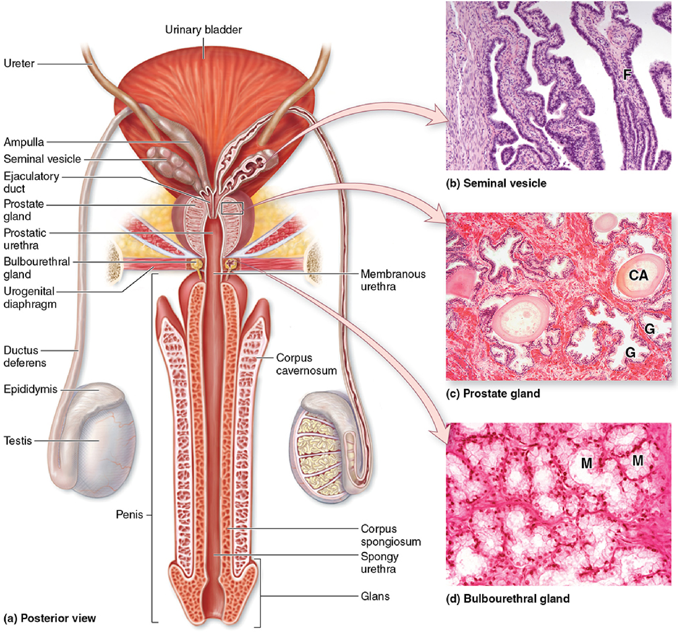
- Junqueira's Basic Histology, 17e (Accessory Glands - Prostate)
- Campbell Walsh Wein Urology (Prostatitis - Histology)
- Smith and Tanagho's General Urology, 19e (Prostatic and Seminal Vesicle Stones)
Corpora Amylacea in the Optic Disc (Ophthalmology)
In ophthalmology, the term is used for drusen-like crystalline deposits on the optic disc surface, seen in chronic optic disc conditions (e.g., chronic papilledema). They appear as calcified concretions and can be associated with optociliary shunt vessels.
- Kanski's Clinical Ophthalmology, 10e
Adult Polyglucosan Body Disease (APBD) - Special Entity
This is a distinct multisystem neurologic disease where corpora amylacea (called polyglucosan bodies in this context) are deposited in massive quantities throughout the nervous system:
- Genetics: Most commonly in Ashkenazi Jewish individuals due to glycogen branching enzyme (GBE) deficiency (same enzyme deficient in glycogenosis type IV / Anderson disease)
- Presentation: Slowly progressive motor + sensory loss in the legs (axonal polyneuropathy), neurogenic bladder, upper motor neuron signs (can mimic ALS), and sometimes dementia
- Biopsy: Sural nerve biopsy demonstrates profuse deposition of polyglucosan bodies in the endoneurium
- Distinction: In normal aging, small numbers of corpora amylacea in the brain are considered innocuous; massive deposition = pathologic
- Adams and Victor's Principles of Neurology, 12e (Polyglucosan Disease)
- Localization in Clinical Neurology, 8e
Analogous Structures
| Body | Location | Composition | Disease |
|---|---|---|---|
| Corpora amylacea (CNS) | Astrocytic end-processes | Polyglucosan + heat-shock proteins + ubiquitin | Normal aging, AD, APBD |
| Lafora bodies | Neurons, hepatocytes, myocytes | Identical structure and biochemistry | Lafora body disease (myoclonic epilepsy) |
| Polyglucosan bodies (APBD) | Nervous tissue, widespread | Same as corpora amylacea | Adult polyglucosan body disease |
| Corpora amylacea (prostate) | Prostatic glandular lumina | Condensed secretions + sloughed cells | Normal; may calcify into prostatic calculi |
Summary Table
| Feature | CNS | Prostate |
|---|---|---|
| Nature | Astrocytic inclusion (degenerative) | Secretory concretion |
| Composition | Polyglucosan + heat-shock proteins | Prostatic secretions + epithelial debris |
| Staining | PAS+, faintly basophilic | Concentrically laminated on H&E |
| Size | 5-50 µm | Variable |
| Significance | Age-related, increased in AD, massive in APBD | Normal; rarely obstructive; may calcify |
This is a shared conversation. Sign in to Orris to start your own chat.