
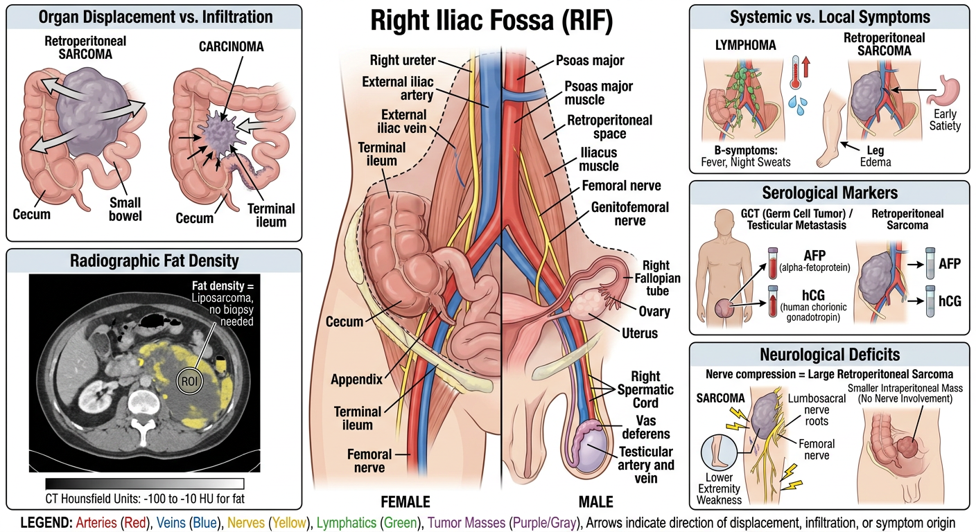
Organ Displacement vs. Infiltration: Sarcomas typically displace adjacent intra-abdominal organs, whereas carcinomas are more likely to invade or infiltrate them. Systemic vs. Local Symptoms: Presence of fevers and night sweats (B-symptoms) suggests lymphoma, while sarcomas primarily cause local compression symptoms (edema, early satiety). Radiographic Fat Density: The detection of variable fat density on CT/MRI is highly specific for liposarcoma, often removing the need for a tissue biopsy. Serological Markers: Elevated AFP and hCG in males distinguish germ cell tumors or testicular metastases from primary sarcomas. Neurological Deficits: Development of lower extremity paresthesia or paresis indicates nerve root compression, a feature more common in large retroperitoneal sarcomas than in intraperitoneal RIF masses. Make a image describing all this with all related structures in rif region
Detailed medical educational diagram of the Right Iliac Fossa (RIF) region showing differential diagnosis concepts for retroperitoneal and intra-abdominal masses. The image should be a clean, labeled anatomical illustration divided into clear sections: 1. CENTRAL ANATOMY: Show the RIF region with labeled structures: cecum, appendix, terminal ileum, right ureter, iliac vessels (external iliac artery and vein), psoas muscle, iliacus muscle, femoral nerve, genitofemoral nerve, right ovary/fallopian tube (female), right spermatic cord structures, retroperitoneal space boundary. 2. TOP LEFT PANEL - "Organ Displacement vs. Infiltration": Show a large retroperitoneal SARCOMA pushing/displacing the cecum and adjacent bowel anteriorly (arrows showing displacement, intact organ borders). Next to it, show a CARCINOMA with irregular spiculated margins invading into adjacent bowel wall (arrows showing infiltration, disrupted borders). Label clearly. 3. TOP RIGHT PANEL - "Systemic vs. Local Symptoms": Show LYMPHOMA with enlarged lymph nodes, fever/sweat icons (B-symptoms: thermometer, sweat drops). Show SARCOMA with compression arrows causing leg edema (swollen leg outline) and stomach compression (early satiety icon). Label clearly. 4. MIDDLE LEFT PANEL - "Radiographic Fat Density": CT cross-section appearance showing liposarcoma with heterogeneous fat density regions (yellow/low-density areas mixed with soft tissue), labeled "Fat density = Liposarcoma, no biopsy needed". Include CT Hounsfield unit values (-100 to -10 HU for fat). 5. MIDDLE RIGHT PANEL - "Serological Markers": Show male figure with testicular germ cell tumor / testicular metastasis, with lab vial icons showing elevated AFP (alpha-fetoprotein) and hCG markers with upward arrows. Compare to sarcoma marker panel showing normal AFP/hCG. Label "GCT vs. Primary Sarcoma". 6. BOTTOM PANEL - "Neurological Deficits": Show large retroperitoneal sarcoma compressing the lumbosacral nerve roots / femoral nerve, with lightning bolt symbols indicating paresthesia, and lower extremity weakness icon. Compare smaller intraperitoneal RIF mass without nerve involvement. Label "Nerve compression = Large Retroperitoneal Sarcoma". Style: Professional medical textbook illustration, white background, anatomical color coding (arteries red, veins blue, nerves yellow, lymphatics green, tumor masses in purple/gray), clear bold labels, arrows for relationships, clean sans-serif font. Include a legend/key at the bottom.
| Panel | Concept Illustrated |
|---|---|
| Top Left | Displacement vs. Infiltration — sarcoma pushes bowel aside with intact margins; carcinoma invades with spiculated, irregular borders |
| Top Right | Systemic vs. Local Symptoms — lymphoma with B-symptom icons (fever, night sweats); sarcoma with compression effects (edema, satiety) |
| Middle Left | Radiographic Fat Density — CT cross-section of liposarcoma showing heterogeneous fat (−100 to −10 HU), removing biopsy need |
| Middle Right | Serological Markers — elevated AFP/hCG in males pointing to GCT/testicular metastasis vs. normal markers in sarcoma |
| Bottom | Neurological Deficits — large retroperitoneal sarcoma compressing femoral/lumbosacral nerves with paresthesia symbols; contrast with smaller intraperitoneal RIF mass sparing nerves |