Anatomy -Layers and internal structure of the eyeball
Reading File
Finding Sources
Finding Sources
Searching Images
eyeball layers internal structure anatomy cross section
Finding Sources
Finding Sources
Reading File
Finding Sources
Reading File
Loading Image
Loading Image
Reading File
Reading File
Loading Image
Reading File
Loading Image
I now have all the content needed for a comprehensive answer. Let me compile it:
Layers and Internal Structure of the Eyeball
The eyeball is a tough, fibrous globe approximately 22 mm in diameter that maintains overall shape while containing transparent refracting media, a photosensitive layer, and a neural processing system. It has three concentric tunics (layers), plus non-layered internal contents.
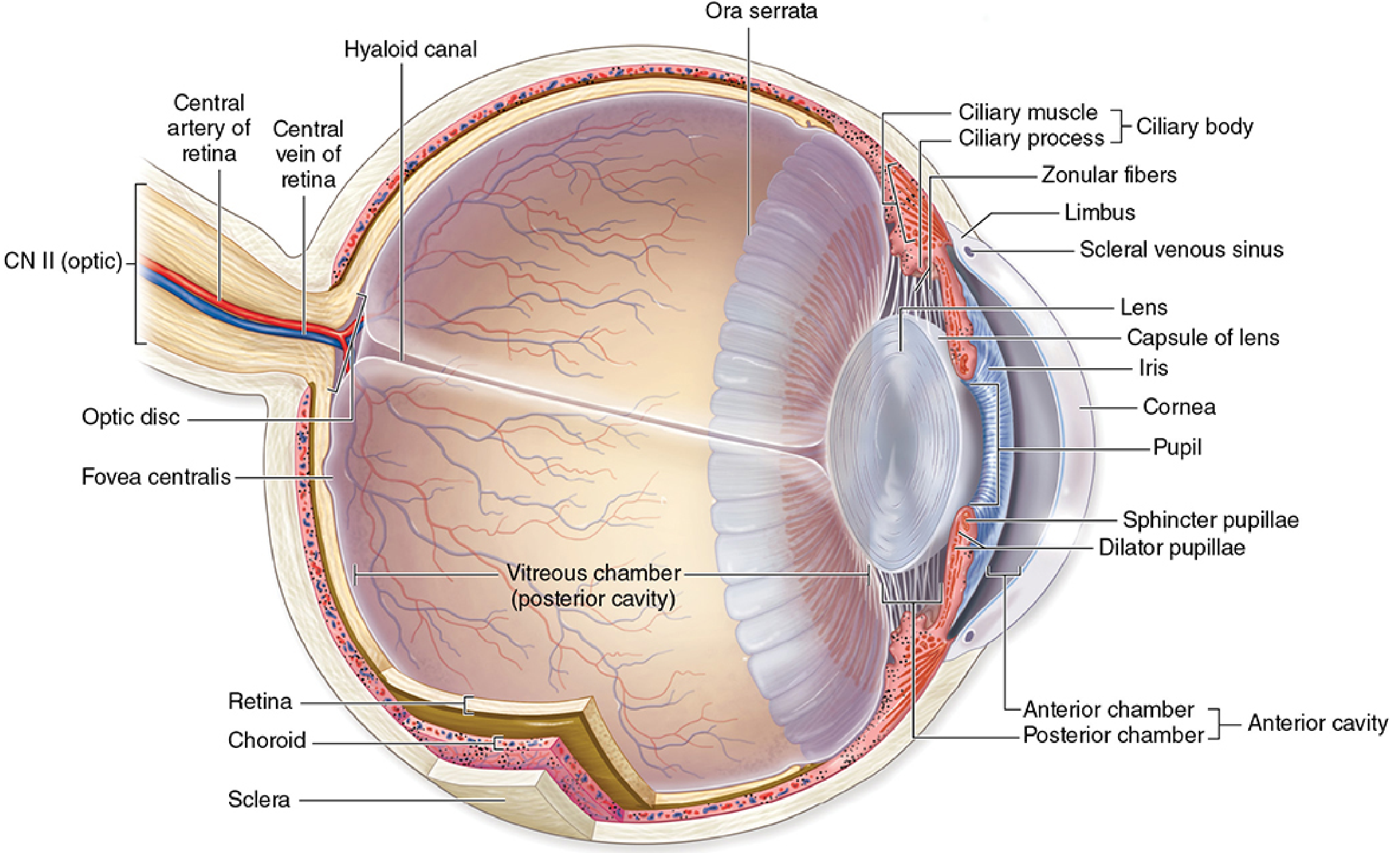
The Three Tunics
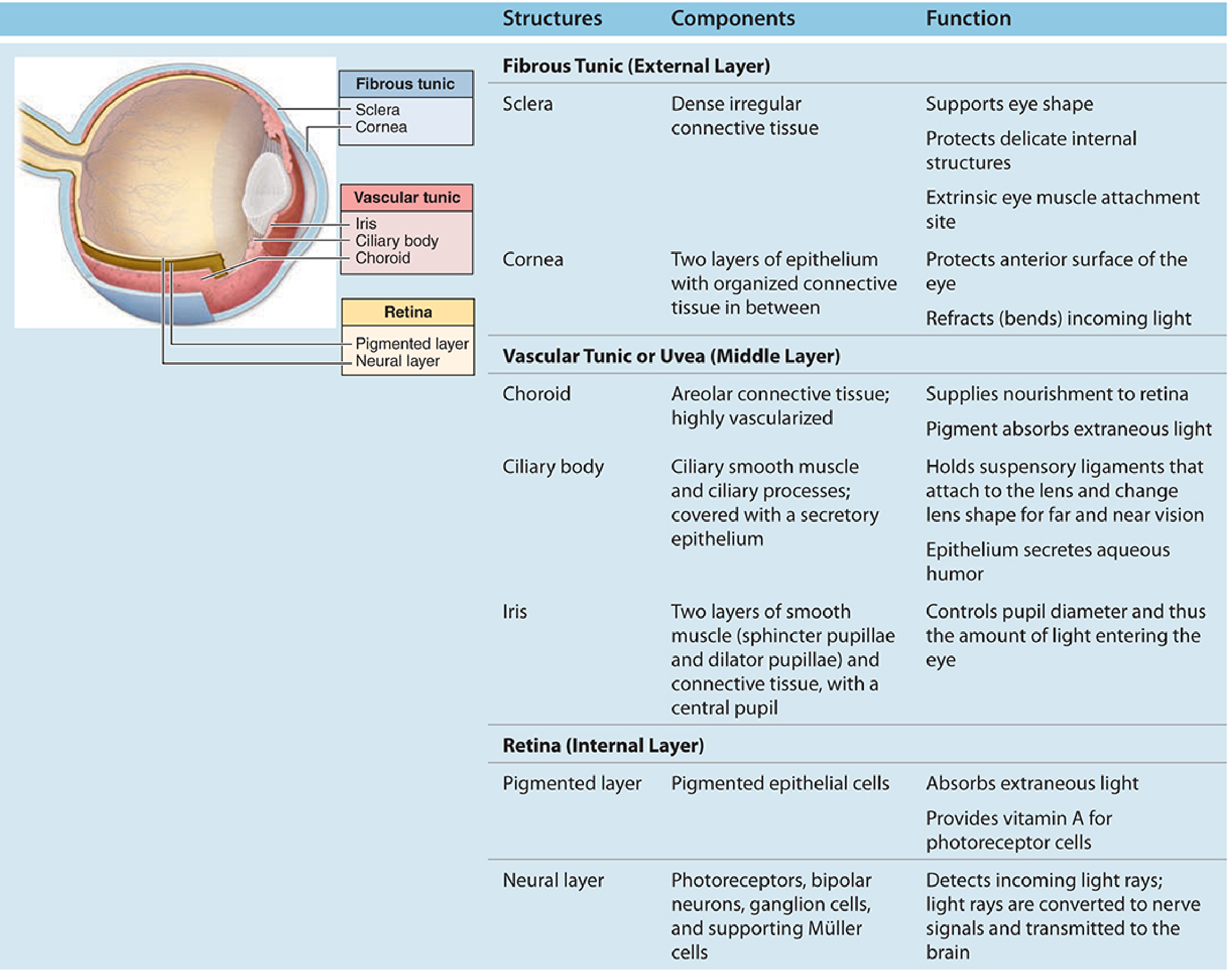
1. Fibrous Tunic (External Layer)
The outermost layer provides structural support and protects delicate internal contents. It has two regions joined at the limbus:
Sclera
- The posterior 5/6 of the fibrous layer
- Opaque, whitish, ~0.5 mm thick (thickens to ~1 mm posteriorly)
- Composed of dense connective tissue — flat bundles of type I collagen oriented parallel to the surface but intersecting in multiple directions
- Microvasculature is mainly in the outer surface (episclera)
- Tendons of the six extraocular muscles insert into the anterior sclera
- Posteriorly joins with the epineurium of the optic nerve
- Inner surface: suprachoroidal lamina — less collagen, more fibroblasts, elastic fibers, and melanocytes
Cornea
- The anterior 1/6 of the eye; completely transparent and avascular
- Has five distinct layers (from anterior to posterior):
| Layer | Description |
|---|---|
| Corneal epithelium | Nonkeratinized stratified squamous; 5–6 cells thick; richly supplied with sensory nerve endings (triggers blink reflex); stem cells replenish from the limbus |
| Bowman's membrane | Thick anterior limiting membrane (basement membrane of the epithelium) |
| Stroma | ~90% of corneal thickness; ~60 alternating layers of type I collagen in a precise orthogonal array; interspersed with flat keratocytes; completely avascular — nutrients diffuse from limbus and aqueous humor |
| Descemet's membrane | Thick posterior limiting membrane (basement membrane of endothelium) |
| Corneal endothelium | Simple squamous epithelium; actively pumps fluid to maintain corneal transparency |
2. Vascular Tunic / Uvea (Middle Layer)
Highly vascular, pigmented; three components from posterior to anterior:
Choroid
- Located in the posterior 2/3 of the eye, between sclera and retina
- Consists of well-vascularized connective tissue with large flattened melanocytes that block excess light penetrating the sclera
- Two main sublayers:
- Choroidocapillary lamina (inner): rich microvasculature that nourishes the outer retinal layers
- Bruch's membrane (basal complex): a layer of elastin sandwiched between two sheets of collagen, continuous with the basement membranes of the capillaries and retinal pigment epithelium
Ciliary Body
- Anterior expansion of the uvea that encircles the lens; lies posterior to the limbus
- Major components:
- Ciliary muscle: smooth muscle in three complementary orientations; contraction/relaxation changes lens curvature (accommodation)
- Ciliary processes: finger-like folds covered by a bilaminar cuboidal epithelium (inner nonpigmented + outer pigmented); they secrete aqueous humor into the posterior chamber
- Ciliary zonule (zonular fibers): microfibrils connecting the ciliary body to the lens capsule — tension regulated by ciliary muscle to change lens shape
- When ciliary muscles relax → zonule taut → lens flattens (distant vision)
- When ciliary muscles contract → zonule loosens → lens rounds up (near vision / accommodation)
Iris
- Opaque, pigmented extension of the uvea anterior to the lens
- Surrounds the central aperture: the pupil
- Contains two layers of smooth muscle:
- Sphincter pupillae: circular muscle — constricts pupil (parasympathetic)
- Dilator pupillae: radial muscle — dilates pupil (sympathetic)
- Separates the anterior chamber (in front) from the posterior chamber (behind)
3. Retina (Inner Sensory Layer)
The innermost tunic, extending from the optic nerve posteriorly to the ora serrata anteriorly. Communicates with the brain via the optic nerve.
The retina has two main parts:
Pigmented Layer (RPE)
- Single layer of cuboidal epithelial cells containing melanin
- Rests on Bruch's membrane of the choroid
- Functions: absorbs stray light, recycles photopigments, provides vitamin A to photoreceptors
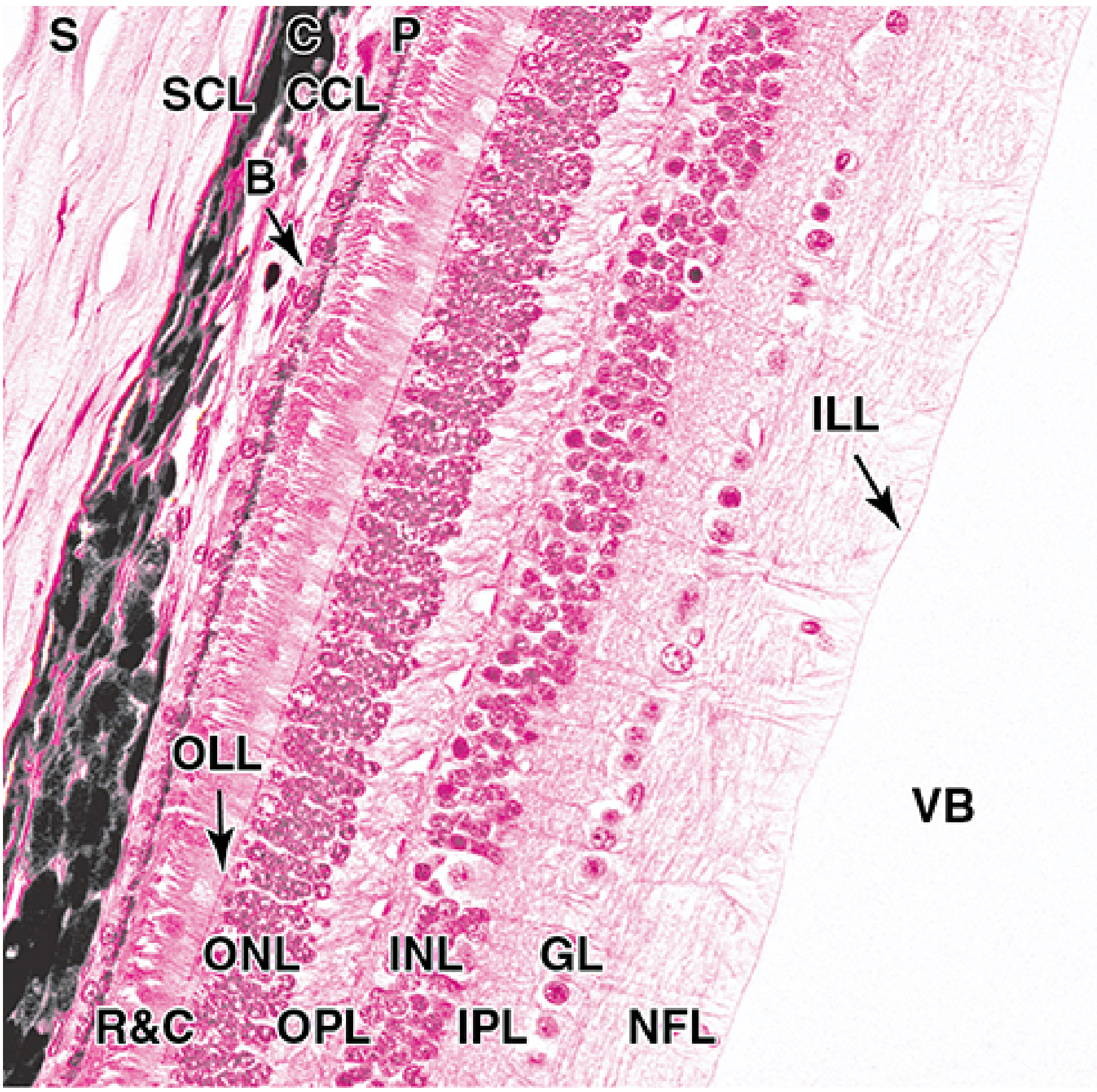
Neural Layer — 9 Distinct Sublayers (outer → inner)
| Layer | Contents |
|---|---|
| Photoreceptor layer (R&C) | Outer segments of rods and cones |
| Outer limiting layer (OLL) | Junctional complexes between photoreceptors and Müller glial cells |
| Outer nuclear layer (ONL) | Cell bodies (nuclei) of rods and cones |
| Outer plexiform layer (OPL) | Synapses between photoreceptor axons and bipolar/horizontal cell dendrites |
| Inner nuclear layer (INL) | Cell bodies of bipolar cells, horizontal cells, amacrine cells, and Müller glial cells |
| Inner plexiform layer (IPL) | Synapses between bipolar/amacrine cell axons and ganglion cell dendrites |
| Ganglionic layer (GL) | Cell bodies of ganglion cells |
| Nerve fiber layer (NFL) | Axons of ganglion cells converging toward the optic disc |
| Inner limiting layer (ILL) | Basement membrane of Müller cells — boundary with vitreous body |
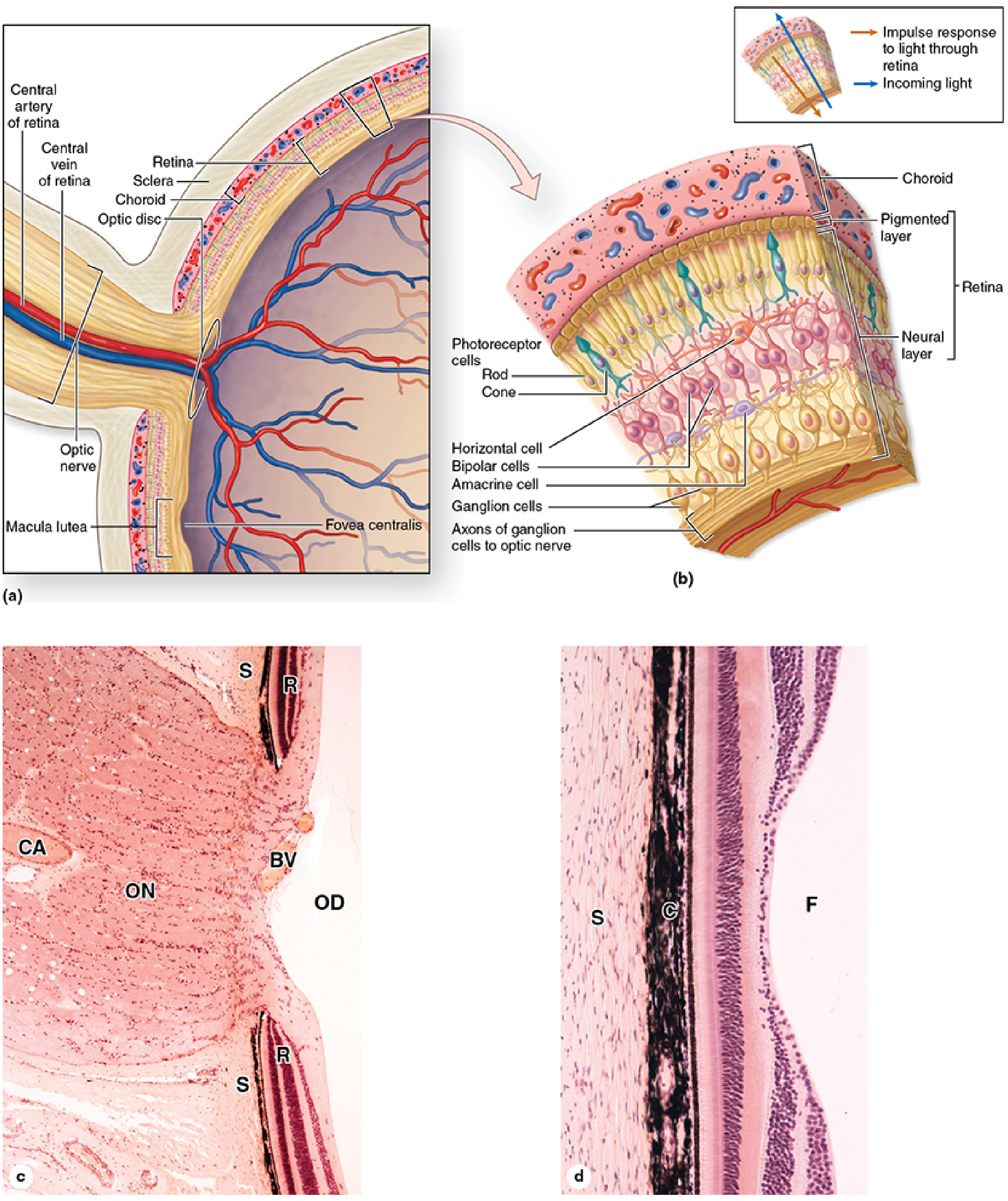
Special Regions of the Retina
| Region | Features |
|---|---|
| Optic disc | Where ganglion cell axons converge and leave as the optic nerve; also where the central retinal artery and vein enter/leave; has no photoreceptors → "blind spot" |
| Macula lutea | Area of central retina with highest cone density and thickest ganglionic layer |
| Fovea centralis | Depression within the macula; contains only cones (no rods); ganglionic and inner nuclear layers are displaced peripherally allowing light to hit cones directly → highest visual acuity |
Internal Contents (Non-layered)
Lens
- Transparent, biconvex, avascular, elastic structure suspended by the ciliary zonule
- Three components:
- Lens capsule: very thick (~10–20 µm) basement membrane of type IV collagen surrounding the entire lens
- Lens epithelium: single layer of cuboidal cells on the anterior surface only; proliferates at the equator to form new lens fibers
- Lens fibers: densely packed, anucleate, elongated cells filled with crystallins — the basis of transparency
- Loses elasticity with age → presbyopia (typically after 4th decade)
Aqueous Humor
- Clear fluid secreted by the ciliary process epithelium
- Fills the anterior chamber (between cornea and iris) and posterior chamber (between iris and lens)
- Flows through the pupil from posterior to anterior chamber, then drains via the scleral venous sinus (canal of Schlemm) at the iridocorneal angle
Vitreous Body
- Large gelatinous mass of transparent connective tissue filling the posterior (vitreous) chamber behind the lens
- Occupies ~80% of the eyeball's volume
- Contains the hyaloid canal — a remnant of the fetal hyaloid artery
Summary Table
| Tunic | Component | Key Structure | Function |
|---|---|---|---|
| Fibrous | Sclera | Type I collagen bundles | Shape, protection, muscle attachment |
| Fibrous | Cornea | Stroma + 5 layers | Transparency, light refraction |
| Vascular | Choroid | Choroidocapillary lamina + Bruch's membrane | Nutrition of retina, light absorption |
| Vascular | Ciliary body | Ciliary muscle + processes | Accommodation, aqueous humor secretion |
| Vascular | Iris | Sphincter/dilator pupillae | Pupil size control, light regulation |
| Retina | RPE | Melanin-filled cuboidal cells | Light absorption, photopigment recycling |
| Retina | Neural layer | Rods, cones, bipolar, ganglion cells | Phototransduction and visual signal processing |
Source: Junqueira's Basic Histology — Text and Atlas, 17e, Chapter 23 (Eyes: The Photoreceptor System)
This is a shared conversation. Sign in to Orris to start your own chat.