
Inflamed uncinate process of nose
Reading File
The uncinate process is a sickle-shaped bony projection of the lateral nasal wall that plays a key role in sinus drainage. When inflamed, it typically refers to uncinitis - inflammation as part of rhinosinusitis or as a primary finding. Let me search the medical library for detailed information.
Searching sources...
Finding Sources
Finding Sources
Reading File
Reading File
Finding Sources
Finding Sources
Reading File
Finding Sources
Finding Sources
Finding Sources
Reading File
Finding Sources
Reading File
Loading Image
Finding Sources
Now I have comprehensive information. Let me compile the full answer.
Inflamed Uncinate Process of the Nose
Anatomy
The uncinate process is a crescent/sickle-shaped, sagittally oriented bony projection of the ethmoid bone located on the lateral nasal wall. Key structural features:
- Measures approximately 3-4 mm wide and 1.5-2 cm in length
- Its posterior margin is free, forming the anterior boundary of the hiatus semilunaris
- It is the medial wall of the ethmoidal infundibulum - the funnel-shaped 3D space that drains the maxillary, frontal, and anterior ethmoid sinuses
- The maxillary sinus natural ostium lies lateral and superior to its inferior aspect
- Anteriorly attaches to the ethmoidal crest of the maxilla and lacrimal bone; posteriorly to the ethmoidal process of the inferior turbinate and palatine bones
Borders of the ethmoidal infundibulum:
- Medial: uncinate process
- Lateral: lamina papyracea
- Posterior: anterior wall of ethmoid bulla
- Anterior/superior: frontal process of maxilla
(K J Lee's Essential Otolaryngology; Cummings Otolaryngology)
Role in the Osteomeatal Complex (OMC)
The uncinate process is the key structural component of the OMC - the final common pathway for drainage and ventilation of the ethmoidal, maxillary, and frontal sinuses. It is the first structure encountered in the middle meatus when the middle turbinate is medialized on endoscopy. Inflammation or obstruction here causes a cascade of sinus disease.
Inflammation of the Uncinate Process (Uncinitis)
Although "uncinitis" as an isolated diagnosis is rarely used, inflammation of the uncinate process is central to the pathophysiology of chronic rhinosinusitis (CRS) and occurs in the following contexts:
1. Causes / Contributing Factors
- Chronic rhinosinusitis - mucosal edema and inflammation affect the uncinate as part of OMC disease
- Acute rhinosinusitis - viral or bacterial infection causes mucosal swelling of the uncinate, obstructing the infundibulum
- Anatomical variants predisposing to inflammation:
- Atelectatic uncinate process - the free edge adheres to the lamina papyracea/orbital floor, obliterating the infundibulum -> causes hypoplastic, chronically opacified maxillary sinus (linked to Silent Sinus Syndrome)
- Pneumatized uncinate - can narrow the ethmoidal infundibulum
- Bifid uncinate - anatomical variation
- Paradoxical curvature - can obstruct the middle meatus
(Cummings Otolaryngology - "Variations in the Uncinate Process")
2. Consequences of Obstruction/Inflammation
When the uncinate process is inflamed or its position is abnormal, drainage of the anterior sinuses is compromised:
- Maxillary sinusitis - most common consequence, as the natural ostium is blocked
- Frontal sinusitis - drainage depends on the uncinate's superior attachment; inflammation disrupts the frontal recess
- Anterior ethmoidal sinusitis
- Silent Sinus Syndrome - atelectatic uncinate leads to chronic occlusion, negative pressure, and progressive maxillary sinus collapse
CT Findings
Coronal CT sinus is the gold standard for evaluating the uncinate:
- Mucosal thickening along the uncinate and infundibulum
- Opacification of the ethmoidal infundibulum
- Associated ipsilateral maxillary or ethmoid sinus opacification
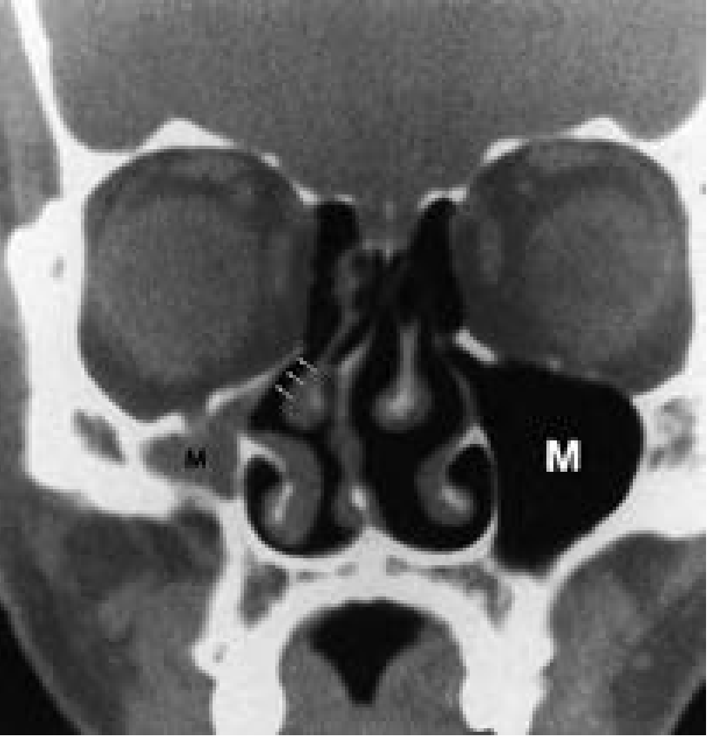
- Atelectatic uncinate: the uncinate is retracted laterally against the lamina papyracea (as shown in the CT image below - right side has normal large maxillary sinus "M", while left shows atelectatic variant)
Management
Medical (first-line)
- Intranasal corticosteroids - reduce mucosal edema of the uncinate and OMC
- Saline irrigation - aids mucociliary clearance
- Antibiotics - if acute bacterial superinfection
- Antihistamines - if allergic component
Surgical - Functional Endoscopic Sinus Surgery (FESS)
When medical therapy fails, the cornerstone of surgery is uncinectomy (resection of the uncinate process):
Technique (Retrograde Uncinectomy - preferred):
- Zero-degree endoscope used; uncinate identified as sickle-shaped structure with a posterior free margin
- A ball-tipped probe is slid into the infundibulum behind the free edge to medialize the uncinate, away from the lamina papyracea
- A pediatric backbiter punch is used to retrogradely incise the uncinate via the hiatus semilunaris (at junction of inferior 1/3 and superior 2/3) - this avoids orbital penetration
- Incision continued anteriorly until the lacrimal bone (anterior maxillary line) is reached - caution: penetration risks lacrimal duct injury
- Middle third is removed with cutting instruments or microdebrider
- Superior third may be preserved initially (to prevent frontal recess scarring)
- Inferior aspect is rotated medially and removed
Uncinectomy then exposes the infundibulum and allows access to the maxillary sinus natural ostium for antrostomy.
(Cummings Otolaryngology - "Resection of the Uncinate Process")
Key Clinical Points Summary
| Feature | Detail |
|---|---|
| Structure | Sickle-shaped ethmoid bone, medial wall of ethmoidal infundibulum |
| Critical role | OMC drainage of maxillary, frontal, anterior ethmoid sinuses |
| Key variant | Atelectatic UP -> Silent Sinus Syndrome |
| Inflammation consequence | OMC obstruction -> multi-sinus disease |
| CT diagnosis | Coronal CT paranasal sinuses |
| Surgery | Uncinectomy (retrograde preferred) as first step of FESS |
This is a shared conversation. Sign in to Orris to start your own chat.