
Rheumatoid Arthritis and ILD
"rheumatoid arthritis" AND "interstitial lung disease"
rheumatoid arthritis interstitial lung disease HRCT UIP pattern
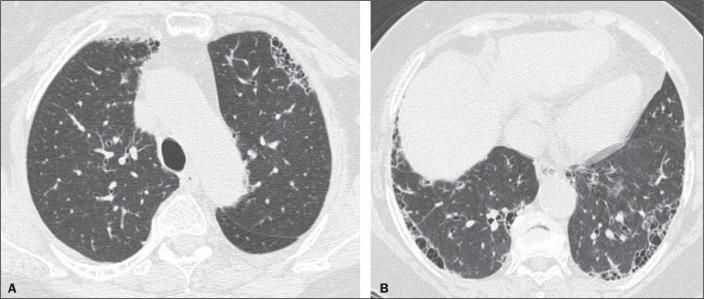
High-resolution computed tomography (HRCT) of the chest in axial views (A and B) demonstrating a typical Usual Interstitial Pneumonia (UIP) pattern. Image A (upper level) and Image B (lower level) reveal characteristic features of fibrosing interstitial lung disease. Key findings include prominent subpleural honeycombing, characterized by clustered, thick-walled cystic airspaces measuring 3-10 mm in diameter. These are associated with reticular opacities and architectural distortion. The distribution is classically peripheral and basilar-predominant, with Image B showing more extensive involvement of the lower lobes compared to the upper zones in Image A. Traction bronchiectasis and subtle ground-glass opacities are also visible within the areas of fibrosis. This pattern is consistent with a multidisciplinary diagnosis of UIP, which in this case is associated with rheumatoid arthritis-related interstitial lung disease.
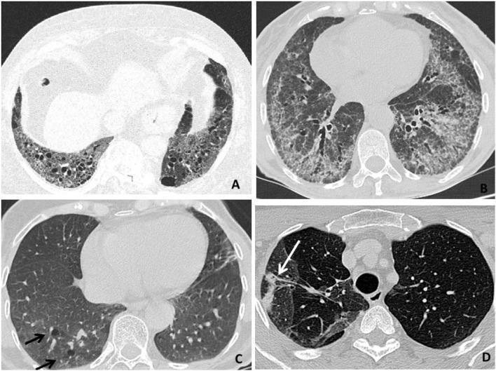
This composite of four axial high-resolution computed tomography (HRCT) scans illustrates various radiologic patterns of Rheumatoid Arthritis-associated Interstitial Lung Disease (RA-ILD). Panel A demonstrates a Usual Interstitial Pneumonia (UIP) pattern, characterized by bibasilar, subpleural honeycombing (clusters of cystic airspaces), reticular opacities, and traction bronchiectasis. Panel B displays a Nonspecific Interstitial Pneumonia (NSIP) pattern, showing diffuse, patchy ground-glass opacities and septal thickening with relative subpleural sparing. Panel C reveals a Lymphocytic Interstitial Pneumonia (LIP) pattern, highlighted by perivascular thin-walled lung cysts (indicated by black arrows). Panel D illustrates an Organizing Pneumonia (OP) pattern, featuring focal areas of parenchymal consolidation (indicated by a white arrow). These images serve as educational benchmarks for differentiating pulmonary manifestations in systemic autoimmune diseases. The radiological findings are critical for determining prognosis and guiding therapeutic strategies, such as distinguishing between predominantly fibrotic (UIP) versus inflammatory (NSIP/OP) processes.
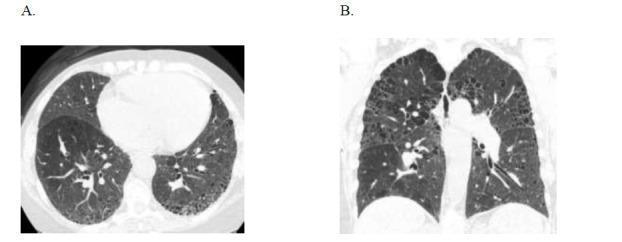
These axial (A) and coronal (B) High-Resolution Computed Tomography (HRCT) images of the chest demonstrate a classic radiographic pattern of Usual Interstitial Pneumonia (UIP). The predominant finding is subpleural honeycombing, characterized by clusters of cystic airspaces with well-defined walls, primarily localized along the costal and fissural pleural surfaces. This honeycombing is most extensive in the lower lung zones and bases. Additional findings include mild biapical paraseptal emphysema, visualized as small subpleural lucencies at the lung apices. The lung architecture is distorted by these fibrotic changes, leading to the replacement of normal parenchyma with cystic spaces. This pattern is characteristic of interstitial lung disease associated with connective tissue disorders, such as rheumatoid arthritis. The imaging is critical for differentiating UIP from other patterns like Non-Specific Interstitial Pneumonitis (NSIP) based on the presence of macrocystic honeycombing and its peripheral, basal-predominant distribution.
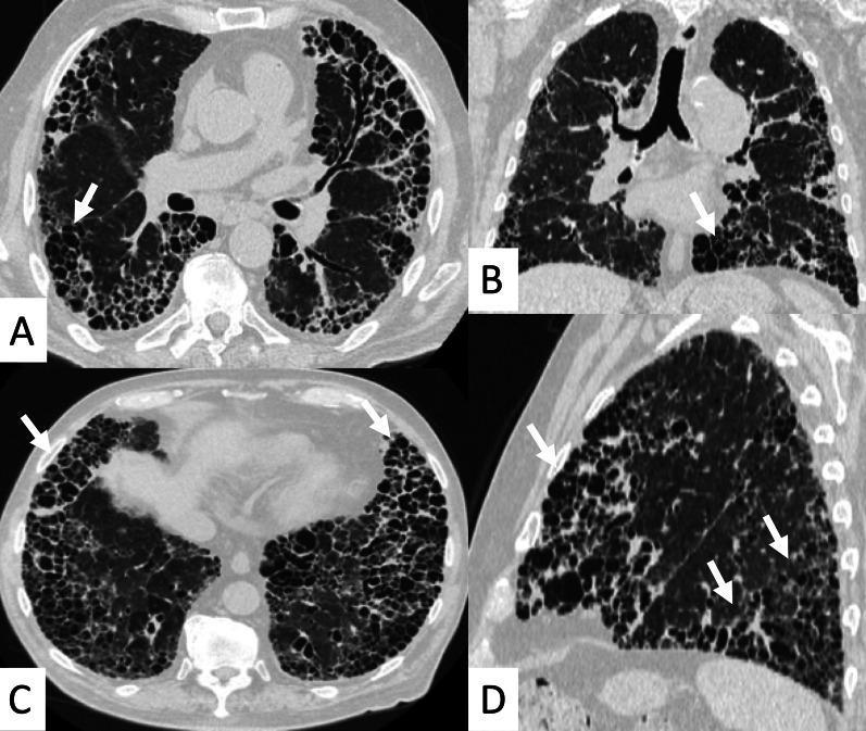
High-resolution computed tomography (HRCT) of the chest in axial (A, C), coronal (B), and sagittal (D) planes demonstrating advanced interstitial lung disease. The images show diffuse, extensive honeycombing involving more than 50% of the lung parenchyma. This finding is characterized by the 'exuberant honeycombing sign,' featuring hypertrophic, well-defined macrocystic spaces (white arrows) that are larger than those seen in typical honeycomb patterns. The cystic changes are most prominent in the peripheral and subpleural regions of both lungs, with a notable basal predominance. These findings represent a Usual Interstitial Pneumonia (UIP) pattern in the context of Rheumatoid Arthritis-associated Interstitial Lung Disease (RA-ILD). Associated features include marked architectural distortion and the absence of significant ground-glass opacities, indicating end-stage fibrotic disease.
Rheumatoid Arthritis-Associated Interstitial Lung Disease (RA-ILD)
Overview
Epidemiology & Prevalence
- Clinically evident RA-ILD: ~10% of RA patients (national registry data); up to 10-30% in referral populations
- Subclinical disease detectable on HRCT: 20-60% of RA patients
- Subclinical RA-ILD is not benign - patients already show respiratory symptoms and functional decrements, and a subset progresses to clinically significant pulmonary fibrosis
- Bimodal incidence: most patients develop ILD ~1 decade after articular disease onset; a minority develop it shortly after joint disease appears; rarely, ILD is the first manifestation of RA
Risk Factors
| Risk Factor | Notes |
|---|---|
| Male sex | Consistently associated with both incidence and worse prognosis |
| Older age | Both a risk factor for development and for mortality |
| Smoking | Strong independent risk factor; may be an initiating site for RA-related autoimmunity |
| High-titer RF | Seropositive disease carries higher risk |
| Anti-CCP antibodies | Higher titers associated with RA-ILD; citrullination in lung tissue may be a disease initiator |
| MUC5B promoter variant | A genetic variant strongly associated with RA-ILD (shared with IPF) |
| UIP pattern | Associated with worse prognosis compared to NSIP |
| Low baseline lung function | Predicts increased mortality |
Pathogenesis
- Autoimmune-mediated inflammation: The lung may be the initiating site of RA-related autoimmune injury. Early inflammatory airway involvement has been found in anti-CCP+ subjects even without clinically evident arthritis
- Citrullination: Anti-CCP antibodies and protein citrullination appear to play a pathogenic role in pulmonary fibrosis
- Pro-fibrotic cytokines: TGF-β, PDGF, and endothelin-1 drive progression from inflammation to fibrosis
- Genetic susceptibility: MUC5B promoter variant, HLA associations
Histopathologic & Radiologic Patterns
| Pattern | Frequency in RA-ILD | Key Features | Prognosis |
|---|---|---|---|
| UIP | Most common (~50-60%) | Peripheral, subpleural honeycombing; reticular opacities; traction bronchiectasis; basal predominance | Worse; progressive fibrosis |
| NSIP | Second most common | Bilateral ground-glass opacities; peribronchovascular distribution; relative subpleural sparing | Better than UIP |
| OP (Organizing Pneumonia) | Less common | Focal/patchy/diffuse alveolar consolidation | Steroid-responsive |
| LIP | Rare; associated with Sjögren overlap | Perivascular thin-walled lung cysts | Variable |
| DAD | Rare, acute | Diffuse alveolar damage pattern | Very poor |
| Follicular bronchiolitis | Rare | Centrilobular nodules on HRCT | Variable |
Key point: UIP is the most common pattern in RA-ILD - more prevalent than in any other connective tissue disease. However, the prognosis for RA-UIP is considered better than idiopathic UIP (IPF), though long-term prognosis remains poor. (Fishman's Pulmonary Diseases; Grainger & Allison's Diagnostic Radiology)
HRCT Imaging - RA-ILD Patterns
Intrathoracic Manifestations of Rheumatoid Disease (Full Spectrum)
- Pleural effusion or thickening - most common chest manifestation; usually small, unilateral or bilateral, most resolve spontaneously; pleuritic pain in ≥20%
- Interstitial fibrosis (UIP-type) - most clinically significant
- Bronchiectasis - seen in up to 30% on chest CT
- Obliterative (constrictive) bronchiolitis - obstructive physiology; associated with gold, penicillamine therapy; severe cases require lung transplant
- Organizing pneumonia - may precede arthritis; alveolar opacities; steroid-responsive
- Follicular bronchiolitis - centrilobular nodules on HRCT
- Rheumatoid nodules - incidental; cavitation can cause hemoptysis or pneumothorax
- Caplan syndrome - rheumatoid nodules + coal miner's pneumoconiosis
- Drug-induced lung disease - especially methotrexate pneumonitis (hypersensitivity pattern)
- Pulmonary hypertension - secondary to hypoxic vasoconstriction in advanced ILD
Clinical Presentation
- Symptoms: Progressive exertional dyspnea, dry cough (often insidious onset)
- Physical exam: Bibasilar fine crackles ("Velcro" crackles), digital clubbing, features of cor pulmonale in advanced disease
- Note: Pulmonary symptoms may precede the articular manifestations by months to years in some patients
Diagnosis & Workup
| Investigation | Findings |
|---|---|
| HRCT chest | Gold standard; identifies pattern (UIP vs NSIP vs OP), distribution, extent |
| PFTs | Restrictive pattern: reduced FVC, TLC, DLCO; serial monitoring for progression |
| BAL | Lymphocytosis (NSIP/LIP); neutrophilia/eosinophilia (UIP); rule out infection |
| Serology | RF, anti-CCP (high titers support diagnosis); ANA, anti-Ro |
| Biomarkers | KL-6, SP-D elevated in active ILD; useful for monitoring |
| Surgical lung biopsy | Reserved for diagnostic uncertainty; confirms histologic pattern |
| Echocardiography | Screen for pulmonary hypertension |
Management
Inflammatory Patterns (NSIP, OP, LIP)
- Corticosteroids - first-line; OP is typically steroid-responsive
- Mycophenolate mofetil (MMF) - effective for inflammatory ILD; first-line maintenance
- Azathioprine - alternative immunosuppressant
- Rituximab (anti-CD20) - emerging evidence for refractory RA-ILD
- Tocilizumab (anti-IL-6) - data emerging
- Cyclophosphamide - reserved for severe/refractory disease
Fibrotic/Progressive Patterns (UIP)
- Nintedanib - antifibrotic; demonstrated to slow decline in FVC in CTD-ILD (INBUILD trial); now a standard of care for progressive RA-ILD
- Pirfenidone - Phase II (TRAIL1, NCT02808871) evaluated pirfenidone in RA-ILD; results were inconclusive for definitive recommendation but showed trends toward benefit
General Principles
- Treat the underlying RA - tight disease control may reduce ILD progression
- Caution with methotrexate - can cause drug-induced pneumonitis (must distinguish from RA-ILD progression); generally avoid in established RA-ILD
- Leflunomide - also associated with pulmonary toxicity; use with caution
- TNF inhibitors - may occasionally trigger drug-induced ILD; conversely, some data suggest benefit in certain RA-ILD phenotypes
- Lung transplantation - for selected patients with progressive, end-stage disease (also indicated in refractory obliterative bronchiolitis)
- Pulmonary rehabilitation - improves exercise tolerance and quality of life
- Oxygen supplementation - for resting/exertional hypoxia
Prognosis
- UIP pattern: 5-year mortality ~35-40%; worse than NSIP but better than idiopathic IPF
- NSIP pattern: More favorable; better response to immunosuppression
- Predictors of mortality: Older age, male sex, UIP pattern, low baseline FVC/DLCO, significant decline in FVC at follow-up
- Subclinical RA-ILD: A meaningful proportion progress to clinically significant disease; warrants monitoring
Recent Evidence (2024-2025)
| Study | Key Finding |
|---|---|
| Prasanna et al., Rheumatol Int 2025 (PMID: 39825929) | Global prevalence ~20%; significant geographic variation |
| Wang et al., Ann Med 2024 (PMID: 38547537) | Pooled prevalence 21%; male sex, smoking, high RF/anti-CCP as consistent risk factors |
| Yu et al., BMJ Open Respir Res 2024 (PMID: 39551575) | Male sex, older age, smoking, high RF, anti-CCP positivity confirmed as independent risk factors for ILD incidence |
| Guo et al., Front Immunol 2024 (PMID: 39534599) | KL-6 and SP-D are the most predictive biomarkers; meta-analysis of 30+ studies |
Summary
- UIP is the dominant pattern in RA-ILD (unlike most other CTDs where NSIP predominates)
- ILD can precede joint disease - always consider RA in a patient with UIP pattern and positive anti-CCP
- The lung may be the site where RA autoimmunity is initiated (anti-CCP-positive pre-RA subjects already have airway inflammation)
- Treatment hinges on pattern: nintedanib for progressive fibrosing disease, immunosuppression for inflammatory patterns
- Methotrexate pneumonitis must always be excluded when RA patients present with new pulmonary infiltrates