Incompetent lower oesophageal sphincter with grade II reflux oesophagitis in the lower end. Resolving, acute erosive gastritis noted in the distal stomach and antrum. The fundus and proximal body appear normal. Samples were collected for H.pylori presence. To await report. Oedematous and congested pylorus. There are multiple small erosions seen on the anterior and inferior walls of the duodenal bulb. No cicatrisation. The rest of the duodenum is normal. H pylori positive ( mild )

Reading Document
Medical Text Search

gastro-oesophageal reflux disease grade II oesophagitis lower oesophageal sphincter management

Medical Text Search

H pylori positive erosive gastritis duodenal erosions treatment eradication

GradeResponse
GradeResponse
Medical Text Search

H pylori eradication triple therapy first line treatment peptic ulcer disease

Medical Text Search

reflux oesophagitis proton pump inhibitor treatment GERD lifestyle modification

Here is a comprehensive interpretation and clinical summary of this upper GI endoscopy report:

Upper GI Endoscopy — Findings Summary & Clinical Interpretation


1. Incompetent Lower Oesophageal Sphincter (LOS) + Grade II Reflux Oesophagitis

What it means: The LOS fails to maintain adequate pressure, allowing gastric contents to reflux into the oesophagus. This has caused Grade II oesophagitis — using the Los Angeles (LA) Classification, this corresponds to mucosal breaks >5 mm that do not extend between the tops of two mucosal folds (Bailey & Love's, p. 1136).
LA GradeDescription
AMucosal breaks ≤5 mm
B (Grade II)Mucosal breaks >5 mm, not confluent between fold tops
CConfluent breaks <75% circumference
DMucosal break ≥75% circumference
Clinical significance: Moderate GERD with mucosal injury. Requires active treatment to prevent progression to Grade C/D, Barrett's oesophagus, or stricture formation.
Management:
  • Proton pump inhibitor (PPI) — e.g., Omeprazole 20–40 mg or Pantoprazole 40 mg once daily before meals, for 8 weeks initially
  • Lifestyle measures: Head-of-bed elevation, avoid late meals, reduce caffeine/alcohol/fatty foods, weight loss if overweight
  • Repeat endoscopy after 8–12 weeks of therapy to confirm healing
  • Long-term maintenance PPI if symptoms recur on stopping

2. Resolving Acute Erosive Gastritis — Distal Stomach & Antrum

What it means: There is mucosal erosion (superficial, not penetrating the muscularis mucosae) in the distal stomach and antrum, but the process is already resolving — a positive sign. The fundus and proximal body are spared, which is a typical pattern for antral-predominant H. pylori gastritis or NSAID-related injury.
Common causes:
  • H. pylori infection (confirmed here — see below)
  • NSAIDs / aspirin
  • Alcohol
  • Stress (critical illness)

3. H. pylori Positive (Mild)

What it means: Helicobacter pylori infection confirmed. Even mild positivity warrants eradication, as it is the primary driver of antral gastritis, peptic ulcers, and duodenal erosions seen in this report.
First-line Eradication (Standard Triple Therapy — 14 days):
DrugDoseFrequency
PPI (e.g., Omeprazole)20–40 mgTwice daily
Amoxicillin1 gTwice daily
Clarithromycin500 mgTwice daily
Where clarithromycin resistance is high (>15–20% locally), Bismuth quadruple therapy is preferred: PPI + Bismuth subsalicylate + Metronidazole + Tetracycline × 10–14 days.
Post-eradication testing: Confirm eradication 4–6 weeks after completing therapy (urea breath test or stool antigen test — not serology).

4. Oedematous and Congested Pylorus

What it means: The pylorus shows inflammatory changes (oedema, congestion), consistent with the surrounding antral gastritis and H. pylori infection extending to the pyloric region. This can cause transient delayed gastric emptying, contributing to bloating, early satiety, or nausea.
Management: Will resolve with H. pylori eradication and PPI therapy. No separate intervention required at this stage unless obstructive symptoms develop.

5. Multiple Small Erosions — Anterior & Inferior Walls of Duodenal Bulb

What it means: These are duodenal erosions (pre-ulcerative lesions) in the bulb, the most common site for H. pylori-related duodenal disease. The fact that there is no cicatrisation (scarring) is reassuring — no prior deep ulceration or fibrosis has occurred. The rest of the duodenum is normal.
Clinical significance: Erosions can progress to frank duodenal ulcers if H. pylori is not eradicated. With successful eradication and PPI cover, these typically heal completely.

Integrated Management Plan

  1. Initiate H. pylori eradication (14-day triple or quadruple therapy as above) — this addresses the gastritis, pyloric congestion, and duodenal erosions simultaneously
  2. Continue PPI throughout and for 4–8 weeks post-eradication to heal oesophagitis and duodenal erosions
  3. Confirm eradication at 4–6 weeks post-therapy (urea breath test preferred)
  4. Lifestyle modification for GERD
  5. Avoid NSAIDs, aspirin, steroids, and alcohol during healing phase
  6. Follow-up endoscopy at 8–12 weeks to reassess Grade II oesophagitis healing and confirm duodenal erosion resolution
  7. If GERD symptoms persist despite PPI, consider oesophageal manometry and 24-hour pH monitoring to guide further management (possible fundoplication in refractory cases)

Prognosis: With H. pylori eradication and appropriate acid suppression, full mucosal healing is expected within 6–12 weeks in the majority of patients. Recurrence rates after successful eradication are very low (<5% per year).

How does h pylori attack ?

Medical Text Search

H pylori pathogenesis mechanism of injury virulence factors CagA VacA

Medical Image Search

H pylori pathogenesis gastric mucosa infection mechanism

GradeResponse
GradeResponse
Medical Text Search

H pylori urease ammonia mucosal damage colonisation gastric epithelium inflammation

GradeResponse
Here is a detailed breakdown of how H. pylori attacks the gastric mucosa:

How H. pylori Attacks

Step 1 — Survival in Acid: The Urease Shield

The stomach is highly acidic (pH ~1–2), which should kill most bacteria. H. pylori survives by producing urease, an enzyme that splits urea (naturally present in gastric juice) into ammonia (NH₃) and CO₂. The ammonia neutralises acid immediately around the bacterium, creating a protective microenvironment. However, ammonia is itself directly toxic to gastric epithelial cells, beginning mucosal damage from the outset.

Step 2 — Colonisation & Burrowing into the Mucus Layer

Using its flagella (corkscrew-like tails), H. pylori drills through the protective mucus layer and adheres directly to gastric epithelial cells. It attaches via specialised adhesins (e.g., BabA, SabA) that bind to receptors on the mucosal surface. This intimate contact is what makes it so pathogenic — it cannot simply be washed away.

Step 3 — Disruption of the Mucosal Barrier

Once attached, H. pylori disrupts the gastric mucous barrier through several enzymes (Bailey & Love's, p. 1176):
Enzyme/ProductEffect
UreaseProduces toxic ammonia; damages epithelium
Proteases & LipasesDegrade mucus glycoproteins, thinning the protective gel
PhospholipasesBreak down the hydrophobic lipid layer lining the stomach wall
This strips away the stomach's first line of defence, exposing the epithelium to acid and pepsin.

Step 4 — Injecting Virulence Proteins (CagA)

Strains carrying the cag pathogenicity island (cag PAI) — a cluster of virulence genes — are the most dangerous. They use a Type IV secretion system (essentially a molecular syringe) to inject the protein CagA directly into gastric epithelial cells (Harrison's, p. 4806).
Inside the host cell, CagA is phosphorylated and hijacks normal cell signalling pathways, causing:
  • Proliferative changes — abnormal cell growth (cancer risk)
  • Cytoskeletal disruption — alters cell shape and tight junctions
  • Pro-inflammatory signalling — triggers release of IL-8 and other cytokines, recruiting neutrophils

Step 5 — VacA Toxin

The VacA (vacuolating cytotoxin A) protein is secreted by H. pylori and:
  • Forms pores in cell membranes
  • Causes vacuolation (bubble-like spaces) inside epithelial cells, leading to cell death
  • Disrupts mitochondrial function
  • Suppresses the local immune response, helping the bacteria evade clearance

Step 6 — Triggering Inflammation

The net result is a classical inflammatory cascade (Bailey & Love's, p. 1176):
  • Acute phase: Neutrophils migrate to the mucosa and degranulate, releasing oxidative species that damage tissue further
  • Chronic phase: Macrophages and lymphocytes accumulate — this is the hallmark of chronic active gastritis (Type B gastritis)
  • Persistent inflammation leads to mucosal erosion, ulceration, and over decades, atrophy

The Disease Spectrum — Where It All Leads
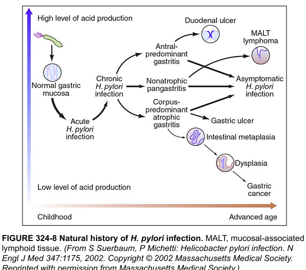
H. pylori natural history and disease progression
Harrison's Principles of Internal Medicine, 21st Edition, p. 8909 — Natural history of H. pylori infection
As shown above, the site and severity of gastritis determines the downstream disease:
PatternConsequence
Antral-predominant gastritisHigh acid → Duodenal ulcer (your patient's pattern)
Corpus-predominant atrophic gastritisLow acid → Gastric ulcer, gastric cancer
PangastritisMALT lymphoma, intestinal metaplasia

Why Your Patient Has Duodenal Erosions

H. pylori in the antrum suppresses somatostatin-producing D cells, which normally inhibit gastrin release. With less somatostatin, gastrin rises → more acid is produced → acid load overwhelms the duodenal buffer → duodenal mucosal erosion and ulceration. This explains the antral gastritis + duodenal bulb erosions seen together in this patient's endoscopy.
This is a shared conversation. Sign in to Orris to start your own chat.