
Wigand fess technique, messenkinger fess technique and other such fess techniques according to standard ENT textbook in simplified manner, also give fess complications in detail
FESS functional endoscopic sinus surgery technique diagram
endoscopic sinus surgery uncinectomy maxillary antrostomy
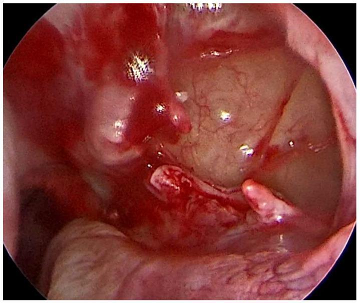
This diagnostic endoscopic clinical photograph captures an intraoperative view of a middle meatal antrostomy performed during Functional Endoscopic Sinus Surgery (FESS). The visual field focuses on the surgically widened natural ostium of the maxillary sinus. Central to the frame is the sinus interior, characterized by a pale, smooth mucosal surface with visible fine vascularity. The periphery shows the margins of the antrostomy, where the tissue appears erythematous, raw, and hemorrhagic due to recent uncinectomy and surgical manipulation. Areas of active bleeding and pooled bright red blood are visible across the mucosal surfaces, reflecting the acute surgical setting. Some fleshy, finger-like projections of inflamed mucosa or polyps are evident near the drainage pathway. The image demonstrates the goal of establishing improved sinus ventilation and drainage while attempting to preserve as much healthy Schneiderian membrane as possible to maintain mucociliary clearance in the treatment of odontogenic sinusitis.
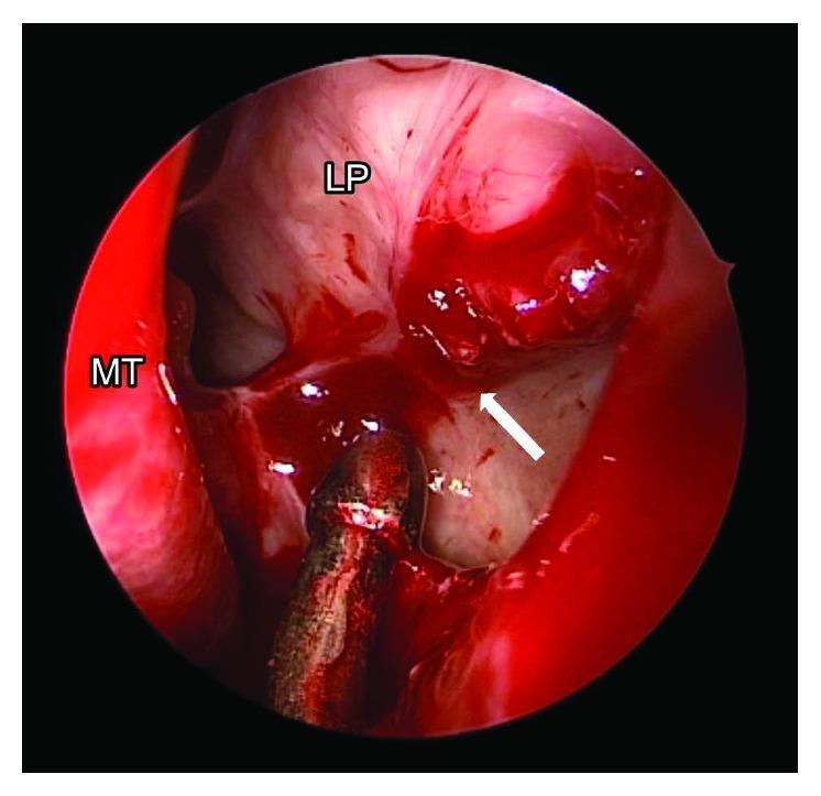
This clinical photograph displays an intraoperative endoscopic view of the left nasal cavity during revision endoscopic sinus surgery (ESS) and uncinectomy. The image captures key anatomical structures including the middle turbinate (MT) and the lamina papyracea (LP). The operative field shows fresh blood and evidence of surgical manipulation. A white arrow points to a distinct bulge in the sinonasal mucosa. This visual finding is significant as it demonstrates a connection to an orbital defect; the bulging occurs when surgical instrumentation is applied through the orbital floor. The image serves as an educational tool for identifying iatrogenic orbital injury and the topographical relationship between the maxillary sinus, ethmoid cavity, and the medial orbital wall. It highlights the importance of precise anatomical localization during antrostomy to avoid injury to the orbital floor and adjacent structures like the inferior rectus muscle.
FESS - Functional Endoscopic Sinus Surgery
Background
The Two Core Techniques
1. Messerklinger Technique (Anterior-to-Posterior Approach)
- Diagnostic nasal endoscopy - Identify landmarks, polyps, pus, anatomical variants
- Uncinectomy - Remove the uncinate process (the key first step; opens access to the infundibulum)
- Maxillary antrostomy - Identify and enlarge the natural maxillary ostium using a 30-45 degree endoscope
- Anterior ethmoidectomy - Open ethmoid bulla and anterior ethmoid cells
- Basal lamella perforation - Enter posterior ethmoid through the inferomedial quadrant of the ground lamella
- Posterior ethmoidectomy - Remove posterior ethmoid cells
- Sphenoidotomy - If needed, via the sphenoethmoidal recess
- Frontal recess - Approached last (most technically difficult)
2. Wigand Technique (Posterior-to-Anterior Approach)
- Sphenoid sinus identification - The sphenoid ostium is located first, medial to the superior turbinate in the sphenoethmoidal recess, at the level of the antral roof
- Posterior ethmoidectomy - Working forward from the sphenoid
- Anterior ethmoidectomy - Continue anteriorly
- Uncinectomy and maxillary antrostomy - Completed last
Comparison Table
| Feature | Messerklinger | Wigand |
|---|---|---|
| Direction | Anterior → Posterior | Posterior → Anterior |
| Starting point | Uncinate process / infundibulum | Sphenoid sinus |
| Landmark anchor | Ostiomeatal complex (OMC) | Sphenoid (fixed reference) |
| Scope of surgery | Targeted (based on disease extent) | Total ethmoidectomy |
| Common use | Primary sinus surgery | Revision / distorted anatomy |
| Mucosal preservation | Strong emphasis | May be more radical |
Stammberger Modification
- Emphasized the importance of mucociliary transport patterns and the role of OMC obstruction in sinus pathogenesis
- Stressed that surgery should be targeted, minimally traumatic, and aimed at restoring physiological drainage - not radical mucosa removal
- Advocated meticulous preservation of the middle turbinate and mucosa
Kennedy Modification (United States)
- The term "Functional Endoscopic Sinus Surgery"
- A more systematic, stepwise approach emphasizing endoscopic visualization with a camera monitor system (rather than looking directly through the scope)
- Emphasis on wide surgical corridors and image-guided surgery
Balloon Sinusotomy (Modern Addition)
- Can be used for maxillary, frontal, or sphenoid ostia
- "Hybrid procedure" when combined with conventional FESS
- Best for mild-moderate disease without polyps or fungal sinusitis
- Avoids mucosal trauma and may reduce scarring
- Contraindicated if there are skull base or orbital dehiscences
Basic Steps of FESS (Universal Sequence)
- Patient positioning (reverse Trendelenburg 10-20 degrees, head turned to surgeon)
- CT review (mandatory - always displayed during surgery)
- Diagnostic nasal endoscopy + topical anesthetic injections
- Medialization of middle turbinate to expose the OMC (optional basal lamella relaxing incision)
- Uncinectomy with 0-degree endoscope
- Maxillary antrostomy - 30 or 45 degree scope; identify floor of orbit, follow medially to lamina papyracea
- Removal of ethmoid bulla; identify lamina papyracea in its medial wall
- Identify basal lamella (horizontal + oblique segments)
- Perforate inferomedial basal lamella → enter posterior ethmoid
- Ethmoidectomy - stay low, between superior turbinate medially and lamina papyracea laterally
- Frontal recess dissection (Draf I / II / III as needed)
- Sphenoidotomy if indicated
- Middle turbinate management (preserve as default; partial resection in severe polyposis only)
Intraoperative Anesthesia and Vasoconstriction
- Pledgets soaked in cocaine/adrenaline (Moffat's solution), co-phenylcaine, or oxymetazoline
- Injection of 1% lidocaine with 1:80,000-1:100,000 adrenaline into the axilla of the middle turbinate, septum, and lateral nasal wall
- Greater palatine block - reduces posterior sinonasal bleeding during posterior ethmoidectomy and sphenoidotomy (38.3% reduction in nasal blood flow with head elevation vs. only 4.7% from palatine injection)
- Allow vasoconstrictors time to work before cutting; systemic epinephrine effects (tachycardia, brief worsening of field) dissipate before mucosal vasoconstriction peaks
FESS Complications
Major Complications
1. Orbital Complications
| Complication | Mechanism | Recognition | Management |
|---|---|---|---|
| Orbital fat herniation | Breach of lamina papyracea | Fat appears in surgical field | Stop surgery, no further manipulation; observe |
| Medial rectus injury | Direct trauma during ethmoidectomy | Prolapse of muscle into field | Urgent ophthalmology; may cause permanent diplopia |
| Orbital haematoma | Bleeding into orbital compartment | Proptosis, periorbital ecchymosis, loss of pupil reactivity | Surgical emergency - lateral canthotomy + cantholysis; decompression |
| Blindness / Vision loss | Optic nerve trauma or vascular injury | Loss of light perception | Ophthalmic emergency; prognosis often poor |
- Resistance to microdebrider that changes with eye pressure (pressing the globe increases resistance)
- Swelling of the eyelid or medial canthus
- Herniation of fat into the field
2. CSF Leak / Skull Base Breach
- Cause: Instrument directed superiorly into the skull base; confusion of skull base with ethmoid partitions; blind use of microdebrider superiorly
- Recognition: Clear watery fluid, "halo sign" on pledgets, positive beta-2-transferrin
- Complication of CSF leak: Ascending meningitis, pneumocephalus
- Management: Small dural tears may be repaired endoscopically with fat/fascia/mucosal grafts and fibrin glue. Large tears may require intracranial repair.
- Prevention: Always perforate the basal lamella in the inferomedial quadrant. Stay low. Review skull base height and asymmetry on preoperative CT (Keros classification of olfactory fossa depth).
3. Intracranial Complications
- Meningitis - from CSF leak with contamination
- Encephalocele / Meningoencephalocele - herniation of brain tissue through dural defect
- Intracranial haemorrhage - from anterior or posterior ethmoidal artery injury with retraction
4. Ethmoidal Artery Injury
- Anterior ethmoidal artery - runs in the supraorbital recess, occasionally within the anterior wall of the ethmoid bulla (can be injured by microdebrider superiorly)
- Posterior ethmoidal artery - lies close to the optic nerve
- Risk: If the artery retracts into the orbit, it can cause an orbital haematoma - a surgical emergency
- Prevention: Identify and avoid the artery; do not blindly use the microdebrider superiorly
Minor / Less Severe Complications
5. Haemorrhage (Intraoperative / Postoperative)
- Most common complication
- Sources: anterior ethmoidal artery, sphenopalatine artery, septal branches
- Management: Bipolar diathermy, hemostatic agents (Surgicel, Gelfoam), nasal packing
- Prevention: Adequate vasoconstriction, controlled hypotension under GA, head-up position, meticulous technique
6. Synechia (Adhesions)
- Middle turbinate lateralizes and adheres to lateral nasal wall → blocks drainage
- Most common cause of surgical failure and need for revision
- Prevention: Preserve middle turbinate attachment, use spacers/stents, meticulous postoperative debridement
- Management: Division in clinic/office under local anesthesia during follow-up
7. Middle Turbinate Destabilization / Loss
- Aggressive manipulation → fracture at the skull base, lateralization, or loss of the turbinate
- Prevention: Gentle medialization; if relaxing incision performed, do it under direct vision
8. Anosmia / Hyposmia
- Injury to olfactory epithelium (roof of olfactory cleft, cribriform area)
- Can be temporary (mucosal edema) or permanent
- Prevention: Avoid manipulation of the olfactory cleft roof; preserve mucosa
9. Nasolacrimal Duct Injury
- During uncinectomy, if the bone anterior to the uncinate (lacrimal bone) is breached
- Presentation: Epiphora (watering eye)
- Prevention: Retrograde uncinectomy stops at the "anterior maxillary line" (junction of uncinate with lacrimal bone). Penetrating harder lacrimal bone = warning sign.
10. Optic Nerve Injury (Onodi Cell)
- An Onodi cell (posterior ethmoid cell) may pneumatize around the optic nerve
- Unrecognized Onodi cell → drilling or instrumentation directly on the optic nerve
- Prevention: Preoperative CT identification of Onodi cells; always know exact location in posterior ethmoid
11. Infection / Toxic Shock Syndrome
- Rare but serious
- Can occur postoperatively, especially with packing
- Prevention: Remove packing within 24-48 hours; perioperative antibiotics in active infection
12. Persistent/Recurrent Sinusitis
- Due to incomplete surgery, scarring, synechia, or residual disease
- Most common "complication" in functional terms
- Requires thorough postoperative care: saline irrigations, topical steroids, office debridement
Intraoperative Orbital Haematoma - Emergency Protocol
- Immediately stop the surgical procedure
- Reverse any controlled hypotension
- Assess eye: increasing proptosis, chemosis, loss of pupillary reaction, visual loss
- Lateral canthotomy and inferior cantholysis - bedside decompression (within minutes)
- IV mannitol and IV dexamethasone
- Emergent ophthalmology consult
- Formal orbital decompression if no improvement
Summary of Complications by Category
| Category | Examples |
|---|---|
| Orbital | Haematoma, fat herniation, medial rectus injury, blindness |
| Intracranial | CSF leak, meningitis, encephalocele, haemorrhage |
| Vascular | Ethmoidal artery injury, epistaxis |
| Functional | Synechia, restenosis, anosmia, epiphora |
| Structural | Middle turbinate destabilization, nasolacrimal injury |
| Infective | Toxic shock syndrome, meningitis |
- Cummings Otolaryngology Head and Neck Surgery - Chapter 44 (Basic Techniques: Messerklinger and Wigand)
- Scott-Brown's Otorhinolaryngology Head and Neck Surgery Vol 1 - Chapter on FESS for rhinosinusitis
- Bailey and Love's Short Practice of Surgery 28th Ed. - Chapter 51 (Management of CRS/polyps)